- Medline/PubMed数据库
- Scopus数据库
- PMC数据库
- CSCD
- 北大核心收录期刊
- 统计源期刊
- 我国高质量科技期刊T1级
- 第6届中国精品科技期刊
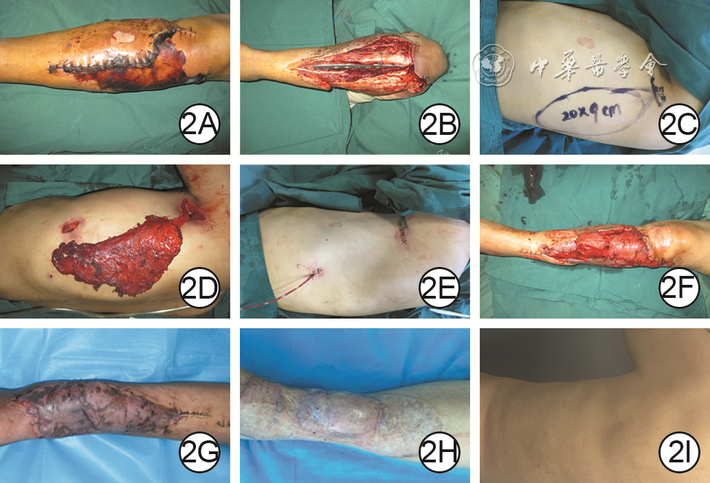
| Citation: | Hou HY,Xie ST,Cao T,et al.Clinical effects of free latissimus dorsi muscle flap harvested through small incision under the assistance of endoscopy in repairing deep wounds in limbs[J].Chin J Burns Wounds,2025,41(8):768-774.DOI: 10.3760/cma.j.cn501225-20240929-00361.

|

| [1] |
ChanPYW,ColonAF,CluneJ,et al.External tissue expansion in complex extremity reconstruction[J].J Hand Surg Am,2021,46(12):1094-1103.DOI: 10.1016/j.jhsa.2021.07.039.
|
| [2] |
MathieuL,PlangS,de l'EscalopierN,et al.Extremity soft tissue coverage in the combat zone: use of pedicled flap transfers by the deployed orthopedic surgeon[J].Mil Med Res,2020,7(1):51.DOI: 10.1186/s40779-020-00281-5.
|
| [3] |
唐举玉,汪华侨,HallockGG,等.关注皮瓣供区问题—减少皮瓣供区损害专家共识[J].中华显微外科杂志,2018,41(1):3-5.DOI: 10.3760/cma.j.issn.1001-2036.2018.01.001.
|
| [4] |
韩军涛,王洪涛,谢松涛,等. 供瓣区选择与修复策略的初步探讨[J].中华烧伤杂志,2020,36(2):85-90. DOI: 10.3760/cma.j.issn.1009-2587.2020.02.002.
|
| [5] |
AkitaS,TokumotoH,YamajiY,et al.Scarless donor site for breast reconstruction by endoscopically assisted extended latissimus dorsi flap plus lipofilling[J].Plast Reconstr Surg,2024,153(6):1209-1219.DOI: 10.1097/PRS.0000000000010698.
|
| [6] |
ChaY,LeeS.Endoscopy-assisted latissimus dorsi muscle flap harvesting technique for immediate breast reconstruction[J].Ann Chir Plast Esthet,2023,68(4):308-314.DOI: 10.1016/j.anplas.2022.09.004.
|
| [7] |
LeeJT,HsuH,LinCM,et al.A comparison study between endoscope-assisted and the standard approach in the harvesting of the free rectus femoris muscle flap[J].Microsurgery,2019,39(1):39-45.DOI: 10.1002/micr.30284.
|
| [8] |
BekenyJC,ZolperEG,SteinbergJS,et al.Free tissue transfer for patients with chronic lower extremity wounds[J].Clin Plast Surg,2021,48(2):321-329.DOI: 10.1016/j.cps.2021.01.004.
|
| [9] |
ProkuskiV,StrohlA.Soft tissue coverage for severe infections[J].Hand Clin,2020,36(3):369-379.DOI: 10.1016/j.hcl.2020.03.011.
|
| [10] |
FilighedduE,ZianiF,ArricaG,et al.Microsurgical reconstruction of extensive lower limb defects: latissimus dorsi free flap for circumferential soft tissue loss following high-energy trauma[J].J Clin Med,2025,14(13):4424.DOI: 10.3390/jcm14134424.
|
| [11] |
邵志恩,巨积辉,祝伟,等.背阔肌皮瓣治疗四肢难治性创面[J].中国美容整形外科杂志,2019,30(7):426-429.DOI: 10.3969/j.issn.1673-7040.2019.07.013.
|
| [12] |
计鹏,曹涛,张智,等.扩张胸背动脉穿支皮瓣游离移植整复烧伤后颈部瘢痕挛缩畸形的临床效果[J].中华烧伤与创面修复杂志,2022,38(4):328-334.DOI: 10.3760/cma.j.cn501120-20211231-00426.
|
| [13] |
EscandónJM,ManriqueOJ,ChristianoJG,et al.Breast reconstruction with latissimus dorsi flap: a comprehensive review and case series[J].Ann Transl Med,2023,11(10):355.DOI: 10.21037/atm-23-469.
|
| [14] |
KangCM,ShimJS,ParkSH,et al.Volume change of muscle and fat portions of latissimus dorsi myocutaneous flap after breast reconstruction[J].Plast Reconstr Surg Glob Open,2021,9(4):e3536.DOI: 10.1097/GOX.0000000000003536.
|
| [15] |
NaallaR,BhattacharyyaS,SahaS,et al.Versatility of the pedicled latissimus dorsi myocutaneous flap in reconstruction of upper limb and trunk soft tissue defects[J].J Hand Microsurg,2020,12(3):168-176.DOI: 10.1055/s-0039-1694293.
|
| [16] |
OngHS,JiT,ZhangCP.The pedicled latissimus dorsi myocutaneous flap in head and neck reconstruction[J].Oral Maxillofac Surg Clin North Am,2014,26(3):427-434.DOI: 10.1016/j.coms.2014.05.011.
|
| [17] |
周健, 郑玉岑, 肖顺娥, 等. 折叠式带蒂背阔肌肌皮瓣在肩背部软组织缺损创面修复的初步应用[J]. 中国修复重建外科杂志,2024,38(1):69-73. DOI: 10.7507/1002-1892.202310013.
|
| [18] |
周健辉,石惠文,王腾彬,等.游离背阔肌双叶皮瓣修复肢体软组织缺损的效果[J].临床骨科杂志,2024,27(3):354-357.DOI: 10.3969/j.issn.1008-0287.2024.03.018.
|
| [19] |
MatsushimaH,IshimineT,TaniguchiN,et al.Treatment of infected thoracic aortic aneurysm with combined abscess debridement and stent-graft wrapping using pedicled latissimus dorsi muscle flaps after thoracic endovascular aortic repair[J].J Cardiothorac Surg,2023,18(1):57.DOI: 10.1186/s13019-023-02155-y.
|
| [20] |
MiuraS,ShichinoheR,IbaY.Successful treatment of recurrent thoracic aortic prosthetic graft infection after omentopexy by free latissimus dorsi and rectus abdominis muscle flap[J].Eur J Cardiothorac Surg,2022,62(4):ezac460.DOI: 10.1093/ejcts/ezac460.
|
| [21] |
张万福,徐婧,胡晓龙,等. 背阔肌肌瓣在电烧伤后肩周肌力重建中的临床应用效果[J].中华烧伤杂志,2021,37(7): 622-628. DOI: 10.3760/cma.j.cn501120-20210329-00107.
|
| [22] |
张盼,李雷,唐林峰,等.不同闭合方式的背阔肌肌皮瓣移植术后供区并发症的分析[J].中国美容整形外科杂志,2024,35(3):133-135.DOI: 10.3969/j.issn.1673-7040.2024.03.002.
|
| [23] |
杜伟力,沈余明,胡骁骅,等.供瓣区美学修复方法的探讨[J].中华烧伤杂志,2020,36(2):97-105.DOI: 10.3760/cma.j.issn.1009-2587.2020.02.004.
|
| [24] |
杨燕文,亓发芝.乳房重建中应用背阔肌肌皮瓣适应证及技术原则[J].中国实用外科杂志,2019,39(11):1161-1164.DOI: 10.19538/j.cjps.issn1005-2208.2019.11.10.
|
| [25] |
冯玉,罗桂林,梁法清,等.腔镜与开放手术获取背阔肌用于乳房重建的比较研究[J].中国普外基础与临床杂志,2025,32(3):293-299.
|
| [26] |
HsiungN,WangXC.Endoscopic-assisted latissimus dorsi muscle flap for chest wall reconstruction in poland syndrome: clinical application and literature review[J].Aesthetic Plast Surg,2025,49(7):1906-1914.DOI: 10.1007/s00266-024-04520-1.
|
| [27] |
EscandónJM,EscandónL,AhmedA,et al.Breast reconstruction using the latissimus dorsi flap and immediate fat transfer (LIFT): a systematic review and meta-analysis[J].J Plast Reconstr Aesthet Surg,2022,75(11):4106-4116.DOI: 10.1016/j.bjps.2022.08.025.
|
| [28] |
LeeJ,JungJH,KimWW,et al.Endoscopy-assisted muscle-sparing latissimus dorsi muscle flap harvesting for partial breast reconstruction[J].BMC Surg,2020,20(1):192.DOI: 10.1186/s12893-020-00853-1.
|
| [29] |
ZhengH,ZhuG,LiX,et al.Partial latissimus dorsi muscle flap with implant for immediate breast reconstruction[J].J Surg Res,2022,269:134-141.DOI: 10.1016/j.jss.2021.08.013.
|
| [30] |
曹晓蔓,梁法清,邬昊,等.背阔肌切取联合腺体切除双腔镜背阔肌乳房重建术技术要点[J].中国实用外科杂志,2024,44(11):1222-1227.DOI: 10.19538/j.cjps.issn1005-2208.2024.11.06.
|
| [31] |
欧阳熠烨,栾杰,刘春军.内窥镜下背阔肌切取术的研究进展[J].中华医学美学美容杂志,2018,24(4):250-252.DOI: 10.3760/cma.j.issn.1671-0290.2018.04.010.
|
| [32] |
马超,陶然,舒军,等.背阔肌肌皮瓣修复较大软组织缺损的方法及供区继发创面的处理[J].中华烧伤杂志,2020,36(12):1199-1203.DOI: 10.3760/cma.j.cn501120-20191121-00439.
|
| [33] |
EscandónJM,NazeraliR,CiudadP,et al.Minimally invasive harvest of the latissimus dorsi flap for breast reconstruction: a systematic review[J].Int J Med Robot,2022,18(6):e2446.DOI: 10.1002/rcs.2446.
|
| [34] |
张盼,李雷,唐林峰,等.改良Constant量表对背阔肌皮瓣供区疗效评价的应用[J].中国美容整形外科杂志,2023,34(3):145-148.DOI: 10.3969/j.issn.1673-7040.2023.03.005.
|
| [35] |
GattoA, ParisiP, BrambillaL, et al. Thoracodorsal artery perforator flap, muscle-sparing latissimus dorsi, and descending branch latissimus dorsi: a multicenter retrospective study on early complications and meta-analysis of the literature[J]. J Plast Reconstr Aesthet Surg, 2022, 75(11):3979-3996. DOI: 10.1016/j.bjps.2022.06.083.
|
| [36] |
WachtelN,GiuntaRE,HellwegM,et al.What about the donor site morbidity - how invasive is the free latissimus dorsi flap?[J].Bone Jt Open,2024,5(12):1114-1119.DOI: 10.1302/2633-1462.512.BJO-2024-0058.R1.
|
| [37] |
夏有辰,李比,陈小迅,等.背阔肌肌皮瓣的血管解剖及其临床应用[J].解剖学报,2020,51(1):93-97.DOI: 10.16098/j.issn.0529-1356.2020.01.016.
|
| [38] |
胡潇丹,徐豪越,李晓,等.超声刀在辅助制备游离腓骨肌皮瓣中的临床应用[J].中国口腔颌面外科杂志,2023,21(2):152-157.DOI: 10.19438/j.cjoms.2023.02.009.
|
| [39] |
赵书明,刘娜,刘学亮,等.彩色多普勒超声辅助下超薄胸背动脉穿支皮瓣的切取方案及临床应用效果[J].中华烧伤与创面修复杂志,2024,40(3):281-288.DOI: 10.3760/cma.j.cn501225-20231012-00111.
|
| [40] |
VourtsisSA,PaspalaA,LykoudisPM,et al.Robotic-assisted harvest of latissimus dorsi muscle flap for breast reconstruction: review of the literature[J].J Robot Surg,2022,16(1):15-19.DOI: 10.1007/s11701-021-01232-5.
|
 侯宏义 8月4日.mp4
|

|

Figures(3)
Copyright © Chinese Journal of Burns京ICP备07035254号-14
E-mail:shaoshangzazhi@163.com
Supported by:
Beijing Renhe Information Technology Co. Ltd


 DownLoad: