2025 Vol. 41, No. 8

Guideline and Consensus
Clinical expert consensus on application of topical growth factors in chronic wounds (2025 edition)
2025, 41(8): 711-724. doi: 10.3760/cma.j.cn501225-20250426-00191
Abstract:
Chronic wounds refer to wounds that fail to achieve complete structural repair within the expected time frame despite standardized systemic treatment, which are characterized by high incidence, complex mechanisms, prolonged clinical course, and difficulty in treatment. Topical growth factor drugs or biological agents, which regulate cell proliferation, migration, and differentiation, thereby effectively promoting the wound healing process, are widely used in the repair of various acute and chronic wounds. However, there is still some controversy over the application of topical growth factors in the clinical practice of treating various chronic wounds. Chinese Burn Association and Editorial Committee of Chinese Journal of Burns and Wounds jointly initiated and organized a multidisciplinary expert group. Based on current evidence-based medical evidence, clinical practice, and authoritative guidelines from other disciplines, this clinical expert consensus was developed for further standardization of the application of topical growth factors in the clinical treatment of chronic wounds.
Expert consensus on enteral nutrition treatment for burned children (2025 edition)
2025, 41(8): 725-733. doi: 10.3760/cma.j.cn501225-20250206-00054
Abstract:
Burns lead to a substantial increase in energy expenditure of body, making timely and effective nutritional support essential for burn management. The metabolic responses to burns in children are significantly different from those in adults, necessitating distinct approaches for nutritional support and treatment. Through processes including literature review, questionnaire survey, and evidence screening, the experts from Pediatric Burn Study Group of Chinese Burn Association, Burn Trauma Professional Committee of Chinese Medical Women's Association, and Pediatric Burn and Plastic Surgery Professional Committee of Beijing Research Society for Human Body Injury Repair conducted in-depth discussions and reached a consensus on enteral nutrition treatment for burned children in terms of nutritional screening, energy calculation, nutritional timing, and nutritional modalities, hoping to provide reference standards for clinical practice.
Expert Forum
Managing refractory wounds in the elderly with the concept of chronic critical illness
Guo Guanghua, Huang Shengyu, Zhu Feng
2025, 41(8): 734-739. doi: 10.3760/cma.j.cn501225-20250314-00130
Abstract:
The skin structure undergoes alterations in the elderly, making the skin soft tissue more susceptible to injury and thereby increasing the risk of developing refractory wounds. Elderly patients exhibit unique pathophysiological characteristics, often accompanied by severe and complex comorbidities (such as organ diseases, psychiatric and psychological issues, geriatric syndromes, etc.) and polypharmacy. Some patients may already be in a state of chronic critical illness for an extended period. Advanced age, refractory wounds, and critical illness often interact clinically, creating a vicious cycle. The management of refractory wounds in elderly patients should be regarded as an essential component of the overall management of geriatric comorbidities. To this end, the author proposes incorporating the concept of chronic critical illness into the management of refractory wounds in the elderly, focusing on the interplay between comorbidities and polypharmacy through multidisciplinary, dynamic, and stratified approaches. While focusing on the diagnosis and treatment of refractory wounds in the elderly, it also takes into account the diagnosis and treatment of geriatric organ diseases, functional maintenance, and actual needs.
Original Article·Elderly and Pediatric Wounds
Repair strategies and efficacy analysis of deep electrical burn wounds in children's fingers
Li Dan, Chen Jigang, Wang Yanni
2025, 41(8): 740-748. doi: 10.3760/cma.j.cn501225-20241031-00421
Abstract:
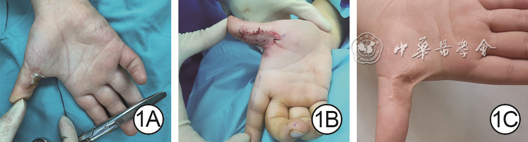
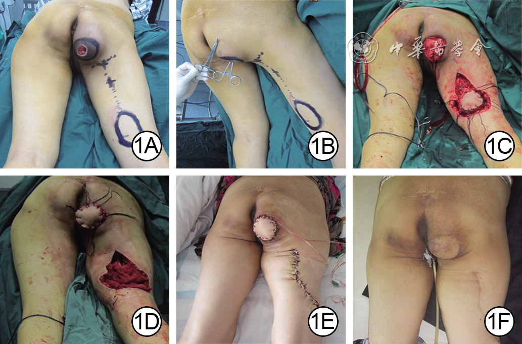
  Objective  To explore the repair strategies for deep electrical burn wounds in children's fingers and analyze their efficacy.  Methods  This study was a retrospective observational study. From January 2008 to January 2024, 80 children with deep electrical burn wounds in fingers meeting the inclusion criteria were admitted to Beijing Children's Hospital Affiliated to Capital Medical University, including 54 males and 26 females, aged 11 months to 12 years and 9 months with a total of 170 fingers affected. Repair strategies were formulated based on wound characteristics after debridement. For wounds with exposed tendons and/or bones, local flaps or abdominal pedicled flaps were preferentially used. For some of the patients who had higher aesthetic requirements for the donor sites, bilayer artificial dermis (AD)+split-thickness skin graft (STSG) was applied for wound repair. For wounds without exposed tendons or bones, full-thickness skin graft (FTSG) was used to repair those with full-thickness skin defects, while STSG alone was employed for wound repair in patients who had higher aesthetic requirements for the donor sites. Bilayer AD+STSG, abdominal pedicled flaps, or local flaps were used to repair the wounds with severe subcutaneous tissue defects located on the volar side of joints. For narrow strip-shaped wounds, direct suturing was performed regardless of tendon and/or bone exposure if tension-free closure was achievable, the incision direction was oblique or parallel to finger creases, and there was no risk of major vascular or nerve injury. The wounds were classified according to whether they were accompanied by tendon and/or bone exposure after debridement, the repair methods, wound area, and survival rate of flaps or skin grafts of the affected fingers were recorded. At the final follow-up, the total active motion (TAM) of affected finger joints was measured with a goniometer to assess function of affected fingers and the excellent and good ratio was calculated, the Vancouver scar scale (VSS) was used to evaluate scar conditions of affected fingers, and a self-designed scale was employed to survey the satisfaction of primary caregivers of the child with treatment outcomes.  Results  Among the 59 affected fingers with wounds accompanied by tendon and/or bone exposure, 31, 9, 10, and 9 of them were treated respectively with abdominal pedicled flaps, local flaps, bilayer AD+STSG, and direct suturing, with the fingers that underwent direct suturing had the smallest wound area of 0.20 (0.20, 0.80) cm2, and the fingers that were transplanted with abdominal pedicled flaps had the largest wound area of 2.00 (2.00, 4.00) cm2. The median survival rate of flaps or skin grafts for the fingers transplanted with abdominal pedicled flaps, local flaps, and bilayer AD+STSG was 90%. The scar VSS score was the lowest 3.0 (2.0, 4.0) for fingers that underwent direct suturing, while the median scar VSS score was 4.0 to 5.0 for fingers that underwent other surgical procedures. The excellent and good ratio of joint TAM was the highest at 9/9 for fingers that underwent direct suturing, followed by 24/31 for fingers that underwent abdominal pedicled flap transplantation and 7/10 for fingers that underwent bilayer AD+STSG transplantation. The median satisfaction score of the primary caregivers of the child with the treatment outcomes was 9.0 points, except for those who underwent local flap transplantation, which was 8 points. Among the 111 affected fingers without tendon or bone exposure, 9, 5, 69, 11, 10, and 7 of them were treated respectively with abdominal pedicled flaps, local flaps, FTSG, STSG alone, bilayer AD+STSG transplantation, and direct suturing, with the fingers that underwent direct suturing had the smallest wound area of 0.25 (0.10, 0.50) cm2, and the fingers that were transplanted with abdominal pedicled flaps had the largest wound area of 2.00 (1.50, 3.00) cm2. The median survival rate of flaps or skin grafts for the fingers transplanted with abdominal pedicled flaps, local flaps, FTSG, and bilayer AD+STSG was 90%. The highest survival rate of skin grafts was 92% (90%, 100%) for fingers that underwent STSG alone transplantation. The scar VSS score was the lowest 3.0 (3.0, 4.0) for fingers that underwent direct suturing, while the scar VSS score was 5.0 (5.0, 7.0) for fingers that underwent bilayer AD+STSG transplantation, which was high. The excellent and good ratio of joint TAM was the highest at 5/5 for fingers that underwent local flap transplantation, while the excellent and good ratio of joint TAM was the lowest at 9/11 for fingers that underwent STSG alone transplantation. The median satisfaction score of the primary caregivers of the child with the treatment outcomes was 9.0 points, except for those who underwent abdominal pedicled flap or local flap transplantation, which was 8.0 points.  Conclusions  For children's fingers with deep electrical burns, different methods can be used for wound repair according to specific circumstances with skin flaps as the preferred for repairing the wounds with exposed tendons and/or bones, skin grafts for repairing the wounds without tendon or bone exposure, and direct suturing for narrow strip-shaped wounds that meet the conditions to obtain the best functional and scar scores. Individualized repair plans can effectively balance the recovery of finger function and the satisfaction of caregivers with the therapeutic outcome.
A multicenter study on the classification and repair effects of stage 4 pressure injury in ischial tuberosity in elderly patients
Chen Lixin, Zhang Xuan, Zhao Zunjiang, Zhang Xiangzhou, Zhang Baode, Lyu Dalun, Duan Longsheng, Peng Kai, Xu Dongwei, Wu Dejin
2025, 41(8): 749-758. doi: 10.3760/cma.j.cn501225-20250421-00185
Abstract:
  Objective  To explore the classification and repair effects of stage 4 pressure injury in ischial tuberosity in elderly patients.  Methods  This study was a retrospective observational study. From January 2021 to December 2022, 45 elderly patients with stage 4 pressure injury in ischial tuberosity of type 4b, 4c, and 4d, who met the inclusion criteria and were admitted to the First Affiliated Hospital of Wannan Medical College, Bengbu Third People's Hospital Affiliated to Bengbu Medical University, and Lu'an Hospital Affiliated to Anhui Medical University (hereinafter referred to as the three hospitals) were selected as the control group, including 28 males and 17 females, aged 60 to 78 years. Fifty elderly patients with stage 4 pressure injury in ischial tuberosity of type 4b, 4c, and 4d, who met the inclusion criteria and were admitted to the three hospitals from January 2023 to December 2024 were selected as the observation group, including 31 males and 19 females, aged 60 to 80 years. The stage 4 pressure injury in ischial tuberosity of patients in observation group were classified on admission, and the targeted surgical methods were selected. For type 4b, the fascial flap was preferred for repair, including fascial flap with skin pedicle (12 cases) and kite flap (14 cases); for type 4c, myocutaneous flap repair was preferred (17 cases); for type 4d, island myocutaneous flap (4 cases) or a combination of muscle flap and flaps (3 cases) was preferred for repair. The surgical methods for repairing the pressure injury of patients in control group were selected based on the surgeon's clinical experience. The type 4b was repaired with fascial flap with skin pedicle in 18 cases and kite flap in 6 cases; the type 4c was repaired with fascial flap with skin pedicle in 6 cases, kite flap in 7 cases, and myocutaneous flap in 3 cases; the type 4d was repaired with kite flap in 2 cases and myocutaneous flap in 3 cases. The sizes of wounds in the two groups of patients were 5.5 cm×4.5 cm to 8.0 cm×7.0 cm after the wound bed was prepared, and the sizes of grafted tissue flaps were 5.0 cm×4.5 cm to 10.0 cm×8.0 cm. The wounds in the flap donor sites were directly sutured or repaired with a relay flap. The following data were recorded, including the wound healing rate after one-time surgical repair, wound healing time, length of hospital stay, hospitalization treatment cost, pressure ulcer scale for healing (PUSH) score at discharge, and recurrence rate of pressure injury during follow-up in two groups of patients.  Results  The wound healing rate after one-time surgical repair of patients in observation group was 88.00% (44/50), which was significantly higher than 66.67% (30/45) in control group (with relative risk of 1.32, 95% confidence interval of 1.08 to 1.61, χ2=6.48, P<0.05). After adjusting the general information using multiple logistic regression analysis, the wound healing rate after one-time surgical repair of patients in observation group was still significantly higher than that in control group (with relative risk of 1.30, 95% confidence interval of 1.05 to 1.59, χ2=6.14, P<0.05). The wound healing time of patients in observation group was (18±5) d, which was significantly shorter than (27±6) d in control group (with mean difference of -9 d, 95% confidence interval of -12 to -7 d, t=7.73, P<0.05). After adjusting the general information using multivariate covariance analysis, the wound healing time of patients in observation group was still significantly shorter than that in control group (with mean difference of -9 d, 95% confidence interval of -11 to -6 d, F=37.10, P<0.05). The length of hospital stay of patients in observation group was significantly shorter than that in control group (t=5.04, P<0.05), the hospitalization treatment cost was significantly lower than that in control group (t=2.11, P<0.05), and the PUSH score at discharge was significantly lower than that in control group (t=3.08, P<0.05). After a follow-up period of 2 to 24 months after discharge, the recurrence rate of pressure injury of patients in observation group was significantly lower than that in control group (χ2=5.02, P<0.05).  Conclusions  Dividing stage 4 pressure injury in ischial tuberosity into multiple subtypes in elderly patients can help guide clinical selection of appropriate repair strategies, increase the wound healing rate after one-time surgical repair, shorten the wound healing time and length of hospital stay, reduce hospitalization treatment cost, and lower the recurrence rate of pressure injury. It is worthy of clinical promotion.
Efficacy of artificial dermis in repairing small to medium-sized deep partial-thickness scalds in infants and young children
Ding Xionghui, Xiao Jun, Li Tianwu, Mao Xiaobo, Zhou Rong, Qiu Lin
2025, 41(8): 759-767. doi: 10.3760/cma.j.cn501225-20250512-00219
Abstract:
  Objective  To investigate the efficacy of artificial dermis in repairing small to medium-sized deep partial-thickness scalds in infants and young children.  Methods  The study was a retrospective observational study. From June 2022 to October 2024, 149 infants and young children with small to medium-sized deep partial-thickness scalds meeting the inclusion criteria were admitted to the Children's Hospital of Chongqing Medical University (hereinafter referred to as our institution). There were 96 males and 53 females, aged from 2 days to 3 years. Based on wound management method, the children were divided into debridement-alone group (n=55), artificial dermis group (n=31), autologous split-thickness skin group (n=36), and combined skin grafting group (n=27). Wounds in the first group of children underwent debridement alone. Wounds in the latter three groups of children were treated with artificial dermis transplantation, autologous split-thickness skin grafts transplantation, or artificial dermis combined with autologous split-thickness skin grafts transplantation, respectively, following debridement. Donor sites for autologous split-thickness skin grafts were the head, back, or lateral thigh. After debridement, wound secretion specimens were collected from the debridement-alone group and artificial dermis group of children for microbial detection, the detection rate was calculated, and wound infection was observed. Wound healing time and length of hospital stay (referring to the length of hospital stay in the Department of Burn and Plastic Surgery of our institution, the same hereafter) were recorded in four groups of children. At the 6-month follow-up after wound healing, the scar (referring to primary wound site scars, the same hereafter) condition of four groups of children was assessed using the modified Vancouver scar scale (mVSS), and both total mVSS score and pliability score were recorded. The total mVSS score of scars was the primary outcome indicator; other indicators were secondary outcome.  Results  After debridement, the microbial detection rate in wound secretion specimens of children in debridement-alone group was 29.1% (16/55), significantly higher than 9.7% (3/31) in artificial dermis group, χ2=4.34, P<0.05. Wound infection occurred in 2 children in debridement-alone group, whereas no wound infection occurred in children in artificial dermis group. Compared with that in debridement-alone group, the wound healing time of children in artificial dermis group, autologous split-thickness skin group, and combined skin grafting group was significantly shortened (P<0.05). Compared with that in artificial dermis group, the wound healing time of children in autologous split-thickness skin group was significantly shortened (P<0.05). Compared with that in autologous split-thickness skin group, the wound healing time of children in combined skin grafting group was significantly prolonged (P<0.05). Compared with that in debridement-alone group, the length of hospital stay of children in artificial dermis group and autologous split-thickness skin group was significantly shortened (P<0.05). Compared with that in artificial dermis group, the length of hospital stay of children in autologous split-thickness skin group was significantly shortened (P<0.05), the length of hospital stay of children in combined skin grafting group was significantly prolonged (P<0.05). Compared with that in autologous split-thickness skin group, the length of hospital stay of children in combined skin grafting group was significantly prolonged (P<0.05). At the 6-month follow-up after wound healing, the total mVSS score of scars of children in debridement-alone group was 8.1±1.1, significantly higher than 6.8±0.9 in artificial dermis group (with mean difference of 1.3, 95% confidence interval of 0.8 to 1.8, P<0.05), and significantly lower than 9.4±1.5 in autologous split-thickness skin group (with mean difference of -1.3, 95% confidence interval of -1.8 to -0.8, P<0.05). Compared with that in artificial dermis group, the total mVSS score of children in autologous split-thickness skin group was significantly increased (with mean difference of -2.6, 95% confidence interval of -3.2 to -2.0, P<0.05). Compared with that in autologous split-thickness skin group, the 7.7±1.0 of total mVSS score of children in combined skin grafting group was significantly decreased (with mean difference of 1.7, 95% confidence interval of 1.1 to 2.3, P<0.05). Compared with that in debridement-alone group, the scar pliability score of children in artificial dermis group was significantly decreased (P<0.05). Compared with that in artificial dermis group, the scar pliability score of children in autologous split-thickness skin group was significantly increased (P<0.05). Compared with that in autologous split-thickness skin group, the scar pliability score of children in combined skin grafting group was significantly decreased (P<0.05).  Conclusions  Artificial dermis used for the repair of small to medium-sized deep partial-thickness scald wounds in infants and young children can improve scar quality, reduce the risk of wound infection, and relatively shorten wound healing time. It is particularly suitable for infants and young children whose aesthetic and functional outcomes are prioritized.
Original Article
Clinical effects of free latissimus dorsi muscle flap harvested through small incision under the assistance of endoscopy in repairing deep wounds in limbs
Hou Hongyi, Xie Songtao, Cao Tao, He Xiang, Zhang Nan, Guan Hao
2025, 41(8): 768-774. doi: 10.3760/cma.j.cn501225-20240929-00361
Abstract:
  Objective  To investigate the clinical effects of free latissimus dorsi muscle flap harvested through small incision under the assistance of endoscopy in repairing deep wounds in limbs.  Methods  This study was a retrospective observational study. From March 2015 to March 2024, 38 patients with deep wounds in limbs, who met the inclusion criteria, were admitted to Xijing Hospital of Air Force Medical University, including 26 males and 12 females, aged 19 to 64 years. The wounds were located in the lower leg in 21 cases, ankle in 9 cases, knee joint in 3 cases, and forearm in 5 cases. After debridement, the wound area required for muscle flap transplantation was 11.0 cm×9.0 cm to 28.0 cm×13.0 cm. The free latissimus dorsi muscle flaps were harvested through a small horizontal incision of 4-5 cm in length along the dermatoglyph in the armpit combined with a secondary incision of about 1 cm in length at the posterior axillary line under the assistance of endoscopy, with area of the muscle flap of 12.0 cm×10.0 cm to 29.0 cm×14.0 cm and length of vascular pedicle of 6-8 cm. The split-thickness skin grafts were harvested to cover the muscle flaps and the residual wounds. The harvesting time and the amount of blood loss during the harvesting process of the free latissimus dorsi muscle flap were recorded. The survivals of the transplanted muscle flap and skin graft, wound healing, and complications in the flap donor and recipient areas were observed after surgery. During follow-up, the appearance in the recipient area, the scar condition in the flap donor area, and the function of the upper limb on the flap donor side were observed.  Results  The time for harvesting the free latissimus dorsi muscle flap was 160-200 minutes, with an average of 185 minutes. The amount of blood loss during the harvesting process of the muscle flap was 10-30 mL, with an average of 25 mL. Two patients developed complications in the recipient area after surgery. Among them, one patient experienced vascular crisis, the muscle flap recovered blood flow after emergency surgery to remove the thrombus and re-anastomosis of the vein, and ultimately the wound healed well; one patient had an infection at the edge of the muscle flap, and the wound healed after treatment such as debridement and dressing change. The transplanted muscle flaps and skin grafts completely survived in the remaining 36 patients. Two patients developed cystic effusion in the flap donor area about one week after surgery, and the flap donor area recovered well after puncture drainage and pressure bandaging treatment three weeks after surgery; the remaining patients had no complications such as incision infection or rupture in the flap donor area. Follow up for 6 to 18 months showed that the transplanted muscle flap and skin graft in the recipient area had good appearance, the scar in the donor area was concealed and mild, and the upper limb movement on the flap donor side was not restricted.  Conclusions  Under the assistance of endoscopy, the free latissimus dorsi muscle flap can be harvested through a small horizontal incision along the dermatoglyph in the armpit, with small damage to the donor area, fast postoperative recovery, mild and concealed scar. It is in line with the goal of minimally invasive surgery and worthy of promotion and application.
Effects and mechanism of electroacupuncture stimulation on the survival of multi-territory perforator flaps in rats
Huo Lei, Xie Jianing, Tan Qi
2025, 41(8): 775-782. doi: 10.3760/cma.j.cn501225-20240401-00115
Abstract:
  Objective  To explore the effects and mechanism of electroacupuncture stimulation on the survival of multi-territory perforator flaps in rats.  Methods  This study was an experimental study. Thirty male Sprague-Dawley rats aged 8-10 weeks were collected and divided into electroacupuncture stimulation group and control group according to the random number table method, with 15 rats in each group. Doppler blood flow detectors were used to explore the positions of the dorsal deep circumflex artery, posterior intercostal artery, and thoracodorsal artery in the two groups of rats, and a multi-territory perforator flap was designed and resected with the dorsal deep circumflex artery as the pedicle after ligation of the posterior intercostal artery and thoracodorsal artery, and the flap was replanted in situ. Before the operation, the skin in the area containing choke vessels at the junction between the angiosomes of the thoracodorsal artery and posterior intercostal artery (i.e. the choke zone Ⅱ) in the flaps of rats in electroacupuncture stimulation group was subjected to electroacupuncture stimulation for 1 hour per day for 7 consecutive days, while the flaps of rats in control group received no electroacupuncture stimulation. Seven days after the operation, the survival status of the flaps of all rats in the two groups was observed and the flap survival rate was calculated; the skin tissue from the choke zone Ⅱ was collected and stained with hematoxylin-eosin to observe the microvascular neogenesis and calculate the microvessel density (with sample number of 3). Immunohistochemical staining was performed to observe the expression and distribution of vascular endothelial growth factor (VEGF), and Western blotting was used to detect the protein expression of VEGF (with sample number of 3).  Results  Seven days after the operation, the flaps of rats in control group were partially blackened and necrotic, while those in electroacupuncture stimulation group survived with almost no necrosis. The flap survival rate of rats in electroacupuncture stimulation group was (92.1±2.1)%, which was significantly higher than (85.2±1.2)% in control group (t=-10.95, P<0.05). Seven days after the operation, compared with those in control group, the number of new microvessels in the skin tissue in the choke zone Ⅱ of the flaps in rats of electroacupuncture stimulation group increased significantly. The microvessel density in the skin tissue in the choke zone Ⅱ of the flaps in rats of electroacupuncture stimulation group was (21.4±3.0) vessels/mm², which was significantly higher than (11.5±3.7) vessels/mm² in control group (t=-7.34, P<0.05). Seven days after the operation, compared with those in control group, the expression of VEGF in the vascular area of the skin tissue in the choke zone Ⅱ of the flaps in rats of electroacupuncture stimulation group was significantly increased, and the protein expression of VEGF was significantly increased (t=12.56, P<0.05).  Conclusions  Electroacupuncture stimulation can increase the expression of VEGF in choke zone Ⅱ of the multi-territory perforator flaps in rats, improve the blood supply at the distal end of flaps through promoting morphological changes of blood vessels in this zone, thus increasing the survival rates of flaps.
Effects and mechanism of retinoic acid on radiation-induced skin injury in mice
Tian Kai, Yi Yuling, Xu Wushuang, Jia Zou, Wu Pingfan, Sheng Lei, Sun Wei, Zhou Xiaozhong, Wu Lijun
2025, 41(8): 783-792. doi: 10.3760/cma.j.cn501225-20240420-00145
Abstract:
  Objective  To investigate the effects and mechanism of retinoic acid on radiation-induced skin injury (RSI) in mice.  Methods  This study was an experimental research. HaCaT cells were obtained and divided into control group (routinely cultured), injury group, treatment group, and antagonism group. The cells in the latter three groups were all exposed to 10 Gy X-ray radiation. The cells in the latter two groups were pretreated with retinoic acid for 12 h before radiation, and the cells in the last group were pre-treated with polyinosinic-polycytidylic acid for 1 h before radiation. Cells from the three irradiated groups at 24 h after radiation and cells from control group at the corresponding time point were collected to detect the reactive oxygen species (ROS) level in the cells by flow cytometry, and measure the protein expressions of interleukin-6 (IL-6), tumor necrosis factor-α (TNF-α), Toll-like receptor 3 (TLR3), and nuclear factor-κB (NF-κB) in the cells by Western blotting, with the sample number being 3. Twenty-four 6-week-old female BALB/c mice were obtained and divided into control group, injury group, treatment group, and antagonism group according to the random number table method (with 6 mice in each group). The right lower limbs of mice in the latter three groups were all exposed to 35 Gy electron beam radiation to induce RSI. Mice in the latter two groups were treated with retinoic acid at 0 (immediately), 7, 14, 21, 28, 35, and 42 days after injury, while mice in the last group were pre-treated with polyinosinic-polycytidylic acid at these time points. Mice in control group were simulated with sham injury. At 42 days after injury, the wound healing rate was calculated, and blood perfusion (denoted as blood flow index) in the skin tissue at the injury site (i.e. wound tissue) was detected by laser speckle flowmetry; wound tissue was collected, the hematoxylin-eosin staining was performed to count inflammatory cells and measure epidermal thickness, the immunohistochemical staining was performed to detect the expression of IL-6 and TNF-α, the immunofluorescence staining was performed to detect the expression of TLR3, and Western blotting was performed to detect the protein expressions of TLR3 and NF-κB.  Results  At 24 h after radiation, the ROS level and protein expressions of IL-6, TNF-α, TLR3, and NF-κB of cells in injury group were significantly higher than those in control group at the corresponding time point (P<0.05). The ROS level and protein expressions of IL-6, TLR3, and NF-κB of cells in treatment group were significantly lower than those in both injury group and antagonism group at 24 hours after radiation (P<0.05). At 42 days after injury, the wound healing rates of mice in control group, injury group, treatment group, and antagonism group were (100.4±2.7)%, (77.5±2.5)%, (89.8±3.2)%, and (70.1±4.8)%, respectively. The wound healing rate of mice in treatment group was significantly higher than that in injury group and antagonism group (both P values <0.05). At 42 days after injury, the blood flow index in the wound tissue of mice in treatment group was significantly lower than that in injury group and antagonism group (both P values <0.05). At 42 days after injury, compared with those in control group, the number of inflammatory cells in the wound tissue of mice in injury group was significantly increased, and the epidermal thickness significantly thickened (P<0.05); compared with those in treatment group, the number of inflammatory cells in the wound tissue of mice in injury group and antagonism group was significantly increased, and the epidermal thickness significantly thickened (P<0.05). At 42 days after injury, the expression levels of IL-6, TNF-α, and TLR3, as well as the protein expressions of TLR3 and NF-κB in the wound tissue of mice in injury group were significantly higher than those in control group (P<0.05), while the expression levels of IL-6, TNF-α, and TLR3, as well as the protein expressions of TLR3 and NF-κB in the wound tissue of mice in treatment group were significantly lower than those in injury group and antagonism group (P<0.05).  Conclusions  Retinoic acid significantly reduces radiation-induced cell injury and promotes the repair of RSI in mice by inhibiting the TLR3/NF-κB signaling pathway and the expression of downstream inflammatory factors.
Effects and mechanism of tannic acid/magnesium nanocomplex on wound healing in rats with full-thickness scald
Li Junda, Jia Jiezhi, Chen Yu, Chen Yajie, Zhang Lijuan, Zuo Fangqing, Yu Yunlong, Yuan Zhiqiang
2025, 41(8): 793-802. doi: 10.3760/cma.j.cn501225-20250126-00041
Abstract:
  Objective  To investigate the effects and mechanism of tannic acid/magnesium nanocomplex (MgTA NC) on wound healing in rats with full-thickness scald.  Methods  This study was an experimental study. The MgTA NC with good biocompatibility was synthesized using the hydrothermal method. Mouse RAW 264.7 cells were divided into endotoxins/lipopolysaccharides (LPS) alone group, low MgTA NC group, medium MgTA NC group, and high MgTA NC group, which were all treated with LPS at final mass concentration of 1 μg/mL, and then cultured respectively with MgTA NC at the final mass concentration of 0 (without), 2.5, 5.0, or 7.5 μg/mL for 24 hours. The protein expressions of M1 type macrophage marker inducible nitric oxide synthase (iNOS), M2 type macrophage marker CD163, as well as glycolysis metabolism-related proteins pyruvate kinase type M2 (PKM2) and hexokinase in cells were detected by Western blotting; the expression levels of succinate dehydrogenase and isocitrate dehydrogenase in cells were detected by enzyme-linked immunosorbent assay method. The sample size in cell experiment was 3. Twelve six-week-old male Sprague-Dawley rats were selected and subjected to full-thickness scald on their backs using a temperature-controlled electrothermal burn device. The rats were assigned to control group, simple hydrogel group, and composite hydrogel group according to the random number table method, with 4 rats in each group. The wounds were treated with phosphate buffered saline, methacrylated gelatin (GelMA) hydrogel, or GelMA hydrogel loaded with MgTA NC, respectively. The wound healing rates were calculated at post-injury day 3, 7, and 14 (with the sample size of 4), and the expression level of inflammatory factor tumor necrosis factor α (TNF-α) in the wound tissue at post-injury day 14 was detected by immunohistochemical staining.  Results  After 24 hours of culture, the protein expressions of iNOS, the M1 type macrophage marker in RAW 264.7 cells decreased successively in LPS alone group, low MgTA NC group, medium MgTA NC group, and high MgTA NC group, while the protein expressions of CD163, the M2 type macrophage marker increased successively, and the protein expressions of PKM2 and hexokinase decreased successively. After 24 hours of culture, compared with those in LPS alone group, the expression levels of succinate dehydrogenase and isocitrate dehydrogenase in RAW 264.7 cells in low MgTA NC group were significantly increased (P<0.05); compared with those in low MgTA NC group, the expression levels of succinate dehydrogenase and isocitrate dehydrogenase in RAW 264.7 cells in medium MgTA NC group were significantly increased (P<0.05); compared with those in medium MgTA NC group, the expression levels of succinate dehydrogenase and isocitrate dehydrogenase in RAW 264.7 cells in high MgTA NC group were significantly increased (P<0.05). At post-injury day 3, there was no statistically significant difference in the wound healing rate among the three groups of rats (P>0.05). At post-injury day 7 and 14, the wound healing rates of rats in simple hydrogel group were (52.28±5.11)% and (81.11±2.09)%, and those in composite hydrogel group were (76.81±2.68)% and (98.93±0.29)%, which were significantly higher than (32.75±6.86)% and (60.10±2.10)% in control group, respectively (P<0.05); the wound healing rates of rats in composite hydrogel group were significantly higher than those in simple hydrogel group (P<0.05). At post-injury day 14, the expression of TNF-α in the wound tissue of rats in simple hydrogel group and composite hydrogel group was significantly reduced compared with that in control group, and the expression of TNF-α in the wound tissue of rats in composite hydrogel group was significantly reduced compared with that in simple hydrogel group.  Conclusions  MgTA NC exhibits excellent biocompatibility, and it is capable of modulating macrophage polarization toward the M2 type, effectively reducing glycolysis level and enhancing oxidative phosphorylation level of macrophages, suppressing excessive inflammatory responses and enhancing the ability of tissue regeneration and repair, therefore significantly accelerating wound healing in rats with full-thickness scald.
Interpretation of Guideline
Interpretation of the 2023 International Society for Burn Injuries "Surviving Sepsis After Burn Campaign"
Zhu Wei, Peng Yizhi, Luo Gaoxing, Yuan Zhiqiang
2025, 41(8): 803-810. doi: 10.3760/cma.j.cn501225-20250403-00164
Abstract:
The Surviving Sepsis After Burn Campaign published by the International Society for Burn Injuries in 2023, are the first international guidelines for the management of sepsis in burn patients. The guidelines systematically integrate the strategies for the diagnosis, prevention, and treatment of burn sepsis for the first time, encompassing the whole processes of sepsis management from early warning indicators (such as dynamic changes in sepsis-related organ failure assessment scores and procalcitonin trends) to personalized interventions (such as wound management and antibiotic de-escalation therapy). More importantly, the guidelines emphasize an organ function-oriented dynamic assessment framework, breaking through the limitations of traditional biomarkers and put forward innovative recommendations specifically for the hypermetabolic state, wound infections, and risks of multidrug-resistant organisms infections uniquely associated with burn sepsis. Based on the progress of international research on burn sepsis, this article provides an in-depth analysis of the guidelines' key points, offering evidence-based insights for clinical practice.