- Medline/PubMed数据库
- Scopus数据库
- PMC数据库
- CSCD
- 北大核心收录期刊
- 统计源期刊
- 我国高质量科技期刊T1级
- 第6届中国精品科技期刊
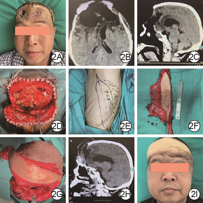
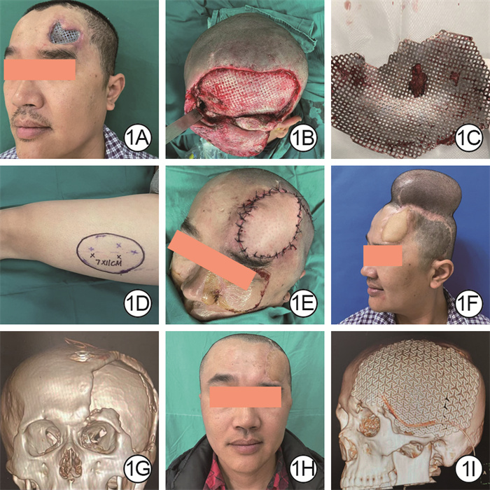
| Citation: | Wang Mengna, Liang Pengfei, Bi Changlong, et al. Repair methods for refractory head wounds involving intracranial structures and their clinical effectiveness[J]. CHINESE JOURNAL OF BURNS AND WOUNDS, 2025, 41(6): 525-533. Doi: 10.3760/cma.j.cn501225-20250106-00008

|

| [1] |
Hutchinson PJ, Adams H, Mohan M, et al. Decompressive craniectomy versus craniotomy for acute subdural hematoma[J]. N Engl J Med, 2023, 388(24): 2219-2229. DOI: 10.1056/NEJMoa2214172.
|
| [2] |
Wang H, Li N, Bao Q, et al. Role of plastic surgery in the treatment of titanium mesh exposure following cranioplasty[J]. J Craniofac Surg, 2024, 35(4): 1080-1083. DOI: 10.1097/SCS.0000000000010145.
|
| [3] |
Zhang P, Fu X, Huang Y, et al. Consensus on the preventionand repair of titanium mesh exposed wound after cranioplasty (2024 edition)[J/OL].Burns Trauma, 2024, 12: tkae055[2025-01-06].
|
| [4] |
杨力, 蔡斌, 薛君荣, 等. 个体化股前外侧皮瓣游离移植修复复杂难愈性创面的临床效果[J]. 中华烧伤杂志, 2020, 36(8): 730-734. DOI: 10.3760/cma.j.cn501120-20190621-00281.
|
| [5] |
Zheng Z, Liu H, Liu S, et al. Mesenchymal stem cells in craniofacial reconstruction: a comprehensive review[J]. Front Mol Biosci, 2024, 11: 1362338. DOI: 10.3389/fmolb.2024.1362338.
|
| [6] |
Yang YH, Jeng SF, Hsieh CH, et al. Vacuum-assisted closure for complicated wounds in head and neck region after reconstruction[J]. J Plast Reconstr Aesthet Surg, 2013, 66(8): e209-216. DOI: 10.1016/j.bjps.2013.03.006.
|
| [7] |
尚银武, 王飞, 刘建雄, 等. 高压电击伤致头皮缺损及脑脓肿1例[J]. 中国神经精神疾病杂志, 2022, 48(2): 105-108. DOI: 10.3969/j.issn.1002-0152.2022.02.008.
|
| [8] |
Dechaene V, Gallet C, Soueges S, et al. Diagnostic, clinical management, and outcome of bone flap-related osteomyelitis after cranioplasty[J]. Int J Infect Dis, 2023, 137: 48-54. DOI: 10.1016/j.ijid.2023.10.008.
|
| [9] |
Zhao YH, Feng YH, Deng HT, et al. Therapeutic strategies for retention of cranioplasty titanium mesh after mesh exposure[J]. Acta Neurochir (Wien), 2022, 164(12): 3101-3106. DOI: 10.1007/s00701-022-05365-w.
|
| [10] |
中国医师协会创面修复专业委员会. 颅骨成形术后钛网外露预防和创面修复全国专家共识(2024版)[J]. 中华烧伤与创面修复杂志, 2024, 40(10): 901-910. DOI: 10.3760/cma.j.cn501225-20240203-00047.
|
| [11] |
Yu LB, Huang Z, Ren ZG, et al. Supraorbital keyhole versus pterional craniotomies for ruptured anterior communicating artery aneurysms: a propensity score-matched analysis[J]. Neurosurg Rev, 2020, 43(2): 547-554. DOI: 10.1007/s10143-018-1053-y.
|
| [12] |
梁鹏飞, 许喜生, 张丕红, 等. 累及鼻窦的面部复杂缺损创面的修复方法及其临床效果[J]. 中华烧伤与创面修复杂志, 2023, 39(3): 221-227. DOI: 10.3760/cma.j.cn501225-20221130-00520.
|
| [13] |
Liao JB, Chen W, Lee HS, et al. Histopathology of fluoroscopy-induced radiation ulcer: a case series study in comparison with morphea[J]. J Dtsch Dermatol Ges, 2020, 18(5): 447-454. DOI: 10.1111/ddg.14092.
|
| [14] |
Zhou Y, Zhang Y. Single- versus 2-stage reconstruction for chronic post-radiation chest wall ulcer: a 10-year retrospective study of chronic radiation-induced ulcers[J]. Medicine (Baltimore), 2019, 98(8): e14567. DOI: 10.1097/MD.0000000000014567.
|
| [15] |
Zhou B, Long Y, Li S, et al. Reconstruction of chronic radiation-induced ulcers in the chest wall using free and pedicle flaps[J]. Front Surg, 2022, 9: 1010990. DOI: 10.3389/fsurg.2022.1010990.
|
| [16] |
Dong W, Zhang X, Luo X, et al. Regional flap: a reliable coverage for post-radiation ulcer[J]. Int Wound J, 2023, 20(6): 2224-2232. DOI: 10.1111/iwj.14103.
|
| [17] |
Yamamura K, Endo Y, Kabashima K. Occipital artery island V-Y flap for the reconstruction of temporal scalp defect[J]. Int J Dermatol, 2020, 59(8): e296-e298. DOI: 10.1111/ijd.14865.
|
| [18] |
Ranjan K, Venkataramu V, Achanti HP, et al. The role of pedicled latissimus dorsi flap in scalp defect reconstruction following tumour excision[J]. Indian J Otolaryngol Head Neck Surg, 2021, 73(1): 129-132. DOI: 10.1007/s12070-020-02071-w.
|
| [19] |
毛小炎, 归来. 颅骨缺损的临床修复进展[J]. 中国美容整形外科杂志, 2017, 28(3): 184-186. DOI: 10.3969/j.issn.1673-7040.2017.03.019.
|
| [20] |
Soto E, Restrepo RD, Grant JH 3rd, et al. Outcomes of cranioplasty strategies for high-risk complex cranial defects: a 10-year experience[J]. Ann Plast Surg, 2022, 88(5 Suppl 5): S449-454. DOI: 10.1097/SAP.0000000000003019.
|
| [21] |
Kwiecien GJ, Aliotta R, Bassiri Gharb B, et al. The timing of alloplastic cranioplasty in the setting of previous osteomyelitis[J]. Plast Reconstr Surg, 2019, 143(3): 853-861. DOI: 10.1097/PRS.0000000000005363.
|
| [22] |
王蕾, 张毅. 现代颅骨修补材料的临床应用分析[J]. 神经损伤与功能重建, 2018, 13(7): 355-357. DOI: 10.16780/j.cnki.sjssgncj.2018.07.009.
|
| [23] |
Yeap MC, Tu PH, Liu ZH, et al. Long-term complications of cranioplasty using stored autologous bone graft, three-dimensional polymethyl methacrylate, or titanium mesh after decompressive craniectomy: a single-center experience after 596 procedures[J]. World Neurosurg, 2019, 128: e841-e850. DOI: 10.1016/j.wneu.2019.05.005.
|
| [24] |
Azzam D, Romiyo P, Nguyen T, et al. Dural repair in cranial surgery is associated with moderate rates of complications with both autologous and nonautologous dural substitutes [J]. World Neurosurg, 2018, 113: 244-248. DOI: 10.1016/j.wneu.2018.01.115.
|
| [25] |
陈政源, 寿雪飞, 沈明, 等. 神经内镜经鼻入路切除颅底肿瘤术中颅底重建的临床疗效[J]. 中华神经外科杂志, 2020, 36(1): 2-6. DOI: 10.3760/cma.j.issn.1001-2346.2020.01.002.
|
| [26] |
Chen W, Wang Y, Zheng J, et al. Characterization of cellular senescence in radiation ulcers and therapeutic effects of mesenchymal stem cell-derived conditioned medium [J/OL]. Burns Trauma, 2023, 11: tkad001[2025-01-06].
|
| [27] |
Li X, Zhang F, Liu X, et al. Staged treatment of chest wall radiation-induced ulcer with negative pressure wound therapy and latissimus dorsi myocutaneous flap transplantation[J]. J Craniofac Surg, 2019, 30(5): e450-e453. DOI: 10.1097/SCS.0000000000005514.
|
| [28] |
Hamada M, Nakahara T, Yazawa M, et al. Radiation-induced osteomyelitis/osteonecrosis of the rib: SPECT/CT imaging for successful surgical management[J]. Plast Reconstr Surg Glob Open, 2019, 7(12): e2536. DOI: 10.1097/GOX.0000000000002536.
|
| [29] |
Borrelli MR, Shen AH, Lee GK, et al. Radiation-induced skin fibrosis: pathogenesis, current treatment options, and emerging therapeutics[J]. Ann Plast Surg, 2019, 83(4S Suppl 1): S59-64. DOI: 10.1097/SAP.0000000000002098.
|
| [30] |
刘鑫, 韩愚弟, 崔蕾, 等. 分阶段游离背阔肌皮瓣移植及颅骨轮廓重建治疗头部钛网外露合并软组织感染[J]. 中国修复重建外科杂志, 2022, 36(7): 828-833. DOI: 10.7507/1002-1892.202202061.
|
| [31] |
郭鹏飞, 王旭, 魏爱周, 等. 基于供区保护理念的游离股前外侧分叶穿支皮瓣在头部电烧伤创面修复中的临床应用效果[J]. 中华烧伤与创面修复杂志, 2022, 38(1): 77-80. DOI: 10.3760/cma.j.cn501120-20201111-00470.
|
| [32] |
于峻懿, 宋达疆, 刘旭, 等. 组合组织瓣移植修复巨大胸壁缺损的临床效果[J]. 中华烧伤与创面修复杂志, 2024, 40(7): 650-656. DOI: 10.3760/cma.j.cn501225-20231120-00199.
|
| [33] |
李文涛, 董中洋, 刘伟, 等. 串联组合皮瓣修复前足负重区及周围大面积软组织缺损[J]. 中华手外科杂志, 2023, 39(2): 147-149. DOI: 10.3760/cma.j.cn311653-20220907-00234.
|
| [34] |
车永琦, 张伟, 程芳斌, 等. 嵌合股前外侧肌皮瓣修复伴有空腔的足部创面10例[J]. 中华显微外科杂志, 2023, 46(2): 190-192. DOI: 10.3760/cma.j.cn441206-20220613-00117.
|
| [35] |
宋达疆, 彭文, 李赞, 等. 股内侧嵌合穿支肌皮瓣的解剖分类和在头颈重建领域的应用[J]. 中华耳鼻咽喉头颈外科杂志, 2020, 55(5): 483-489. DOI: 10.3760/cma.j.cn115330-20190711-00436.
|
| [36] |
Dong L, Dong Y, Liu C, et al. Latissimus dorsi-myocutaneous flap in the repair of titanium mesh exposure and scalp defect after cranioplasty[J]. J Craniofac Surg, 2020, 31(2): 351-354. DOI: 10.1097/SCS.0000000000006016.
|
| [37] |
Vargo JD, Przylecki W, Camarata PJ, et al. Classification and microvascular flap selection for anterior cranial fossa reconstruction[J]. J Reconstr Microsurg, 2018, 34(8): 590-600. DOI: 10.1055/s-0038-1649520.
|
| [38] |
Vuola J, Ohman J, Mäkitie AA. Microvascular free flap reconstruction of skull base penetrating tumors[J]. J Reconstr Microsurg, 2011, 27(5): 313-320. DOI: 10.1055/s-0031-1278715.
|
| [39] |
Nwaba A, Ho A, Ellis MF. Microvascular reconstruction of the anterior skull base[J]. J Craniofac Surg, 2022, 33(8): e886-e890. DOI: 10.1097/SCS.0000000000008930.
|
| [40] |
Neamonitou F, Kotrotsiou M, Stavrianos S. Microvascular reconstruction of the anterior skull base tumors; our experience[J]. J Plast Reconstr Aesthet Surg, 2021, 74(6): 1355-1401. DOI: 10.1016/j.bjps.2020.11.036.
|
| [41] |
Han DH, Park MC, Park DH, et al. Role of muscle free flap in the salvage of complicated scalp wounds and infected prosthetic dura[J]. Arch Plast Surg, 2013, 40(6): 735-741. DOI: 10.5999/aps.2013.40.6.735.
|
| [42] |
Golub VM, Reddy DS. Post-traumatic epilepsy and comorbidities: advanced models, molecular mechanisms, biomarkers, and novel therapeutic interventions[J]. Pharmacol Rev, 2022, 74(2): 387-438. DOI: 10.1124/pharmrev.121.000375.
|
| [43] |
Bader ER, Kobets AJ, Ammar A, et al. Factors predicting complications following cranioplasty[J]. J Craniomaxillofac Surg, 2022, 50(2): 134-139. DOI: 10.1016/j.jcms.2021.08.001.
|
 王梦娜 6月3日.mp4
|

|
Figures(2)
Copyright © Chinese Journal of Burns京ICP备07035254号-14
E-mail:shaoshangzazhi@163.com
Supported by:
Beijing Renhe Information Technology Co. Ltd


 DownLoad: