2025 Vol. 41, No. 6

Expert Forum
Application of iceberg theory in the diagnosis and treatment of refractory wounds at the surgical site after craniotomy
Zhang Pihong, Zhang Yikun, Liang Pengfei
2025, 41(6): 509-515. doi: 10.3760/cma.j.cn501225-20250119-00028
Abstract:
After craniotomy, the surgical site is prone to develop refractory wounds, which seriously affects the patient's quality of life and significantly increases the medical cost. Clinical studies indicate that the pathogenic factors of such wounds are extremely diverse, their formation mechanism is complex, and their clinical treatment is extremely difficult. In this paper, iceberg analysis is applied to the diagnosis and treatment of refractory wounds after craniotomy. Based on domestic and international literature and clinical practice, combined with preoperative image examination results and intraoperative exploration findings, we provided an in-depth discussion on the causes of wound formation, comprehensively analyzed the pathogenic factors affecting wound healing, and shared the clinical diagnosis and treatment experience in this aspect. Based on the iceberg theory, we propose precise debridement measures and targeted effective repair strategies for this type of refractory wounds.
An overview of integrated surgical management for secondary lower limb lymphedema guided by algorithms
Deng Chengliang, Chen Junzhe, Xiao Shun'e, Wu Xiangkui, Li Hai, Wu Bihua, Wei Zairong
2025, 41(6): 516-524. doi: 10.3760/cma.j.cn501225-20250217-00067
Abstract:
Secondary lymphedema is a chronic progressive disease caused by lymphatic flow obstruction due to surgery, trauma, infection, etc., leading to inflammation, edema, adipose deposition, and fibrosis in subcutaneous tissue. While early, standardized, integrated, and sustained treatment is the principle for managing secondary lower limb lymphedema, and the goal is to provide a personalized and standardized treatment. Based on single-center treatment experience, this article summarizes an integrated surgical treatment algorithm for secondary lower limb lymphedema. It proposes that personalized treatment requires comprehensive assessment of lymphedema severity, pathological components, and lymphatic function, incorporating the International Society of Lymphology staging and imaging findings. This algorithm is based on the principles of regionalization (body regions), staged approach, and integrated treatment to achieve precise personalized intervention, which demonstrates certain value for clinical promotion.
Original Article·Treatment of Refractory Wound and Lymphedema
Repair methods for refractory head wounds involving intracranial structures and their clinical effectiveness
Wang Mengna, Liang Pengfei, Bi Changlong, Huang Mitao, He Zhiyou, Zhang Pihong, Zhou Jie, Zeng Jizhang, Lan Song, Liu Jinfang
2025, 41(6): 525-533. doi: 10.3760/cma.j.cn501225-20250106-00008
Abstract:
  Objective  To investigate the repair methods for refractory head wounds involving intracranial structures and their clinical effectiveness.  Methods  This study was a retrospective observational study. From September 2020 to July 2024, 68 patients with refractory head wounds involving intracranial structures who met the inclusion criteria were admitted to the Department of Burns and Plastic Surgery of Xiangya Hospital of Central South University (hereinafter referred to as our hospital) and were co-managed with neurosurgeons from our hospital. Among them, 38 were male and 30 were female, aged 1 to 76 years. Based on the causes of difficult wound healing, the refractory head wounds involving intracranial structures were classified into 5 categories: simple tissue defect wounds, simple infectious wounds, implant-related wounds, wounds communicating with paranasal sinuses, and radiation-damaged wounds. Corresponding management plans were adopted according to the wound condition. After wound bed preparation was completed, according to factors such as wound location, size, blood supply condition, need for soft tissue filling, and the patient's general condition, and also following the principle of minimizing damage, patients with no obvious scalp soft tissue defect were sutured directly. For patients with large defects that could not be sutured directly (with wound area of 8 cm×3 cm to 28 cm×13 cm), the most suitable tissue flaps (including pedicled scalp flaps and free tissue flaps) were designed to repair the wounds. The donor site wounds of scalp flaps were directly sutured or repaired by full-thickness skin grafting and the donor site wounds of free tissue flaps were directly sutured. Before surgery, the types of refractory wounds and the microbial culture results of wound exudate specimens were recorded. During surgery, the wound repair methods, types of free tissue flaps, recipient vessels, and vascular anastomosis methods between donor and recipient sites were recorded. After surgery, the recovery of the head wounds and the tissue flap donor sites was observed. The recipient site appearance, blood supply, wound recurrence, and subsequent management were followed up.  Results  Among 68 patients, 2 cases had simple tissue defect wounds, 15 cases had simple infectious wounds, 43 cases had implant-related wounds, 4 cases had wounds communicating with paranasal sinuses, and 4 cases had radiation-damaged wounds. Before surgery, the microbial culture results of wound exudate specimens were positive in 28 cases. After wound bed preparation was completed, the wounds of 17 patients were sutured directly, the wounds of 31 patients were repaired with pedicled scalp flap transfer, and the wounds of 20 patients were repaired with free tissue flap transplantation. Of the 20 patients who underwent free tissue flap transplantation for wound repair, 12 patients had the superficial temporal arteries and veins as the recipient vessels and 8 patients had the facial arteries and veins as the recipient vessels. Among them, 2 patients had their blood vessels anastomosed using a flow-through technique, while the remaining 18 patients underwent end-to-end anastomosis between donor and recipient vessels. After surgery, the head wounds of 66 patients healed, and the head wounds of 2 patients did not heal, which healed after undergoing debridement surgery again. All tissue flap donor sites recovered well. During follow-up of 6 to 32 months, all patients had good blood supply in the recipient sites, acceptable head shape, and no wound recurrence. Among them, 4 patients underwent titanium mesh reimplantation after scalp expansion at a later stage, and 2 patients developed new-onset epilepsy which was controlled with medication.  Conclusions  Based on an adequate assessment of the causes of difficult wound healing, targeted removal of factors affecting wound healing, and use of direct suture, pedicled scalp flap transfer, or free tissue flap transplantation to repair complex refractory head wounds involving intracranial structures can achieve favorable clinical treatment outcomes.
Clinical efficacy of vascularized lymph node transfer combined with lymphatico-venous anastomosis in treating unilateral upper limb lymphedema after radical mastectomy for breast cancer
Chen Zongcan, Chen Junzhe, Wu Xiangkui, Xiao Shun'e, Li Hai, Wu Bihua, Deng Chengliang
2025, 41(6): 534-542. doi: 10.3760/cma.j.cn501225-20250228-00105
Abstract:
  Objective  To investigate the clinical efficacy of vascularized lymph node transfer (VLNT) combined with lymphatico-venous anastomosis (LVA) in treating unilateral upper limb lymphedema after radical mastectomy for breast cancer.  Methods  This study was a retrospective cohort study. Forty female patients aged 35-75 years with unilateral upper limb lymphedema after radical mastectomy for breast cancer who met the inclusion criteria were admitted to the Affiliated Hospital of Zunyi Medical University from April 2021 to January 2024. Based on the treatment method, patients were divided into LVA group (18 cases) who underwent LVA treatment only and VLNT+LVA group (22 cases) who underwent VLNT combined with LVA treatment. The volumes of the affected limbs in both groups of patients were measured before treatment and 3, 6, and 12 months after treatment. The differences in affected limb volumes at each time point before and after treatment (i.e., the volume differences of affected limbs at each time point after treatment) were calculated. Analysis of covariance (ANCOVA) was used to control the influence of confounding factors such as baseline data on the volume differences of affected limbs at each time point after treatment. The quality of life was assessed using the Lymphedema Quality of Life Questionnaire before treatment and 12 months after treatment. During the follow-up period, the occurrence of infectious complications in patients of both groups were monitored. Ultrasonography or radionuclide imaging was performed 12 months after treatment to evaluate the survival of lymph nodes in patients in VLNT+LVA group.  Results  The volume differences of affected limbs in patients in VLNT+LVA group 6 and 12 months after treatment were (521±193) and (694±355) cm3, respectively, which were significantly greater than (377±92) and (452±229) cm3 in LVA group (with mean differences of 144 and 242 cm3, respectively, 95% confidence intervals of 44-244 and 46-438 cm3, respectively, t values of 2.90 and 2.49, respectively, both P values < 0.05). ANCOVA showed that after adjusting for baseline data, the volume difference of affected limb of patients in VLNT+LVA group 6 and 12 months after treatment were significantly greater than those in LVA group (with F values of 6.52 and 5.26, respectively, P < 0.05). Twelve months after treatment, the quality of life scores of patients in LVA group and VLNT+LVA group were 8 (4, 9) and 7 (4, 9), respectively, which were significantly higher than 6 (3, 7) and 5 (2, 7) before treatment (with Z values of -2.97 and -3.46, respectively, both P values < 0.05). However, there was no statistically significant difference in quality of life score of patients between the two groups 12 months after treatment (P > 0.05). During the 12-month post-treatment follow-up, patients in neither group developed infectious complications such as erysipelas, cellulitis, or lymphangitis. Transplanted lymph nodes of patients survived well in VLNT+LVA group 12 months after treatment.  Conclusions  Compared with LVA alone, VLNT combined with LVA also improves the volume of affected limb and the quality of life of patients with unilateral upper limb lymphedema following radical mastectomy for breast cancer with better mid- and long-term efficacy, which is worthy of clinical promotion.
Clinical effects of extracorporeal shock wave combined with complex decongestive therapy in the treatment of lower limb lymphedema after cervical cancer surgery
Zhou Jiping, Lin Yuyi, Zhu Minfei, Situ Xingxian, Wang Ji
2025, 41(6): 543-551. doi: 10.3760/cma.j.cn501225-20250205-00051
Abstract:
  Objective  To explore the clinical effects of extracorporeal shock wave therapy (ESWT) combined with complex decongestive therapy (CDT) in the treatment of lower limb lymphedema after cervical cancer surgery.  Methods  This study was a prospective randomized controlled trial. From April 2023 to December 2024, 64 patients were admitted to the Department of Oncology Rehabilitation of Nan'ao People's Hospital of Dapeng New District of Shenzhen. All patients were female, aged 33-75 years. The patients were divided into control group treated with CDT alone and combined treatment group treated with ESWT and CDT according to the random number table method, with 32 patients in each group. Four patients withdrew in the research process, and 30 patients were included in each group finally. Before treatment and at the end of 4 weeks of treatment (hereinafter referred to as after treatment), the skin stiffness was measured using a portable MyotonPRO device at the superficial projection position of vastus lateralis, vastus medialis, vastus intermedius, medial gastrocnemius, and lateral gastrocnemius to reflect the degree of skin fibrosis of lower limb, the circumferences were measured at the mid-knee, 10 cm and 20 cm above the superior patellar border, and the mid-ankle, 10 cm and 20 cm below the inferior patellar border to reflect the severity degree of edema of lower limb, the life quality was scored using the simplified Chinese version of the lower limb lymphedema functioning, disability, and health questionnaire (Lymph-ICF-LL), and the pain intensity was scored using the visual analogue scale (VAS). The differences of the above indexes were calculated between before and after treatment.  Results  The skin stiffness at the superficial projection position of vastus lateralis, vastus medialis, vastus intermedius, medial gastrocnemius, and lateral gastrocnemius of patients in combined treatment group after treatment was significantly smaller than that in control group (with t values of 2.78, 2.04, 3.12, 2.01, and 2.35, respectively, P < 0.05). The differences in skin stiffness between before and after treatment at the superficial projection position of the above-mentioned muscles of patients in combined treatment group was (65±23), (24±8), (25±8), (65±27), and (69±34) N/m, respectively, which were significantly larger than (49±23), (16±19), (8±9), (45±39), and (43±42) N/m in control group (with t values of -2.75 -2.35, -7.47, -2.33, and -2.64, respectively, P < 0.05), and the mean differences between groups (95% confidence intervals) were 16 (5 to 28), 9 (1 to 17), 17 (12 to 21), 20 (3 to 36), and 26 (4 to 49) N/m, respectively. The circumferences at 10 cm and 20 cm below the inferior patellar border, mid-knee, and 10 cm and 20 cm above the superior patellar border of patients in combined treatment group after treatment were significantly smaller than those in control group (with t values of -2.41, -2.49, -2.44, -2.21, and -2.36, respectively, P < 0.05). The circumference differences between before and after treatment at the above-mentioned locations of patients in combined treatment group were significantly larger than those in control group (with t values of 2.21, 3.62, 3.35, 4.14, and 3.89, respectively, P < 0.05), and the mean differences between groups (95% confidence intervals) were 2.3 (0.1 to 4.6), 2.4 (1.0 to 3.8), 2.1 (0.8 to 3.4), 3.5 (1.6 to 5.4), and 3.4 (1.5 to 5.2) cm, respectively. The VAS scores of lower limb and the total Lymph-ICF-LL scores of patients in combined treatment group after treatment were significantly lower than those in control group (with t values of -2.46 and -2.63, respectively, P < 0.05); the differences of VAS scores of lower limb and the total Lymph-ICF-LL scores between before and after treatment of patients in combined treatment group were significantly higher than those in control group (with t values of 2.34 and 3.32, respectively, P < 0.05), and the mean differences between groups (95% confidence intervals) were 0.5 (0 to 0.9) and 6 (2 to 9), respectively.  Conclusions  Combined application of ESWT and CDT shows superior efficacy in alleviating lower limb lymphedema and skin fibrosis after cervical cancer surgery, and can relieve pain and improve patients' quality of life.
Application efficacy of lymph pads in complete decongestive therapy after lymphatico-venous anastomosis combined with liposuction in patients with lower limb lymphedema
Yang Chen, Ma Gejia, Zhou Xuchuan, Qin Aoshuang, Qi Xi, Liu Bin
2025, 41(6): 552-558. doi: 10.3760/cma.j.cn501225-20241209-00481
Abstract:
  Objective  To evaluate the application efficacy of lymph pads in complete decongestive therapy after lymphatico-venous anastomosis (LVA) combined with liposuction in patients with lower limb lymphedema.  Methods  This study was a historical control study. From June 2021 to January 2023, a total of 23 patients who underwent LVA combined with liposuction and then complete decongestive therapy without application of lymph pads at the Department of Burn, Plastic and Cosmetic Surgery of Xi'an Central Hospital of Xi'an Jiaotong University (hereinafter referred to as our department) were included as control group. There were 2 males and 21 females in this group, with the age of 58±10 years. From February 2023 to January 2024, another 23 patients who underwent LVA combined with liposuction and then complete decongestive therapy with additional application of lymph pads at our department were included as lymph pad group. There were 3 males and 20 females in this group, with the age of 59±11 years. Before treatment and at 6 and 12 months of treatment, the circumferences of the dorsum of the foot, ankle joint, and upper edge of the knee joint of the affected limbs were measured, and the total score of lymphedema functioning, disability and health questionnaire for lower limb lymphedema (Lymph-ICF-LL) and score of lower limb function in Lymph-ICF-LL were recorded in patients in the two groups.  Results  At 6 and 12 months of treatment, the circumferences of the dorsum of the foot, ankle joint, and upper edge of the knee joint in patients in lymph pad group were (22.9±1.7), (26±3), (44±8) cm and (20.7±1.7), (25±3), (42±6) cm, respectively, which were significantly smaller than (24.3±2.3), (29±4), (49±10) cm and (23.9±2.2), (29±4), (48±12) cm in control group (with t values of 2.18, 2.29, 2.09, and 5.84, 3.92, 2.31, respectively, P < 0.05), and the mean differences (95% confidence intervals) between the two groups were 1.3 (0.1 to 2.5), 2 (0 to 4), 5 (0 to 10) cm and 3.3 (2.2 to 4.5), 4 (2 to 6), 6 (1 to 12) cm, respectively. There were no statistically significant differences in the total Lymph-ICF-LL scores of the affected limbs between the two groups of patients before treatment and at 6 and 12 months of treatment (P > 0.05). At 6 and 12 months of treatment, the lower limb function scores in Lymph-ICF-LL of the affected limbs in lymph pad group of patients were significantly lower than those in control group (with t values of 2.24 and 2.44, respectively, P < 0.05), and the mean differences (95% confidence intervals) between the two groups were 5 (1 to 9) and 5 (1 to 9) cm, respectively.  Conclusions  During complete decongestive therapy following LVA combined with liposuction, the application of lymph pads can significantly reduce the circumference of the dorsum of the foot, ankle joint, and knee joint in patients with lower limb lymphedema, and also improve their lower limb function.
Original Article
Influence and mechanism of extracellular vesicles derived from human dermal papilla cells on skin fibrosis in mice
Wang Yunwei, Cai Feiyu, Shi Ao, Kang Yuchen, Zhao Ruomei, Hu Zhihan, Di Xiaoyu, Liu Yi
2025, 41(6): 559-568. doi: 10.3760/cma.j.cn501225-20240925-00348
Abstract:
  Objective  To explore the influence and mechanism of extracellular vesicles (EVs) derived from human dermal papilla cells (hDPCs), i. e. hDPC-EVs on skin fibrosis in mice.  Methods  This study was an experimental research. One hundred discarded hair follicle units from 2 male patients aged 25 years and 40 years who underwent hair transplantation surgery at the Second Hospital of Lanzhou University in September 2024 were collected, and primary hDPCs were extracted and successfully identified. After hDPCs of passage 3 to 5 were taken and cultured, the hDPC-EVs were extracted and successfully identified. The expression of microRNA-182-5p (miRNA-182-5p) in hDPCs and hDPC-EVs was detected by real-time fluorescence quantitative reverse transcription polymerase chain reaction (RT-PCR, n=4). Thirty 6-week-old male C57BL/6J mice were taken and injected intradermal bleomycin for 4 weeks to establish mouse skin fibrosis models. Six mice after modeling were selected according to the random number table method (the same grouping method applied hereafter), and another 6 healthy untreated 6-week-old male C57BL/6J mice were taken. The protein expression of transforming growth factor β1 (TGF-β1) in normal skin tissue and fibrotic skin tissue of mice was detected by Western blotting (n=3). The remaining 24 mice after modeling were divided into phosphate buffered solution (PBS)+miRNA mimic control group, EV+miRNA mimic control group, EV+miRNA inhibitor group, and miRNA mimic group (n=6). Two weeks after injection of the reagents corresponding to the group names, the protein expressions of α-smooth muscle actin (α-SMA) and type Ⅰ collagen in fibrotic skin tissue was detected by Western blotting (n=3), and the expression of miRNA-182-5p and the mRNA expression of TGF-β1 in fibrotic skin tissue was detected by real-time fluorescence quantitative RT-PCR (n=4). Human hypertrophic scar fibroblasts (HSFs) were taken and divided into miRNA-182-5p mimic+wild-type TGF-β1 group, miRNA-182-5p control+wild-type TGF-β1 group, miRNA-182-5p mimic+mutant-type TGF-β1 group, and miRNA-182-5p control+mutant-type TGF-β1 group. Cells in each group were transfected with the corresponding plasmids and cultured for 36 h. Double luciferase reporter gene assay was performed to detect the interaction between miRNA-182-5p and TGF-β1 (denoted as relative luciferase activity, n=5).  Results  The expression of miRNA-182-5p in hDPC-EVs was significantly higher than that in hDPCs (t=5.48, P < 0.05). Compared with that in normal skin tissue of mice, the protein expression of TGF-β1 was increased in fibrotic skin tissue of mice. After 2 weeks of treatment, compared with those in PBS+miRNA mimic control group, the protein expressions of α-SMA and type Ⅰ collagen in the fibrotic skin tissue of mice in EV+miRNA mimic control group were significantly decreased (P < 0.05); compared with those in EV+miRNA mimic control group, the protein expressions of α-SMA and type Ⅰ collagen in the fibrotic skin tissue of mice in EV+miRNA inhibitor group were significantly increased (P < 0.05); compared with those in EV+miRNA inhibitor group, the protein expressions of α-SMA and type Ⅰ collagen in the fibrotic skin tissue of mice in miRNA mimic group were significantly decreased (P < 0.05). After 2 weeks of treatment, compared with those in EV+miRNA mimic control group, the expression of miRNA-182-5p in the fibrotic skin tissue of mice in PBS+miRNA mimic control group and EV+miRNA inhibitor group was significantly decreased (P < 0.05), while the mRNA expression of TGF-β1 was significantly increased (P < 0.05). Compared with those in EV+miRNA inhibitor group, the expression of miRNA-182-5p in fibrotic skin tissue of mice in PBS+miRNA mimic control was significantly increased (P < 0.05); the expression of miRNA-182-5p in the fibrotic skin tissue of mice was significantly increased (P < 0.05), while the mRNA expression of TGF-β1 was significantly decreased in miRNA mimic group (P < 0.05). After 36 h of culture, the relative luciferase activity of HSFs in miRNA-182-5p mimic+wild-type TGF-β1 group was 0.594±0.019, which was significantly lower than 1.000±0.153 in miRNA-182-5p control+wild-type TGF-β1 group (t=5.87, P < 0.05); the relative luciferase activity of HSFs in miRNA-182-5p mimic+mutant-type TGF-β1 group was 0.911±0.085, which has no statistically significant difference with 0.934±0.027 of miRNA-182-5p control+mutant-type TGF-β1 group (P > 0.05), indicating that miRNA-182-5p could exerted targeted regulation of TGF-β1.  Conclusions  hDPC-EVs alleviate bleomycin-induced skin fibrosis in mice by delivering miRNA-182-5p to inhibit the TGF-β1 signal pathway.
Effects of acellular allogeneic dermis combined with autologous split-thickness skin grafts in repairing deep burn wounds in head, face, neck, and torso in children
Zhang Jinli, Liu Shuhua, Wang Deyun, Jiang Meijun, Xie Weiguo, Xi Maomao
2025, 41(6): 569-576. doi: 10.3760/cma.j.cn501225-20240615-00231
Abstract:
  Objective  To analyze the effects of acellular allogeneic dermis combined with autologous split-thickness skin grafts in repairing deep burn wounds in head, face, neck, and torso in children.  Methods  This study was a historical control study. Thirty children who were admitted to Tongren Hospital of Wuhan University & Wuhan Third Hospital (hereinafter referred to as our hospital) from January 2013 to May 2018 and underwent autologous split-thickness skin grafting alone for repairing deep burn wounds were selected as control group, including 15 males and 15 females, aged from 7 months to 13 years. Thirty-one children who were admitted to our hospital from June 2018 to May 2021 and underwent acellular allogeneic dermis combined with autologous split-thickness skin grafting for repairing deep burn wounds were selected as observation group, including 17 males and 14 females, aged 6 months to 13 years. The wound healing time of children in the two groups was recorded, the survival rates of skin grafts on 15 d after operation were calculated, and the incidences of adverse reactions such as pain and pruritus after wound healing were calculated. One year after operation, the Vancouver scar scale (VSS) was used to score the scars in the grafted area in the two groups of children in terms of color, thickness, softness, and vascular distribution, and the total VSS score was calculated; the satisfaction of one family member of children with the curative effect was investigated by using the efficacy satisfaction questionnaire developed by our hospital, and the satisfaction rate was calculated.  Results  The wound healing time of children in control group was significantly shorter than that in observation group (t=8.86, P < 0.05). There were no statistically significant differences in the survival rates of skin grafts on 15 d after operation, and the incidences of pain and pruritus after wound healing in children between the two groups (P > 0.05). One year after operation, the scores of thickness, softness, color, and vascular distribution and the total score of VSS of scars in the skin grafting areas in children in observation group were 1 (0, 1), 2 (1, 2), 1 (0, 1), 1 (0, 1), and 4 (3, 5), which were significantly lower than 3 (2, 4), 3 (3, 4), 2 (2, 3), 2 (2, 3), and 11 (10, 12), respectively in control group (with Z values of 6.20, 6.10, 6.42, 6.16, and 6.73, respectively, P < 0.05). One year after operation, the satisfaction rate with the curative effect of one family member of children in observation group was 96.77% (30/31), which was significantly higher than 76.67% (23/30) in control group (P < 0.05).  Conclusions  Acellular allogeneic dermis combined with autologous split-thickness skin grafts for repairing deep burn wounds in head, face, neck, and torso in children can improve the wound healing quality, alleviate scar, and increase the satisfaction degree of children's family members. It is worthy of promotion and clinical application.
Analysis of the number, type, and functional heterogeneity of senescent cells in the radiation-induced skin wounds in mice
Chen Yan, Cheng Zhuo, Ma Le, Shi Chunmeng
2025, 41(6): 577-586. doi: 10.3760/cma.j.cn501225-20240604-00209
Abstract:
  Objective  To investigate the number, type, and functional heterogeneity of senescent cells in the radiation-induced skin wounds in mice.  Methods  The study was an experimental study. Forty male p16-diphtheria toxin receptor-tdTomato (p16DTR/Tom) transgenic mice aged 6-8 weeks, which could be used to trace senescent cells, were divided into 35 Gy group and 50 Gy group (with 20 mice in each group) according to the random number table method, and 35 or 50 Gy X-ray irradiation was applied to the skin of the right hind limb of the mice to establish 3 or 4 degree of radiation-induced skin injury model, respectively. The positive area percentage of senescent cells in the wound tissue of mice in two groups was detected before irradiation and at 10, 20, and 30 d after irradiation; at 10 d after irradiation, the co-localization of endothelial cells (ECs), mononuclear macrophages (MMs), keratinocytes (KCs), fibroblasts (Fbs) and senescent cells in the wound tissue of mice in 50 Gy group was observed by immunofluorescence method. Nine male p16DTR/Tom transgenic mice aged 6-8 weeks were divided into unirradiated group without any treatment and 35 Gy group and 50 Gy group with the same treatment as above (with 3 mice in each group) according to the random number table method. The wound tissue of mice in 35 Gy group and 50 Gy group at 10 d after irradiation and the normal skin tissue of mice in unirradiated group at the corresponding time point was taken, and the senescence percentages of KCs, Fbs, ECs, and MMs were detected by flow cytometry. Bioinformatics analysis was performed on publicly available single-cell transcriptome sequencing data from normal skin tissue of healthy rats (setting as control group) and mixed wound tissue of rats for 7 and 14 d after irradiation with 30 Gy electron beams (setting as irradiated group), and the two groups of cells were subjected to senescence assessment to screen for senescent cells, the correlation between the expression profiles of senescence-associated secretory phenotypes (SASPs) of various types of senescent cells in irradiated group was analyzed, and the differentially expressed genes (DEGs) with significantly differential expression between the senescent cells in irradiated group and the corresponding normal cells in control group were screened for gene ontology (GO) enrichment analysis.  Results  The positive area percentage of senescent cells in the wound tissue of mice in 50 Gy group was significantly higher than that in 35 Gy group at 20 and 30 d after irradiation (with t values of -5.56 and -5.48, respectively, P < 0.05). ECs, MMs, KCs, and Fbs co-localized with senescent cells in the wound tissue of mice in 50 Gy group at 10 d after irradiation. The senescence percentages of KCs, Fbs, ECs, and MMs in the wound tissue of mice in 50 Gy group at 10 d after irradiation were (21.07±9.49)%, (16.10±3.27)%, (16.90±5.29)%, and (34.13±8.76)%, respectively, which were significantly higher than (3.58±1.13)%, (4.13±0.19)%, (3.86±1.28)%, and (10.14±4.95)% in the normal skin tissue of mice in unirradiated group at the corresponding time point, with P values all < 0.05. Bioinformatics analysis showed that the senescence scores of ECs, Fbs, KCs, macrophages, monocytes, and Schwann cells in the wound tissue of rats in irradiated group were significantly higher than those in the normal skin tissue of rats in control group (with Z values of -8.71, -9.58, -7.19, -8.82, -6.66, and -2.70, respectively, P < 0.05), i.e., 6 types of senescent cells were screened. The SASPs expression profiles of monocytes and macrophages in the wound tissue of rats in irradiated group were significantly correlated (r=0.83, P < 0.05), but there was no statistically significant correlation between the SASPs expression profiles of the remaining types of senescent cells (P > 0.05). GO enrichment analysis showed that, compared with the corresponding normal cells in control group, the significantly up-regulated DEGs of the 6 types of senescent cells in the wound tissue of rats in irradiated group were significantly enriched in the regulation of apoptosis signaling pathway, the significantly up-regulated DEGs of multiple senescent cells were significantly enriched in the myeloid cell differentiation pathway, and the significantly down-regulated DEGs of multiple senescent cells were significantly enriched in the cell division-related pathway, with P values all < 0.05.  Conclusions  The number of senescent cells in the wounds of mice with radiation-induced skin injury is up-regulated, and the accumulation of senescent cells is radiation dose- and time-dependent; multiple types of cells including ECs, Fbs, KCs, and MMs can undergo senescence, and there are obvious differences in the function and SASP expression profiles among various types of senescent cells.
Analysis of the misdiagnosis causes and treatment strategies for burns combined with alcohol withdrawal syndrome
Shang Xinzhi, Ding Mingxia, Lin Guo'an, Wang Chao, Lin Zhichen, Hu Dongsheng, Li Song, Meng Jinsong, Xiao Rong
2025, 41(6): 587-593. doi: 10.3760/cma.j.cn501225-20240604-00211
Abstract:
  Objective  To investigate the misdiagnosis causes and treatment strategies for burns combined with alcohol withdrawal syndrome (AWS).  Methods  This study was a retrospective observational study. From January 2014 to December 2023, 334 male burn patients aged 29-90 (53±11) years and combined with alcohol dependence were admitted to the PLA Burn Center of the 990th Hospital of Joint Logistics Support Force. Patients were divided into AWS group (29 cases) and non-AWS group (305 cases) based on whether AWS developed or not. The misdiagnosis causes and treatment outcomes of AWS of patients in AWS group were analyzed. The total burn surface area, full-thickness burn area, burn index, hospitalization day, and mortality were compared between patients in the two groups. The 334 patients were further stratified by burn severity into mild-moderate burn patients (306 cases), severe burn patients (11 cases), and critically severe burn patients (17 cases), and the correlation between AWS incidence and burn severity in patients with burns combined with alcohol dependence was analyzed.  Results  The misdiagnosis causes of AWS in AWS group of patients were primarily insufficient clinician awareness of AWS (48.3%, 14/29) and then overlapping symptoms between AWS and burn-related complications, such as shock, electrolyte imbalance, stress-induced mental disorders, and epilepsy (51.7%, 15/29), which influenced judgement of the clinicians. After active treatment, the AWS symptoms alleviated obviously or disappeared in 27 patients, while 2 patients died of multiple organ failure. Compared with those in non-AWS group, the total burn surface area, full-thickness burn area, burn index, and hospitalization day of patients in AWS group were significantly increased (with Z values of -8.35, -6.98, -8.32, and -4.56, respectively, P < 0.05). The mortality of patients in AWS group was 6.9% (2/29), which was significantly higher than 0.7% (2/305) in non-AWS group (P < 0.05). The AWS incidences of patients with mild-moderate burn, severe burn, and critically severe burn were 4/306, 8/11, and 17/17, respectively, and the AWS incidences of burn patients combined with alcohol dependence had strongly positive correlation with burn severity (r=0.87, P < 0.05).  Conclusions  The AWS incidence in burn patients is low, and its incidence is closely correlated with burn severity, predominantly affecting males over 40 years old with prolonged alcohol use and severe burns. The AWS symptoms appear later and are easily masked by symptoms of burn complications, making it easy to misdiagnose or even miss the diagnosis. Therefore, clinicians should strengthen the screening of alcohol drinking history in male severe burn patients over 40 years old, and consult with physicians of relevant disciplines in a timely manner to ensure early diagnosis and intervention to reduce the risks of misdiagnosis or missed diagnosis and to improve prognosis.
Multi-omics Mendelian randomization study on the causality between non-ionizing radiation and facial aging
He Zhanchen, Shang Yuxuan, Xu Xiangping, Jia Chiyu, Wang Yiping
2025, 41(6): 594-603. doi: 10.3760/cma.j.cn501225-20240830-00320
Abstract:
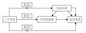
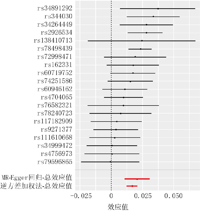
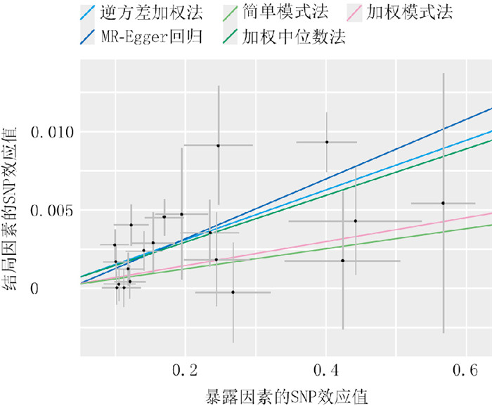
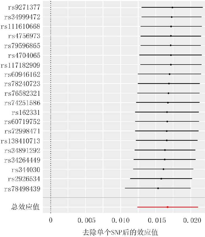
  Objective  To investigate the causality between non-ionizing radiation and facial aging, and to identify potential genes associated with facial aging.  Methods  This study employed a method of analysis based on multiple Mendelian randomization (MR). Genome-wide association study data of non-ionizing radiation (FinnGen database, n=218 281) and facial aging (UK Biobank database, n=423 999) were retrieved. Single nucleotide polymorphisms (SNPs) were used as instrumental variables, with a significance threshold (P < 5×10⁻⁶) applied and further linkage disequilibrium analysis performed to select SNPs associated with non-ionizing radiation. Two-sample MR (TSMR) analysis was conducted to assess the causality between non-ionizing radiation and facial aging, using inverse variance weighting (IVW) method as the primary analytical method and supplementing with MR-Egger regression, weighted median, weighted mode, and simple mode methods for validation. For the selected non-ionizing radiation-associated SNPs, heterogeneity was tested by Cochran Q test, horizontal pleiotropy was assessed by the MR-Egger intercept test and MR-PRESSO test, and robustness was evaluated via leave-one-out analysis. Multivariable MR (MVMR) analysis was performed to adjust for confounding factors affecting facial aging including smoking frequency, blood alcohol concentration, exercise frequency, body mass index, and systolic and diastolic blood pressure. Summary-data-based MR (SMR) analysis using expression quantitative trait loci (eQTL) data was conducted to screen candidate genes of facial aging, which were then validated by TSMR analysis. Protein quantitative trait loci (pQTL) and methylation quantitative trait loci (mQTL) data were analyzed by TSMR analysis to examine the causal role of MED1 gene with facial aging from multi-omics aspect. The genetic association of MED1 gene with facial aging was verified by colocalization analysis (posterior probability H4 > 50%).  Results  Twenty non-ionizing radiation-related SNPs that reached the significance threshold were screened out, with F values being all > 10. IVW analysis demonstrated a positive causality between non-ionizing radiation and facial aging (with odds ratio of 1.02, with 95% confidence interval of 1.01-1.02, P < 0.05). The analysis results of MR-Egger regression, weighted median, simple mode method, and weighted mode method (with odds ratios of 1.02, 1.02, 1.01, and 1.01, respectively, with 95% confidence intervals of 1.01-1.03, 1.01-1.02, 0.99-1.02, respectively, P < 0.05) were consistent with IVW method. For these 20 non-ionizing radiation-related SNPs, Cochran Q test under IVW method and MR-Egger showed no significant heterogeneity (with Q values of 23.20 and 22.59, respectively, P > 0.05); the MR-Egger intercept test (with intercept absolute value of 0.01, with standard error of 0.01, P > 0.05) and MR-PRESSO test (P > 0.05) indicated no horizontal pleiotropy. Leave-one-out analysis further confirmed that no individual SNP had a significant effect on the results. After correction of confounding factors such as systolic blood pressure, diastolic blood pressure, smoking frequency, blood alcohol concentration, body mass index, and exercise frequency, MVMR analysis showed that non-ionizing radiation remained a risk factor for facial aging (with odds ratios of 1.01, 1.01, 1.02, 1.02, 1.01, and 1.04, respectively, with 95% confidence intervals of 1.01-1.02, 1.01-1.02, 1.01-1.02, 1.01-1.02, 1.00-1.01, and 1.03-1.05, respectively, all P values < 0.05). SMR analysis identified 12 potential facial aging-related genes (SENP7, CCND1, LTBP2, IKZF3, MED1, ORMDL3, ZBTB7B, LOX, NEBL, EXOSC6, PSMA4, and EIF2B2, with odds ratios of 1.01, 1.03, 1.04, 0.99, 1.04, 1.01, 1.06, 0.88, 1.01, 0.99, 1.04, and 0.99, respectively, all P values < 0.05). Subsequent TSMR analysis retained 6 risk genes (ZBTB7B, SENP7, NEBL, MED1, PSMA4, and ORMDL3, with odds ratios of 1.04, 1.01, 1.00, 1.02, 1.03, and 1.01, respectively, with 95% confidence intervals of 1.02-1.05, 1.00-1.01, 1.00-1.01, 1.01-1.03, 1.01-1.04, and 1.00-1.01, respectively, all P values < 0.05) for facial aging and 4 protective genes (LOX, EIF2B2, EXOSC6, and IKZF3, with odds ratios of 0.92, 0.99, 0.99, and 0.99, respectively, with 95% confidence intervals of 0.90-0.94, 0.99-0.99, 0.99-1.00, and 0.99-1.00, respectively, all P values < 0.05). TSMR analysis based on pQTL data showed the MED1 protein was positively associated with facial aging (with odds ratio of 1.04, P < 0.05), which was consistent with the causal direction observed in eQTL-based SMR and TSMR analyses. TSMR analysis based on mQTL data indicated MED1 gene methylation (with probes of cg15445000 and cg03013999) had a protective effect on facial aging (with odds ratios of 0.99 and 0.99, respectively, both P values < 0.05). Colocalization analysis yielded a posterior probability H4=58.4%, suggesting that MED1 gene and facial aging likely shared the same causal genetic variant.  Conclusions  Through multi-omics MR analyses, it has confirmed that there is a causality between non-ionizing radiation and facial aging, which remained highly significant after correcting for potential confounders such as smoking frequency, blood alcohol concentration, exercise frequency, and the others. Clearly, 10 genes including SENP7, NEBL, EIF2B2, PSMA4, EXOSC6, IKZF3, ORMDL3, ZBTB7B, LOX, and MED1, particularly the MED1, may be involved in the process of facial aging.
Review
Research advances on the mechanism and clinical application of cold atmospheric plasma in promoting wound healing
Zhao Ziyue, Cai Chenghao, Liu Huan, Liu Shuwen, Wang Xingang
2025, 41(6): 604-608. doi: 10.3760/cma.j.cn501225-20241117-00448
Abstract:
Wound healing is a complex biological process involving multiple stages, including hemostasis, inflammation, proliferation, and remodeling. Cold atmospheric plasma (CAP), as an efficient, non-invasive, and innovative therapy for wound care, has significant application prospects in this field. It has been demonstrated that CAP can promote wound cell proliferation and reduce microbial load on wounds through various mechanisms. Through a comprehensive analysis of recent domestic and international literature on the biological effects of CAP, this paper reviews how CAP promotes the repair of acute and chronic wounds from the perspectives of mechanism and clinical application and the research progress in the application methods of CAP in recent years.
2025, 41(6): 501-502. doi: 10.3760/cma.j.cn501225-20250606-00259
Abstract:
2025, 41(6): 503-504. doi: 10.3760/cma.j.cn501225-20250606-00261
Abstract:
2025, 41(6): 505-506. doi: 10.3760/cma.j.cn501225-20250606-00260
Abstract:
2025, 41(6): 507-508. doi: 10.3760/cma.j.cn501225-20250606-00258
Abstract: