- Medline/PubMed数据库
- Scopus数据库
- PMC数据库
- CSCD
- 北大核心收录期刊
- 统计源期刊
- 我国高质量科技期刊T1级
- 第6届中国精品科技期刊
| Citation: | Wu CL,Zhang Y,Cheng JN,et al.Clinical application effects of the "three-source and four-line method" in the design and harvest of anterolateral lower leg perforator flaps[J].Chin J Burns Wounds,2026,42(3):252-260.DOI: 10.3760/cma.j.cn501225-20250328-00148.

|

| [1] |
石岩,王腾,徐永清,等.数字化技术在胫前动脉穿支皮瓣修复手部创面中的应用[J].中华显微外科杂志,2022,45(2):167-170.DOI: 10.3760/cma.j.cn441206-20211116-00276.
|
| [2] |
张韬,程俊楠,杨林,等.多穿支腓浅动脉穿支皮瓣修复手足创面的疗效[J].中华烧伤与创面修复杂志,2023,39(3):234-240.DOI: 10.3760/cma.j.cn501225-20220723-00305.
|
| [3] |
余少校,周望高,陈国荣,等.显微削薄腓动脉穿支皮瓣修复手指背侧软组织缺损[J].中华显微外科杂志,2022,45(6):617-621.DOI: 10.3760/cma.j.cn441206-20220411-00071.
|
| [4] |
石岩,徐永清,王腾,等.数字重建技术虚拟规划游离胫前动脉穿支皮瓣修复足踝创面[J].中华显微外科杂志,2024,47(6):630-634.DOI: 10.3760/cma.j.cn441206-20240513-00117.
|
| [5] |
熊胜,巨积辉,金光哲,等.彩超定位下游离腓浅动脉穿支皮瓣修复手足部创面[J].中华手外科杂志,2018,34(6):418-420.DOI: 10.3760/cma.j.issn.1005-054X.2018.06.007.
|
| [6] |
赵英波,胡琪,王兆庆,等.腓动脉穿支皮瓣联合阔筋膜游离移植修复手背复合组织缺损[J].中华手外科杂志,2025,41(2):120-123.DOI: 10.3760/cma.j.cn311653-20241008-00260.
|
| [7] |
杨晓东,刘杨武,杨锦,等.腓浅动脉穿支皮瓣游离移植修复手部创面[J].中华手外科杂志,2014,30(2):84-86.DOI: 10.3760/cma.j.issn.1005-054X.2014.02.002.
|
| [8] |
胡雷鸣,欧学海,魏登科,等.应用游离腓动脉穿支皮瓣修复足部小面积感染性溃疡创面[J].中华显微外科杂志,2019,42(3):246-249.DOI: 10.3760/cma.j.issn.1001-2036.2019.03.009.
|
| [9] |
陈步国,张松,潘勇,等.腓动脉近端穿支皮瓣修复手足部中小型创面的临床治疗效果[J].中华手外科杂志,2023,39(6):565-568.DOI: 10.3760/cma.j.cn311653-20230104-00004.
|
| [10] |
袁辉宗,史增元,尹维刚,等. 胫前动脉前肌间隔穿支为蒂的岛状皮瓣的应用解剖[J]. 中国临床解剖学杂志,2011,29(3):249-251,255. DOI: 10.13418/j.issn.1001-165x.2011.03.016.
|
| [11] |
李贤海,周建东,许亚军.带胫前动脉穿支的腓动脉穿支皮瓣修复前中足背创面[J].中华显微外科杂志,2020,43(5):464-468.DOI: 10.3760/cma.j.cn441206-20191215-00382.
|
| [12] |
张伟,梁高峰,贾宗海,等.应用游离腓动脉穿支皮瓣修复老年患者指背感染性溃疡创面[J].中华显微外科杂志,2025,48(1):45-49.DOI: 10.3760/cma.j.cn441206-20240701-00163.
|
| [13] |
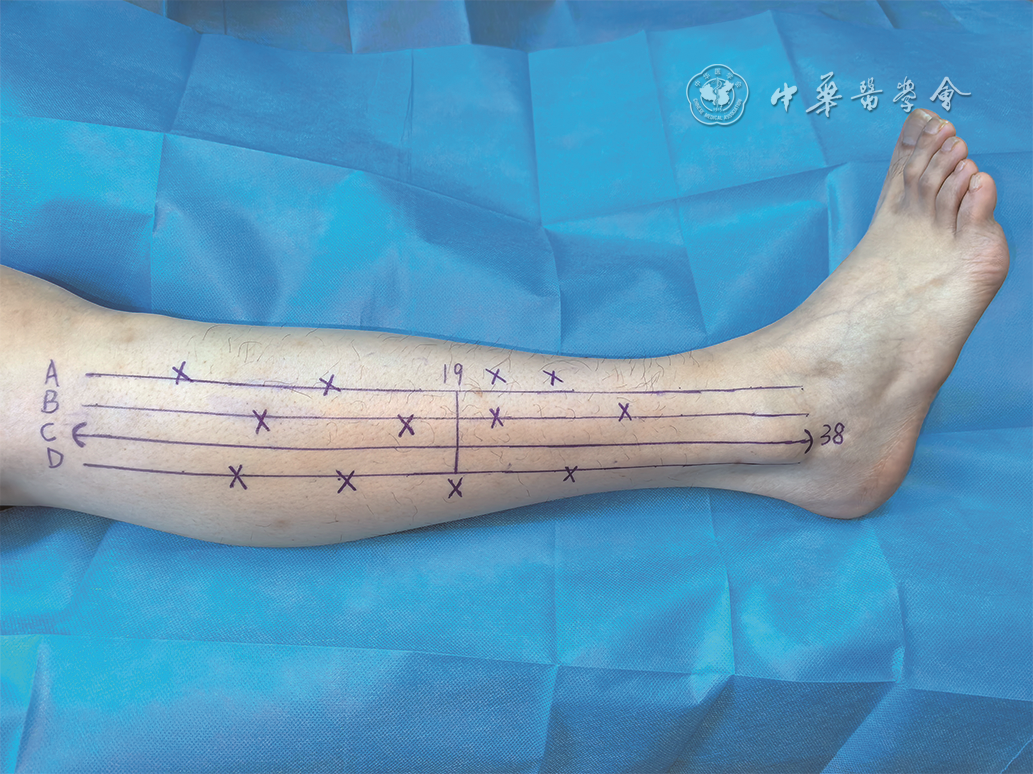
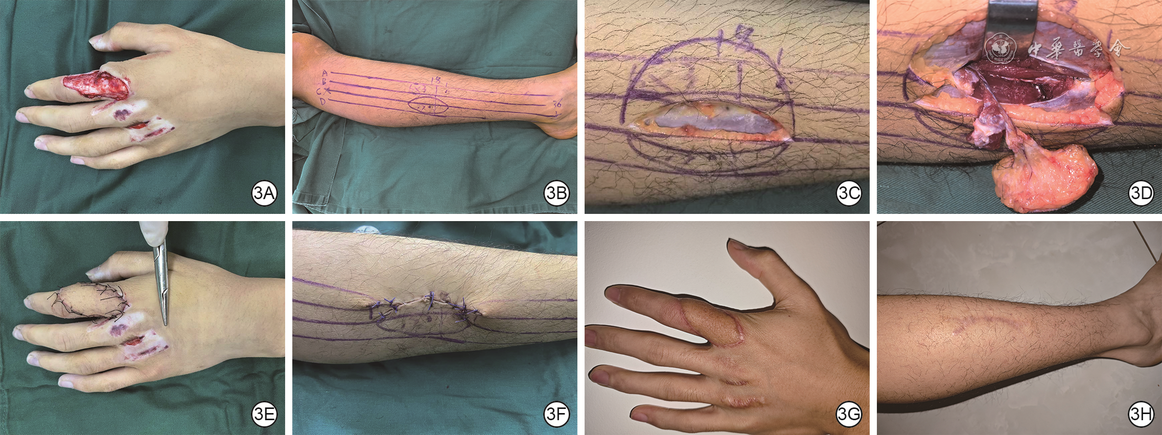
张韬,杨林,程俊楠,等.“ABC”三线定位穿支方法在游离小腿前外侧穿支皮瓣的应用[J].中华显微外科杂志,2023,46(1):70-75.DOI: 10.3760/cma.j.cn441206-20220530-00107.
|
| [14] |
肖飞鹏,柳志锦,刘胜哲,等.综合评价量表在股前外侧皮瓣修复术后疗效评价中的应用[J].中国美容整形外科杂志,2021,32(6):348-351.DOI: 10.3969/j.issn.1673-7040.2021.06.010.
|
| [15] |
石岩,何晓清,杨曦,等.CT血管造影检查结合三维重建辅助设计腓浅动脉穿支皮瓣修复手部创面的疗效[J].中华创伤杂志,2021,37(6):501-507.DOI: 10.3760/cma.j.cn501098-20210202-00105.
|
| [16] |
LiSJ,ChengH,FangX,et al.Modified reversed superficial peroneal artery flap in the reconstruction of ankle and foot defects following severe burns or trauma[J].Burns,2017,43(4):839-845.DOI: 10.1016/j.burns.2016.12.003.
|
| [17] |
ChenYP,ZhongHY,YangR,et al.Optimizing localization accuracy in peroneal artery perforator sequential flap transplantation with digital subtraction angiography and high-frequency ultrasound[J].J Plast Surg Hand Surg,2025,60:46-50.DOI: 10.2340/jphs.v60.42954.
|
| [18] |
DeeringAJ3rd, HarrahPA, WonBW, et al. Chimeric osteocutaneous free flap solution to variation in vascular anatomy[J]. Plast Reconstr Surg Glob Open,2025,13(1):e6443. DOI: 10.1097/GOX.0000000000006443.
|
| [19] |
谢志平,庄跃宏,郑和平,等.腓血管蒂腓骨嵌合组织瓣设计的解剖学基础[J].中国临床解剖学杂志,2014,32(3):259-263.DOI: 10.13418/j.issn.1001-165x.2014.03.006.
|
| [20] |
HaY, YeoKK, PiaoY, et al. Peroneal flap: clinical application and cadaveric study[J]. Arch Plast Surg,2017,44(2):136-143. DOI: 10.5999/aps.2017.44.2.136.
|
| [21] |
LykoudisEG,KoutsourisM,LykissasMG.Vascular anatomy of the integument of the lateral lower leg: an anatomical study focused on cutaneous perforators and their clinical importance[J].Plast Reconstr Surg,2011,128(1):188-198.DOI: 10.1097/PRS.0b013e3182174303.
|
| [22] |
李匡文,唐举玉,刘昌雄,等. 腓动脉穿支皮瓣的应用解剖[J]. 中国临床解剖学杂志,2011,29(4):382-385.
|
| [23] |
PapadimasD,ParaskeuopoulosT,AnagnostopoulouS.Cutaneous perforators of the peroneal artery: cadaveric study with implications in the design of the osteocutaneous free fibular flap[J].Clin Anat,2009,22(7):826-833.DOI: 10.1002/ca.20847.
|
| [24] |
KazemiK,SaadiF,JavanshirK,et al.Reliability of musculoskeletal ultrasonography for peri-ankle muscles in subjects with unilateral chronic ankle instability[J].J Bodyw Mov Ther,2021,27:565-572.DOI: 10.1016/j.jbmt.2021.04.018.
|
| [25] |
李友,唐林峰,熊胜,等.游离多块小腿前外侧穿支皮瓣一期修复单手多指创面[J].中华显微外科杂志,2024,47(3):307-311.DOI: 10.3760/cma.j.cn441206-20230909-00031.
|
| [26] |
郭全伟,赵强,程贺云,等.游离小腿同一供区多块腓浅动脉穿支皮瓣修复手部多处热压伤狭长创面的效果观察[J].中华手外科杂志,2024,40(4):356-359.DOI: 10.3760/cma.j.cn311653-20231207-00095.
|
| [27] |
程贺云,巨积辉,赵强,等. CDU精准定位下游离一侧下肢多个小腿外侧穿支皮瓣修复手指不同部位多个创面的效果[J]. 中华烧伤与创面修复杂志,2026,42(2):182-188. DOI: 10.3760/cma.j.cn501225-20241210-00484.
|
| [28] |
StoccoC,MurphyDC,GargiuloM,et al.Superficial peroneal nerve accessory artery (SPNAA) flap for head and neck reconstruction: a cadaveric anatomical study and retrospective case series review[J].J Plast Reconstr Aesthet Surg,2021,74(7):1524-1533.DOI: 10.1016/j.bjps.2020.11.012.
|
| [29] |
KimJS,SonDG,ChoiTH,et al.An anatomic study of the superficial peroneal nerve accessory artery and its perforators, and clinical application of superficial peroneal nerve accessory artery perforator flaps[J].Ann Plast Surg,2008,60(6):710-716.DOI: 10.1097/SAP.0b013e3180cab77f.
|
| [30] |
程贺云,张韬,巨积辉,等.Flow-through腓浅动脉皮瓣修复手指伴指掌侧固有动脉缺损创面[J].中华显微外科杂志,2021,44(4):369-373.DOI: 10.3760/cma.j.cn441206-20210111-00011.
|
| [31] |
徐永清,何晓清,陈雪松,等.手和足部创面修复时四种吻合血管的皮神经营养血管穿支皮瓣的选择[J].中华显微外科杂志,2020,43(4):331-337.DOI: 10.3760/cma.j.cn441206-20200218-00068.
|
| [32] |
ScaglioniMF,KuoYR,ChenYC.Reconstruction of distal hand and foot defects with the free proximal peroneal artery perforator flap[J].Microsurgery,2016,36(3):183-190.DOI: 10.1002/micr.22364.
|
| [33] |
PouletV,PrevostA,CavallierZ,et al.Fibula free flap perforasomes: vascular anatomical study and clinical applications[J].Surg Radiol Anat,2022,44(5):637-644.DOI: 10.1007/s00276-022-02953-4.
|
| [34] |
KimNG,LeeKS,ChoiTH,et al.Aesthetic reconstruction of lower leg defects using a new anterolateral lower leg perforator flap[J].J Plast Reconstr Aesthet Surg,2008,61(8):934-938.DOI: 10.1016/j.bjps.2007.06.006.
|
| [35] |
杨晓东,刘杨武,杨锦,等.削薄腓浅动脉穿支皮瓣游离移植修复前足创面[J].中华显微外科杂志,2013,36(2):123-125.DOI: 10.3760/cma.j.issn.1001-2036.2013.02.008.
|
| [36] |
程贺云,巨积辉,赵强,等.腓浅动脉穿支皮瓣在拇指和手指瓦合再造术中的临床应用[J].中华显微外科杂志,2023,46(6):631-636.DOI: 10.3760/cma.j.cn441206-20230714-00120.
|
| [37] |
徐永清.小腿穿支皮瓣在中国的临床应用及进展[J].中华显微外科杂志,2020,43(6):521-534.DOI: 10.3760/cma.j.cn441206-20200527-00243.
|
| [38] |
YangX,ZhangG,LiuY,et al.Vascular anatomy and clinical application of anterolateral leg perforator flaps[J].Plast Reconstr Surg,2013,131(4):534e-543e.DOI: 10.1097/PRS.0b013e3182827675.
|
| [39] |
ShiY, XuY, FanX, et al. Three-dimensional digitalized virtual planning of free anterior tibial artery perforator flap for repairing soft tissue defects in extremities[J]. World J Surg,2023,47(7):1821-1827. DOI: 10.1007/s00268-023-06970-1.
|
| [40] |
周健,郑玉岑,陈伟,等.携带神经的腓动脉穿支皮瓣修复腕部电烧伤后期神经缺损的临床效果[J].中华烧伤与创面修复杂志,2024,40(9):835-841.DOI: 10.3760/cma.j.cn501225-20240520-00187.
|
 吴成龙.mp4
|

|

Figures(5)
Copyright © Chinese Journal of Burns京ICP备07035254号-14
E-mail:shaoshangzazhi@163.com
Supported by:
Beijing Renhe Information Technology Co. Ltd


 DownLoad: