- Medline/PubMed数据库
- Scopus数据库
- PMC数据库
- CSCD
- 北大核心收录期刊
- 统计源期刊
- 我国高质量科技期刊T1级
- 第6届中国精品科技期刊
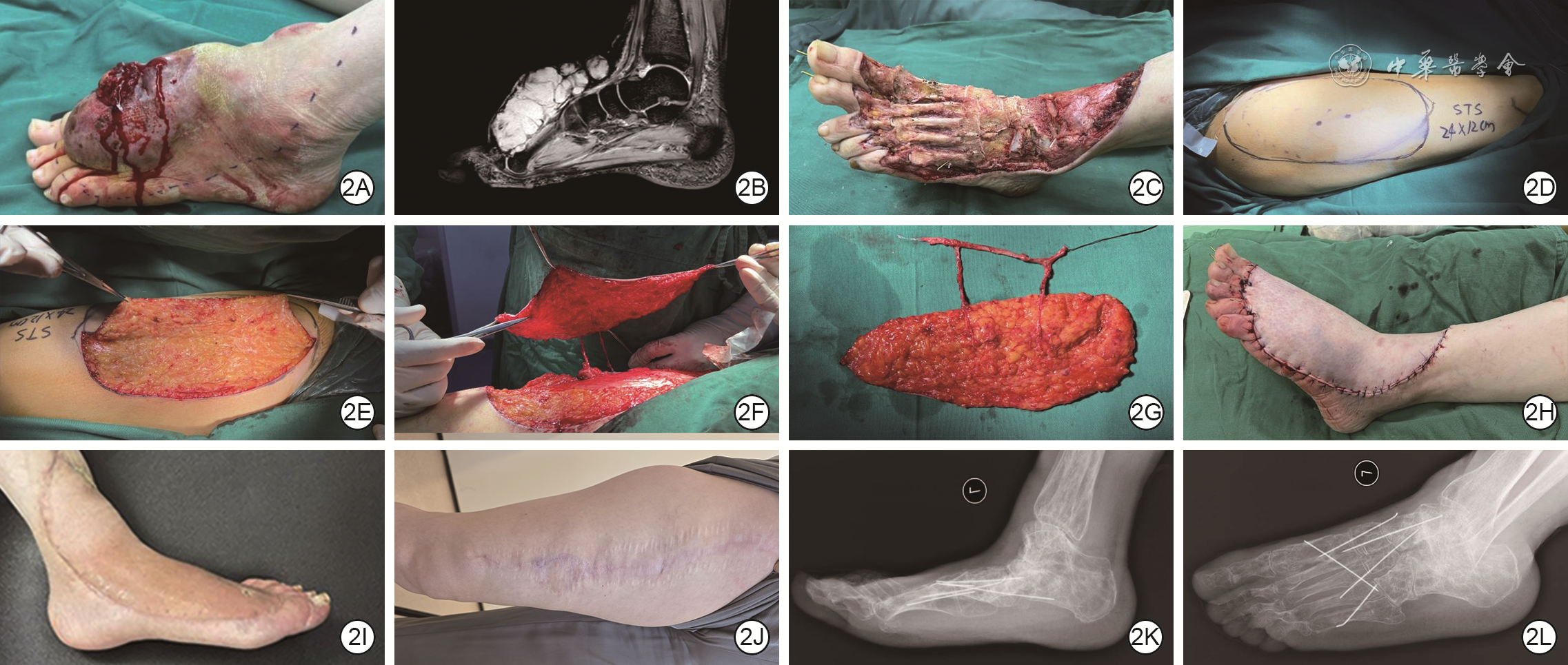
| Citation: | Li B,Yang Y,Li PC,et al.Clinical efficacy of thinned free flaps in reconstructing the wounds following excision of malignant extremity tumors[J].Chin J Burns Wounds,2026,42(8):759-766.DOI: 10.3760/cma.j.cn501225-20260426-00167.

|

| [1] |
KawaguchiN, AhmedAR, MatsumotoS, et al. The concept of curative margin in surgery for bone and soft tissue sarcoma[J]. Clin Orthop Relat Res, 2004(419):165-172. DOI: 10.1097/00003086-200402000-00027.
|
| [2] |
EnnekingWF, SpanierSS, GoodmanMA. A system for the surgical staging of musculoskeletal sarcoma[J]. Clin Orthop Relat Res, 1980(153):106-120.
|
| [3] |
KarakawaR, YoshimatsuH, FuseY, et al. Impact of technological advancements on short-term outcomes in flap reconstruction after soft tissue sarcoma resection: a retrospective comparative analysis[J]. J Reconstr Microsurg, 2025,41(9):761-771. DOI: 10.1055/a-2508-6628.
|
| [4] |
LucattelliE, LusettiIL, CiprianiF, et al. Reconstruction of upper limb soft-tissue defects after sarcoma resection with free flaps: a systematic review[J]. J Plast Reconstr Aesthet Surg, 2021,74(4):755-767. DOI: 10.1016/j.bjps.2020.10.065.
|
| [5] |
HaldemanPB, HarfoucheC, RosalesR, et al. Immediate and delayed flap reconstruction have equivalent outcomes and associated costs following soft tissue sarcoma surgery[J]. J Surg Oncol, 2024, 130(3):562-568. DOI: 10.1002/jso.27770.
|
| [6] |
KrishnanCK, RamanujanV, RamachandranV, et al. Single-stage free flap reconstruction following extremity sarcoma resection: a regional cancer center experience[J]. Indian J Surg Oncol, 2024, 15(Suppl 1):S45-S51. DOI: 10.1007/s13193-023-01713-z.
|
| [7] |
HyakusokuH, GaoJH. The "super-thin" flap[J]. Br J Plast Surg, 1994,47(7):457-464. DOI: 10.1016/0007-1226(94)90027-2.
|
| [8] |
DiamondS, SethAK, ChatthaAS, et al. Outcomes of subfascial, suprafascial, and super-thin anterolateral thigh flaps: tailoring thickness without added morbidity[J]. J Reconstr Microsurg, 2018,34(3):176-184. DOI: 10.1055/s-0037-1607426.
|
| [9] |
杨勇, 李斌, 李金勇, 等. 基于CDU和DSA穿支定位的分层切取方案在削薄股前外侧皮瓣中的应用[J].中华显微外科杂志,2024,47(3):248-253. DOI: 10.3760/cma.j.cn441206-20231215-00103.
|
| [10] |
von MehrenM, RandallRL, BenjaminRS, et al. Soft tissue sarcoma, version 2.2018, NCCN clinical practice guidelines in oncology[J]. J Natl Compr Canc Netw, 2018,16(5):536-563. DOI: 10.6004/jnccn.2018.0025.
|
| [11] |
ElmorsiR, HancockM, CamachoL, et al. Perforator versus traditional flaps in extremity soft-tissue sarcoma reconstruction: a comparative cohort study[J]. J Plast Reconstr Aesthet Surg, 2026,115:427-434. DOI: 10.1016/j.bjps.2026.02.024.
|
| [12] |
AbouarabMH, SalemIL, DegheidyMM, et al. Therapeutic options and postoperative wound complications after extremity soft tissue sarcoma resection and postoperative external beam radiotherapy[J]. Int Wound J, 2018,15(1):148-158. DOI: 10.1111/iwj.12851.
|
| [13] |
TownleyWA, MahE, O'NeillAC, et al. Reconstruction of sarcoma defects following pre-operative radiation: free tissue transfer is safe and reliable[J]. J Plast Reconstr Aesthet Surg, 2013,66(11):1575-1579. DOI: 10.1016/j.bjps.2013.06.029.
|
| [14] |
KarakawaR, YoshimatsuH, FuseY, et al. Comparison of outcomes following pedicled and free flap transfers for the defect after shoulder sarcoma resection[J]. J Plast Reconstr Aesthet Surg, 2023, 83:373-379. DOI: 10.1016/j.bjps.2023.04.049.
|
| [15] |
LeseI, BaesuC, HoyosIA, et al. Flap reconstruction outcome following surgical resection of soft tissue and bone sarcoma in the setting of (neo)adjuvant therapy: a sarcoma center experience[J]. Cancers (Basel), 2023,15(9):2423.DOI: 10.3390/cancers15092423.
|
| [16] |
SureshV, GaoJ, JungSH, et al. The role of reconstructive surgery after skeletal and soft tissue sarcoma resection[J]. Ann Plast Surg, 2018,80(6S Suppl 6):S372-S376. DOI: 10.1097/SAP.0000000000001419.
|
| [17] |
QiaoJ, MaoH, WenL, et al. Reconstruction of soft tissue defect with a free vascularized anterolateral thigh flap after resection of soft tissue sarcoma in extremities[J]. Orthop Surg, 2022, 14(2):215-220. DOI: 10.1111/os.12840.
|
| [18] |
SanniecKJ, VelazcoCS, BryantLA, et al. Immediate versus delayed sarcoma reconstruction: impact on outcomes[J]. Sarcoma, 2016,2016:7972318. DOI: 10.1155/2016/7972318.
|
| [19] |
AbeY, NagasakaS, YamashitaY, et al. Functional outcomes of reconstructive flap surgery for soft tissue sarcoma: long-term outcomes of functional restoration using innervated muscle transplantation[J]. J Plast Reconstr Aesthet Surg, 2024, 91:312-321. DOI: 10.1016/j.bjps.2024.02.018.
|
| [20] |
KimuraN, SatohK. Consideration of a thin flap as an entity and clinical applications of the thin anterolateral thigh flap[J]. Plast Reconstr Surg, 1996,97(5):985-992. DOI: 10.1097/00006534-199604001-00016.
|
| [21] |
KimuraN, SatohK, HasumiT, et al. Clinical application of the free thin anterolateral thigh flap in 31 consecutive patients[J]. Plast Reconstr Surg, 2001,108(5):1197-1208; discussion 1209-1210. DOI: 10.1097/00006534-200110000-00015.
|
| [22] |
HongJP, ChoiDH, SuhH, et al. A new plane of elevation: the superficial fascial plane for perforator flap elevation[J]. J Reconstr Microsurg, 2014,30(7):491-496. DOI: 10.1055/s-0034-1369807.
|
| [23] |
张丕红. 浅谈薄型穿支皮瓣切取与穿支血管探测和皮瓣血运评估[J].中华烧伤与创面修复杂志,2023,39(10):911-918. DOI: 10.3760/cma.j.cn501225-20230812-00047.
|
| [24] |
VivianoSL, LiuFC, TherattilPJ, et al. Peripheral pruning: a safe approach to thinning extra-large anterolateral thigh flaps[J]. Ann Plast Surg, 2018,80(4 Suppl 4):S164-S167. DOI: 10.1097/SAP.0000000000001401.
|
| [25] |
赵书明, 刘娜, 刘学亮, 等. 彩色多普勒超声辅助下超薄胸背动脉穿支皮瓣的切取方案及临床应用效果[J].中华烧伤与创面修复杂志,2024,40(3):281-288. DOI: 10.3760/cma.j.cn501225-20231012-00111.
|
| [26] |
董帅, 王石, 滕志成, 等. 吲哚菁绿血管造影联合彩色多普勒超声在股前外侧穿支皮瓣穿支定位中的临床应用效果[J].中华烧伤与创面修复杂志,2025,41(11):1091-1100. DOI: 10.3760/cma.j.cn501225-20240629-00254.
|
| [27] |
郑炜, 魏在荣, 马琳, 等. 高频彩色多普勒超声在旋股外侧动脉降支穿支分叶皮瓣术前导航中的应用效果[J].中华烧伤杂志,2017,33(10):616-618. DOI: 10.3760/cma.j.issn.1009-2587.2017.10.006.
|
| [28] |
孙丰坤, 王立民, 宋兴华, 等. 彩色多普勒超声探测下应用以旋股外侧动脉斜支为蒂的ALTPF修复四肢皮肤软组织缺损的临床效果[J].中华烧伤与创面修复杂志,2026,42(1):81-90. DOI: 10.3760/cma.j.cn501225-20240623-00247.
|
| [29] |
王珏, 刘海峰, 王旭东, 等. 彩色多普勒超声和CT血管造影技术在下肢穿支皮瓣临床应用中的对比研究[J].组织工程与重建外科杂志,2021,17(5):400-404. DOI: 10.3969/j.issn.1673-0364.2021.05.007.
|
| [30] |
王晓冰, 纪旭, 马春燕. 彩色多普勒超声在股前外侧穿支皮瓣移植术前穿支动脉评估中的作用[J].中国医科大学学报,2022,51(6):513-517. DOI: 10.12007/j.issn.0258-4646.2022.06.007.
|
| [31] |
唐举玉, 卿黎明, 贺继强, 等. 数字化技术辅助旋股外侧动脉降支分叶穿支皮瓣设计的初步应用[J].中华显微外科杂志,2016,39(2):123-126. DOI: 10.3760/cma.j.issn.1001-2036.2016.02.006
|
| [32] |
李雪栋,田惠玲,冯小辉,等.CTA联合增强现实技术在股前外侧皮瓣术中的应用[J].中国矫形外科杂志, 2026,34(6):558-562. DOI: 10.20184/j.cnki.Issn1005-8478.120075.
|
| [33] |
ZhangA, DongY, YuanS, et al. Preoperative perforator mapping of anterolateral thigh flaps using computed tomographic angiography and visual coordinate system for the reconstruction of head and neck defects[J]. Ear Nose Throat J,2025,104(4):NP195-NP202. DOI: 10.1177/01455613221108367.
|
| [34] |
KapoorDA, ChattopadhyayDD, ChauhanDU. Perforator anastomosis of the sural artery system and its application in extended gastrocnemius flap[J]. Injury, 2023,54(6): 1804-1808.DOI: 10.1016/j.injury.2023.02.018.
|
| [35] |
杨林, 程俊楠, 刘胜哲, 等. 基于DSA的旋股外侧动脉斜支的形态学研究及其临床意义[J].中华显微外科杂志,2021,44(6):642-646. DOI: 10.3760/cma.j.cn441206-20210604-00142.
|
| [36] |
YangL, ChengJ, LiuZ, et al. Morphological study of branches of lateral femoral circumflex artery based on digital subtraction angiography[J]. J Plast Reconstr Aesthet Surg, 2023,80:18-24. DOI: 10.1016/j.bjps.2022.08.075.
|
| [37] |
ParkSO, ChangH, ImanishiN. Anatomic basis for flap thinning[J]. Arch Plast Surg, 2018,45(4):298-303. DOI: 10.5999/aps.2017.01543.
|
| [38] |
ParkBY. Flap thinning: Defatting after conventional elevation[J]. Arch Plast Surg, 2018,45(4):314-318. DOI: 10.5999/aps.2018.00787.
|
| [39] |
SethAK, IorioML. Super-thin and suprafascial anterolateral thigh perforator flaps for extremity reconstruction[J]. J Reconstr Microsurg, 2017,33(7):466-473. DOI: 10.1055/s-0037-1601422.
|
Figures(3)
Copyright © Chinese Journal of Burns京ICP备07035254号-14
E-mail:shaoshangzazhi@163.com
Supported by:
Beijing Renhe Information Technology Co. Ltd



 DownLoad: