Abstract:
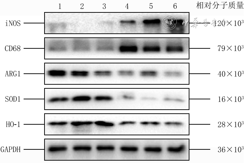
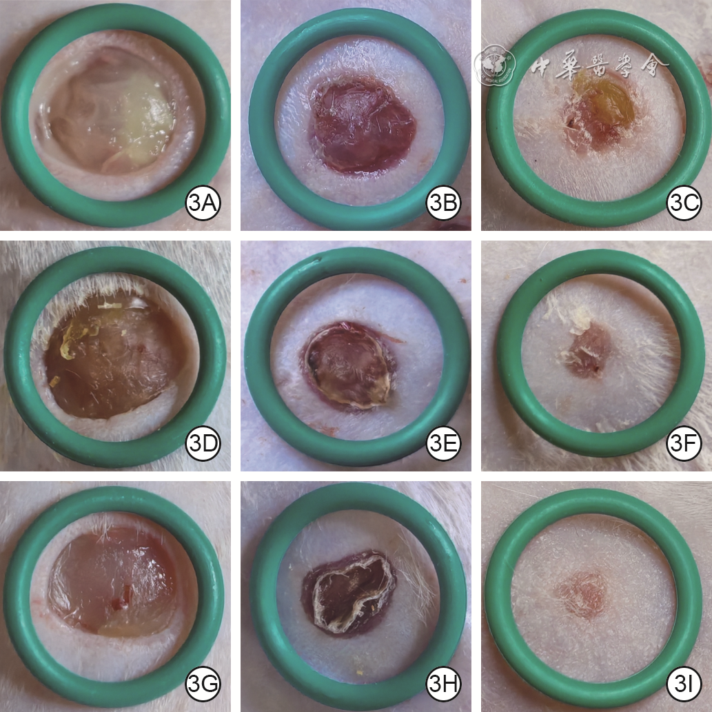
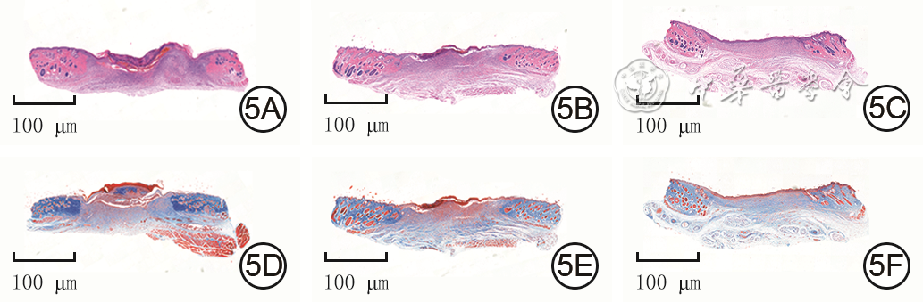
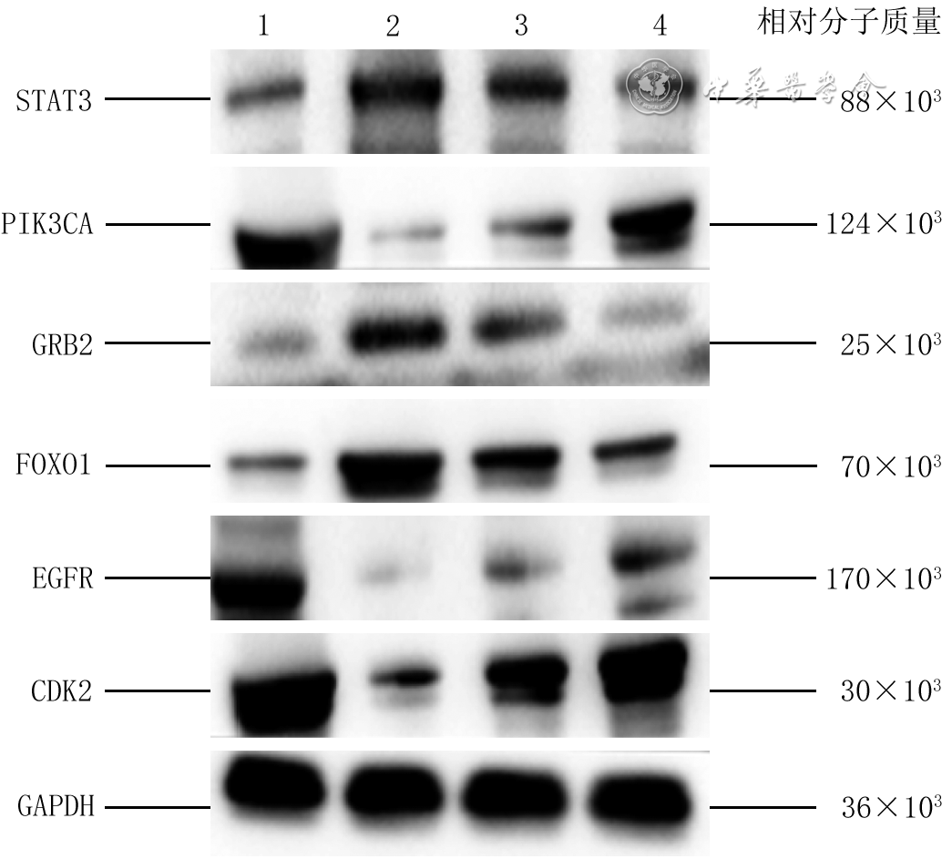
Objective To investigate the effects of astragaloside Ⅰ (AS-Ⅰ) on full-thickness skin defects in diabetic rats and its underlying mechanism. Methods This study was an experimental study with grouped design and repeated measures design. Twenty-four male Sprague-Dawley rats aged 6-8 weeks were divided into control group (n=6) and modeling group (n=18) using a random number table method (the same grouping method was used throughout). After successful induction of diabetes mellitus, the modeling group was further divided into model group, low-dose AS-Ⅰ group, and high-dose AS-Ⅰ group, with 6 rats in each group. A full-thickness skin defect wound of 10 mm in diameter was created on the back of each rat. Wounds in control group and model group were treated with normal saline, while wounds in low-dose AS-Ⅰ group and high-dose AS-Ⅰ group were treated with 1.15 and 11.50 mmol/L AS-Ⅰ, respectively, for 3 consecutive days. Wound healing rates of rats were calculated at 3, 5, 7, 9, 13, and 15 days after injury. At 15 days after injury, Masson's trichrome staining was used to assess the deposition of collagen fibers in wound tissue; immunohistochemical staining was performed to detect Ki-67 positive expression in wound tissue, and the expression level was quantified. Network pharmacology analysis was applied to screen for potential core targets of AS-Ⅰ in treatment of diabetic wounds; molecular docking simulation was utilized to validate the binding affinity of AS-Ⅰ to the core targets, and molecular dynamics simulation was performed to verify the dynamic stability of the interaction between AS-Ⅰ and forkhead box O1 (FOXO1). At 15 days after injury, Western blotting was used to determine the protein expression levels of epidermal growth factor receptor (EGFR), cyclin-dependent kinase 2 (CDK2), growth factor receptor-bound protein 2 (GRB2), phosphatidylinositol-4,5-bisphosphate 3-kinase catalytic subunit alpha (PIK3CA), signal transducer and activator of transcription 3 (STAT3), and FOXO1 in wound tissue of rats in four groups. Results Compared with those in control group, the wound healing rates of rats in model group at 3 and 15 days after injury were significantly decreased (P<0.05); whereas the wound healing rates of rats in low-dose AS-Ⅰ group at 7 and 13 days after injury, and in high-dose AS-Ⅰ group at 5, 7, 9, 13, and 15 days after injury were significantly increased (P<0.05). Compared with those in model group, the wound healing rates of rats in both low-dose AS-Ⅰ group and high-dose AS-Ⅰ group at 3, 9, 13, and 15 days after injury were significantly increased (P<0.05). At 15 days after injury, collagen fibers in the wound tissue of rats in control group were arranged in an orderly and dense manner; whereas those in the wound tissue of model group were sparse and disorganized; the deposition of collagen fibers in the wound tissue of rats in low-dose AS-Ⅰ group was improved compared with model group; collagen fibers in the wound tissue of rats in high-dose AS-Ⅰ group exhibited dense and orderly arrangement with a high degree of maturity. At 15 days after injury, the expression level of Ki-67 in wound tissue of rats in model group was significantly lower than that in control group (P<0.05); compared with that in model group, the Ki-67 expression level in wound tissue of rats in low-dose AS-Ⅰ group was significantly higher (P<0.05); compared with those in control group, model group, and low-dose AS-Ⅰ group, the Ki-67 expression level in wound tissue of rats in high-dose AS-Ⅰ group was significantly higher (with P values all <0.05). The results of network pharmacology analysis identified 63 overlapping targets between AS-Ⅰ-associated targets and diabetic wound-related targets, involving the FOXO signaling pathway (with target protein FOXO1) and target proteins including EGFR, CDK2, GRB2, PIK3CA, and STAT3. The results of molecular docking simulation showed that AS-Ⅰ exhibited strong binding affinity to the target proteins. The results of molecular dynamics simulation demonstrated that the conformation of the complex formed by AS-Ⅰ and FOXO1 protein could maintain dynamic stability. At 15 days after injury, compared with those in control group, the protein expression levels of STAT3, GRB2, and FOXO1 in wound tissue of rats in model group were significantly upregulated (with P values all <0.05), while the protein expression levels of PIK3CA, EGFR, and CDK2 were significantly downregulated (with P values all <0.05). Compared with those in model group, the protein expression levels of STAT3, GRB2, and FOXO1 in wound tissue of rats in low-dose AS-Ⅰ group and high-dose AS-Ⅰ group were significantly downregulated (with P values all <0.05), while the protein expression levels of PIK3CA, EGFR, and CDK2 were significantly upregulated (with P values all <0.05). Compared with those in low-dose AS-Ⅰ group, the protein expression levels of STAT3, GRB2, and FOXO1 in wound tissue of rats in high-dose AS-Ⅰ group were significantly downregulated (with P values all <0.05), while the protein expression levels of PIK3CA, EGFR, and CDK2 were significantly upregulated (with P values all <0.05). Conclusions AS-Ⅰ may promote wound healing of full-thickness skin defects and improve collagen fiber arrangement in diabetic rats by downregulating the protein expression of STAT3, GRB2, and FOXO1 and upregulating that of PIK3CA, EGFR, and CDK2.
Li Y,Zheng YY,Wang YH,et al.Effects of astragaloside Ⅰ on full-thickness skin defects in diabetic rats and its underlying mechanism[J].Chin J Burns Wounds,2026,42(8):793-802.DOI: 10.3760/cma.j.cn501225-20260116-00026.



 Abstract
 PDF
PDF