2021 Vol. 37, No. 10

Expert Forum
Past, present, and future of critical burn treatment
Guo Guanghua, Jiang Zhengying
2021, 37(10): 905-910. doi: 10.3760/cma.j.cn501120-20210209-00055
Abstract:
The fatality rate of patients with critical burns is extremely high, and the clinical treatment is challenging. By reviewing the history on treatment of critically ill burns patients, this article elaborates and analyzes the advanced concepts and technologies at home and abroad about the critical burn treatment in the areas including shock and fluid resuscitation, hypermetabolism and nutrition, inhalation injury and respiratory support, acute kidney injury and continuous renal replacement therapy, wound assessment and management, infection and control, coagulopathy and its prevention and treatment, etc. Furthermore, some thoughts on the future development trend of critical burn treatment are put forward as reference for people in the same field.
Original Articles · Severe Burns and Complications
Clinical application of extracorporeal membrane oxygenation in the treatment of burn patients with acute respiratory distress syndrome: a retrospective analysis and systematic review
Li Haisheng, Yuan Zhiqiang, Song Huapei, Luo Qizhi, Xiang Fei, Ma Siyuan, Zhou Junyi, Tan Jianglin, Zhou Ling, Peng Yizhi, Luo Gaoxing
2021, 37(10): 911-920. doi: 10.3760/cma.j.cn501120-20210803-00266
Abstract:
  Objective  To analyze the clinical effect of extracorporeal membrane oxygenation (ECMO) in the treatment of burn patients with acute respiratory distress syndrome (ARDS).  Methods  The retrospective observational study and the systematic review were applied. From March 2014 to July 2020, five burn patients with ARDS received ECMO treatment in the First Affiliated Hospital of Army Medical University (the Third Military Medical University). All the five patients were male, aged from 40 to 62 years. The average total burn surface area was 58.8% total body surface area (TBSA) and four cases had severe inhalation injury. Patient's ECMO starting time, duration and mode, and whether successfully weaned or the cause of death, and others. were recorded. Furthermore, the changes of oxygenation and infection before, during, and after utilizing ECMO were analyzed. PubMed and Web of Science from the establishment of each database to August 2021 were searched using "Extracorporeal Membrane Oxygenation", "ECMO", "burn", "inhalation" as the search terms and "Title/Abstract" as the field to retrieve the clinical articles that meet the selection criteria . Basic information were extracted from the articles, including sample size, gender, age, total burn area, inhalation injury, the indication of ECMO, the start and lasting time of ECMO, ECMO mode, rate of successful weaning, complications of ECMO, mortality, the combined application of continuous renal replacement therapy (CRRT).  Results  Five patients started venovenous ECMO on an average of 10.2 days after injury and lasted an average of 180.4 hours. Three out of 5 patients were weaned successfully with one patient survived. Four patients died of multiple organ dysfunction syndrome (MODS) and septic shock. Compared with those before ECMO treatment, the arterial oxygen partial pressure (PaO2) and oxygen saturation in arterial blood (SaO2) of three successfully weaned patients obviously increased during and after ECMO treatment. The fraction of inspired oxygen (FiO2) decreased below 50% and PaO2/FiO2 ratio increased above 200 mmHg (1 mmHg=0.133 kPa) during and after ECMO. Furthermore, lactic acid and respiratory rate decreased, basically. Compared with those before ECMO, PaO2 and SaO2 in the other two patients during ECMO, who failed to be weaned, continuously decreased while lactic acid increased. Before and during ECMO, the PaO2/FiO2 ratios of unsuccessfullg weaned cases were less than 200 mmHg, and partial pressure of carbon dioxide in arterial blood (PaCO2) were more than 40 mmHg. Compared with those before ECMO, there were no significant changes in body temperature during and after ECMO, which were less than 38 ℃. Compared with those before ECMO, the leucocyte number (the index without this in unsuccessfully weaned cases was omitted, the same as below) in four patients showed a significant decrease during ECMO, but rose after removal of ECMO. The proportion of neutrophils in three patients were slightly higher during ECMO than before ECMO, and did not change significantly after removal of ECMO. Compared with those before ECMO, platelet counts in three patients were significantly reduced during ECMO, and all five patients during ECMO were below normal levels. Compared with those before ECMO, the procalcitonin levels in four deaths were significantly increased during ECMO. Catheter culture of microorganism was performed in three successfully weaned patients, all of which were negative. A total of 13 literature were included, ranging from 1990 to 2019. The sample size in 6 studies was less than 10, and the sample size in 4 studies was between 10 and 20, and only 2 literatures had a sample size larger than 50. ECMO was applied in 295 burn patients with overall mortality of 48.8% (144/295), including 157 adults and 138 children. The most common indication of ECMO was severe ARDS. Among 157 adult burn patients (95 males and 65 females), 36 cases had inhalation injury. The average burn area was 27%-37%TBSA in 5 reported studies and was more than 50%TBSA in 2 reported studies. The most common mode was venovenous ECMO. ECMO treatment began 26.5 hours to 7.4 days after injury and lasted from 90 hours to 18 days, and the rate of successful weaning ranged from 50% to 100%. The most common complications were bleeding and infection. The mortality was 52.9% (83/157). MODS and sepsis were the leading causes of death. Among 138 pediatric burn patients (77 boys and 61 girls), 29 patients had inhalation injury. The average burn area was 17%-50.2%TBSA in 3 studies. ECMO treatment lasted from 165.2 hours to 324.4 hours. Bleeding was the most common complication. The mortality was 44.2% (61/138).  Conclusions  ECMO is an effective strategy for the salvage treatment of burns complicated with ARDS. Furthermore, the prevention and treatment of bleeding, infection and organ dysfunction should be emphasized during the use of ECMO. More importantly, evidence-based guidelines for burns are urgently needed to further improve the clinical effect of ECMO.
The role of first-aid network construction in the early treatment of patients with critically severe hydrofluoric acid burns
Zhang Yuanhai, Tian Pengfei, Zhang Wei, Ye Chunjiang, Mao Shulei, Han Chunmao, Zhang Jianfen, Wang Xingang
2021, 37(10): 921-928. doi: 10.3760/cma.j.cn501120-20210707-00237
Abstract:
      Objective     To explore the role of first-aid network construction in the early treatment of patients with critically severe hydrofluoric acid burns.      Methods     Twenty-seven fluorine chemical enterprises distributed in Zhejiang province, Jiangxi Province, Fujian Province, and Inner Mongolia Autonomous Region and 22 hospitals with burn/plastic department or professional burn treatment group in Zhejiang province, including Zhejiang Quhua Hospital, and 5 hospitals outside Zhejiang province were involved in the first-aid network construction as member units. As the main unit, Zhejiang Quhua Hospital was responsible for the daily maintenance and technical guidance of the first-aid network. Zhejiang Quhua Hospital was assigned as the designated emergency hospital for 20 fluorine chemical enterprises, a near emergency hospital to the other 7 fluorine chemical enterprises was assigned as the designated hospital for them. Medical records of 56 patients (all males) with critically severe hydrofluoric acid burns who admitted to 5 first-aid network hospitals from January 2006 to June 2021, meeting the inclusion criteria, were involved in the retrospective cohort study. Based on whether the enterprise belonging to the first-aid network construction or not, the patients were divided into first-aid network group (27 cases, aged (41±9) years) and non first-aid network group (29 cases, aged (42±10) years). After the patients in the first-aid network group were injured, the enterprises and hospitals linked up immediately. The hospital where the patient was treated mobilize the treatment force, equipment, materials, and drugs in advance by the first-aid network, thereby realizing seamless joint between pre-hospital first-aid and in-hospital treatment. The hospital started the first-aid process and temporarily mobilized the rescue forces, equipment, materials, and drug after patients in non first-aid network group arrived at the department of emergency of the hospital. The time from injury to medical service,  the first detection time of serum calcium, the time staying in department of emergency, the duration of hypocalcemia and hypomagnesemia, and the treatment outcome of patients in the two groups were recorded. Data were statistically analyzed with chi-square test, Fisher's exact probability test, independent-sample t test, and Wilcoxon rank-sum test.      Results     The time from injury to medical service, the first detection time of serum calcium, and the time staying in department of emergency of patients in first-aid network group were 40.0 (30.0, 55.0), 23.0 (17.5, 37.5), and 42.0 (37.0, 53.0) min, which were significantly shorter than 180.0 (120.0, 240.0), 31.0 (22.5, 47.5), 61.0 (52.0, 65.5) min in non first-aid network group (Z=-6.17, -1.98, -4.15, P<0.05 or P<0.01). The duration of hypocalcemia and hypo- magnesemia of patients in first-aid network group were 1.2 (1.1, 1.6) and 1.9 (1.7, 2.1) h, which were significantly shorter than 4.6 (3.1, 6.2) and 3.2 (2.5, 4.6) h in non first-aid network group (Z=-5.80, -4.81, P<0.01). Three patients (11.1%) in first-aid network group died, among whom 2 patients died at 40 min after injury and 1 patient died 9.0 h after injury. Four patients (13.8%) died in non first-aid network group at 3.0, 3.0, 4.5, and 7.0 h after injury, respectively. The mortality rates of patients in the two groups were similar (P>0.05).      Conclusions     Critically severe hydrofluoric acid burn is an extremely urgent situation encountered in clinical practice. The construction of a first-aid network creates condition for on-site treatment of patients and improves the first-aid efficiency, thereby gaining time to save lives.
Fluid resuscitation strategy and efficacy evaluation in shock stage in severely burned children with different burn areas in different age groups
Yang Meng, Dai Xiaohua, Guo Guanghua, Min Dinghong, Liao Xincheng, Zhang Hongyan, Fu Zhonghua, Liu Mingzhuo
2021, 37(10): 929-936. doi: 10.3760/cma.j.cn501120-20210408-00119
Abstract:
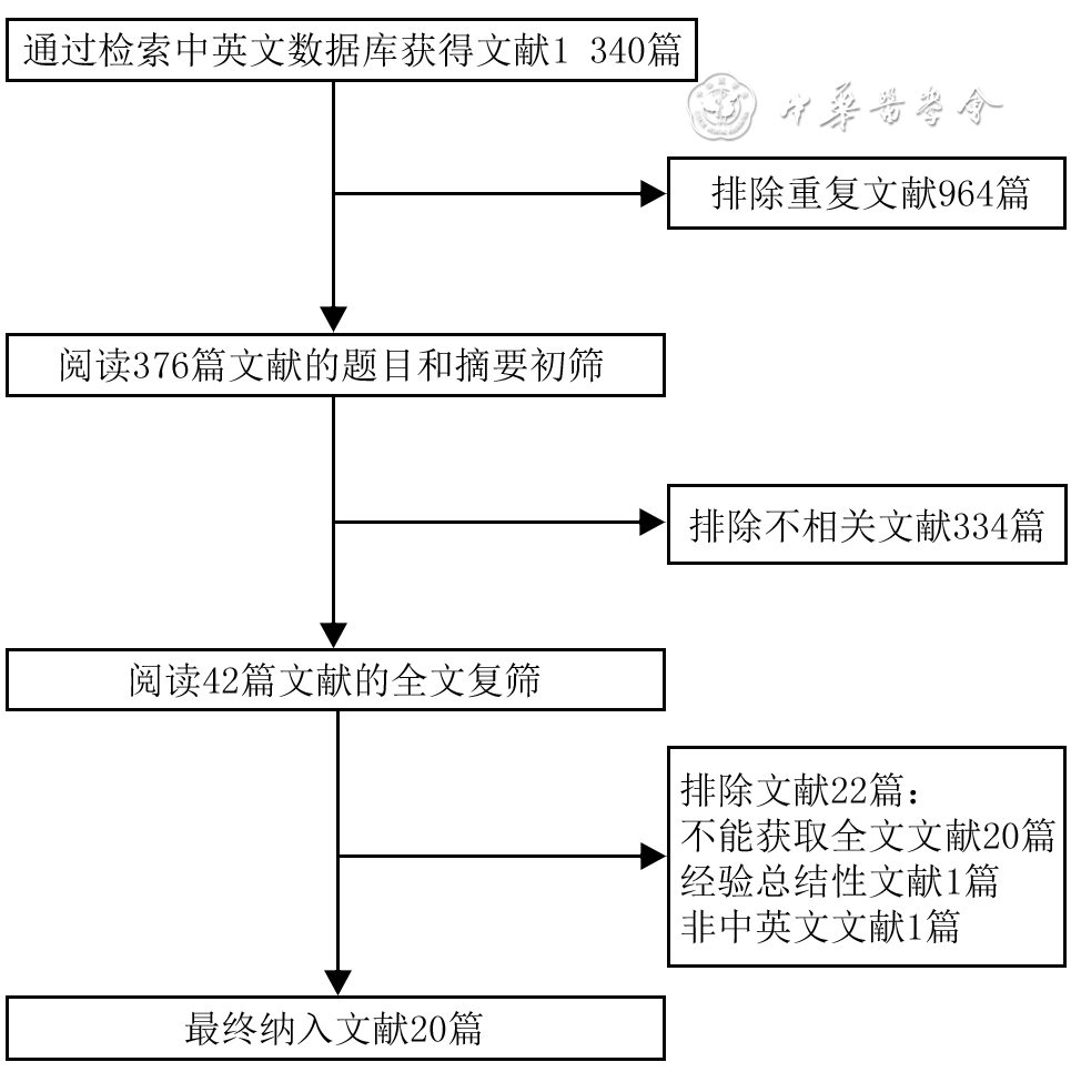
    Objective   To explore the fluid resuscitation strategy in shock stage in severely burned children with different burn areas in different age groups, and to evaluate the curative effect.    Methods   A retrospective cohort study was conducted. From January 2015 to June 2020, 235 children with severe and above burns who met the inclusion criteria were hospitalized in the First Affiliated Hospital of Nanchang University, including 150 males and 85 females, aged 3 months to 12 years. After admission, it was planned to rehydrate the children with electrolyte, colloid, and water according to the domestic rehydration formula for pediatric burn shock, and the rehydration volume and speed were adjusted according to the children's mental state, peripheral circulation, heart rate, blood pressure, and urine output, etc. The actual input volume and planned input volume of electrolyte, colloid, water, and total fluid of all the children were recorded during the 8 hours since fluid replacement and the first and second 24 hours after injury. According to urine output during the 8 hours since fluid replacement, all the children were divided into satisfactory urine output maintenance group (119 cases) with urine output ≥1 mL·kg-1·h-1 and unsatisfactory urine output maintenance group (116 cases) with urine output<1 mL·kg-1·h-1, and the electrolyte coefficient, colloid coefficient, and water coefficient of the children were calculated during the 8 hours since fluid replacement. According to the total burn area, children aged<3 80="" 155="" years="" and="" 3-12="" were="" divided="" into="" total="" body="" surface="" area="" group="">25%TBSA group, respectively. The electrolyte coefficient, colloid coefficient, water coefficient, and urine output of the children were calculated or counted during the first and second 24 hours after injury, and the non-invasive monitoring indicators of body temperature, heart rate, respiratory rate, and percutaneous arterial oxygen saturation and efficacy indicators of hematocrit, platelet count, hemoglobin, albumin, creatinine, and alanine aminotransferase (ALT) of the children were recorded 48 hours after injury. The prognosis and outcome indicators of all the children during the treatment were counted, including complications, cure, improvement and discharge, automatic discharge, and death. Data were statistically analyzed with independent sample or paired sample t test, Mann-Whitney U test, chi-square test, and Fisher's exact probability test.    Results   During the 8 hours since fluid replacement, the actual input volume of electrolyte of all the children was significantly more than the planned input volume, and the actual input volumes of colloid, water, and total fluid were significantly less than the planned input volumes (Z=13.094, 5.096, 13.256, 7.742, P<0.01). During the first and second 24 hours after injury, the actual input volumes of electrolyte of all the children were significantly more than the planned input volumes, and the actual input volumes of water and total fluid were significantly less than the planned input volumes (Z=13.288, -13.252, 3.867, 13.183, -13.191, 10.091, P<0.01), while the actual input volumes of colloid were close to the planned input volumes (P>0.05). During the 8 hours since fluid replacement, compared with those in unsatisfactory urine output maintenance group, there was no significant change in electrolyte coefficient or colloid coefficient of children in satisfactory urine output maintenance group (P>0.05), while the water coefficient was significantly increased (Z=2.574, P<0.05). Among children <3 years="" compared="" with="" those="" in="">25%TBSA group, the electrolyte coefficient and water coefficient of children were significantly increased and the urine output of children was significantly decreased in 15%-25%TBSA group during the first and second 24 hours after injury (Z=-3.867, -6.993, -3.417, -5.396, -5.062, 1.503, P<0.05 or P<0.01), while the colloid coefficient did not change significantly (P>0.05); the levels of efficacy indicators of hematocrit, platelet count, and hemoglobin at 48 h after injury were significantly increased, while ALT level was significantly decreased (Z=-2.720, -3.099, -2.063, -2.481, P<0.05 or P<0.01); the levels of the rest of the efficacy indicators and non-invasive monitoring indicators at 48 h after injury did not change significantly (P>0.05). Among children aged 3-12 years, compared with those in >25%TBSA group, the electrolyte coefficient and water coefficient of children in 15%-25%TBSA group were significantly increased during the first and second 24 hours after injury, the colloid coefficient during the second 24 h was significantly decreased (Z=-2.042, -4.884, -2.297, -3.448, -2.480, P<0.05 or P<0.01), while the colloid coefficient during the first 24 hours after injury, urine output during the first and second 24 hours after injury, and the non-invasive monitoring indicators and efficacy indicators at 48 hours after injury did not change significantly (P>0.05). Complications occurred in 17 children during the treatment. Among the 235 children, 211 cases were cured, accounting for 89.79%, 5 cases were improved and discharged, accounting for 2.13%, 16 cases were discharged automatically, accounting for 6.81%, and 3 cases died, accounting for 1.28%.    Conclusions   The electrolyte volume in early fluid resuscitation in severely burned children exceeding the volume calculated by the formula can obtain a good therapeutic effect. Among children<3 years old, the volume of fluid resuscitation should be appropriately increased in children with extremely severe burns compared with children with severe burns during fluid resuscitation; among children aged 3-12 years, the colloid volume should be appropriately increased in children with extremely severe burns compared with children with severe burns during fluid resuscitation; non-invasive monitoring indicators can be used to monitor hemodynamics and guide fluid resuscitation in severely burned children.
Original Articles
Effects and mechanism of age on the stiffness and the fibrotic phenotype of fibroblasts of human hypertrophic scar
Zhu Dongzhen, Yao Bin, Cui Xiaoli, Huang Sha, Fu Xiaobing
2021, 37(10): 937-945. doi: 10.3760/cma.j.cn501120-20200810-00374
Abstract:
  Objective  To explore the effects and potential molecular mechanism of age on the stiffness and the fibrotic phenotype of fibroblasts (Fbs) of human hypertrophic scar.  Methods  The experimental research method was used. From January to June 2020, the surgically removed hypertrophic scar tissue of 10 scar patients (4 males and 6 females) and residual full-thickness normal skin tissue of 10 cases (5 males and 5 females, aged 7-41 years) were collected after operation in Department of Burns and Plastic Surgery of the Fourth Medical Center of the PLA General Hospital. The hypertrophic scar tissue of 6 patients aged (10.7±1.6) years was included into the young group and the hypertrophic scar tissue of 4 patients aged (40.0±2.2) years was included into the elderly group according to the age of patients. For the normal skin tissue and scar tissue in the two groups, hematoxylin eosin (HE) staining was performed to observe the tissue morphology, Masson staining was performed to observe the morphology and arrangement of collagen and quantify the content of collagen, and scanning electron microscope was used to observe the microscopic difference of dermal collagen fibers after the samples were freeze-dried and metal coated. The stiffness of scar tissue in the two groups was measured by atomic force microscope under the liquid phase. The scar tissue in the two groups was collected and the Fbs were isolated and cultured. The morphological differences of the Fbs were observed under the inverted phase contrast microscope, and the protein expression of paxillin was detected with cellular immunofluorescence to reflect the morphology of the Fbs. Cellular immunofluorescence was used to detect the expressions of pro-fibrosis protein α-smooth actin (α-SMA), transforming growth factor-β1 (TGF-β1), and type Ⅰ collagen, mechanotransduction-related protein Yes-associated protein (YAP), and the proliferation-related protein Ki67. Real-time fluorescent quantitative reverse transcription polymerase chain reaction was used to detect the mRNA expressions of pro-fibrosis genes of TGF-β1, α-SMA, and type Ⅰ collagen, fibrosis inhibiting gene of TGF-β3, and mechanotransduction-related genes of Rho-associated protein 1 (ROCK1) and YAP. Data were statistically analyzed with one-way analysis of variance and least significant difference t test.  Results  HE staining showed that the epidermal layer of normal skin was uneven, and blood vessels and sweat glands could be seen in the dermal layer; the epidermal layer of the scar tissue in the two groups was relatively flat, and blood vessels and sweat glands were rare. Masson staining and scanning electron microscopy showed that the collagen fibers in normal skin arranged loosely and disorderly, while the collagen fibers in scar tissue of the two groups arranged densely and orderly, and the collagen fibers in scar tissue of the young group were denser than those of the elderly group. The collagen content in scar tissue of the young group and the elderly group was significantly higher than that of the normal skin tissue (t=8.02, 3.15, P<0.05 or P<0.01), and the collagen content in scar tissue of the elderly group was significantly lower than that of the young group (t=4.84, P<0.05). The dermal stiffness of scar tissue in the elderly group was (50.3±1.1) kPa, significantly higher than (35.2±0.8) kPa in the young group (t=11.43, P<0.05). There were no obvious differences in the morphology of scar Fbs in the two groups observed under inverted phase contrast microscope and by cellular immunofluorescence. The expressions of type Ⅰ collagen and TGF-β1 in scar Fbs cytoplasm of the elderly group were significantly higher than those in the young group, while the expressions of α-SMA in scar Fbs cytoplasm were close in the two groups. The expressions of YAP in cytoplasm and nucleus of scar Fbs in the elderly group were significantly higher than those in the young group, while the expressions of Ki67 in scar Fbs nucleus of the two groups were close. The mRNA expressions of TGF-β1 and type Ⅰ collagen in scar Fbs of the elderly group were significantly higher than those in the young group (t=2.87, 4.85, P<0.05 or P<0.01), the mRNA expression of TGF-β3 in scar Fbs of the elderly group was significantly lower than that in the young group (t=3.36, P<0.05), and the mRNA expressions of α-SMA in scar Fbs of the two groups were close (t=1.14, P>0.05). The mRNA expressions of ROCK1 and YAP in scar Fbs of the elderly group were significantly higher than those in the young group (t=2.98, 7.60, P<0.05 or P<0.01).  Conclusions  The elderly are prone to scar healing after skin injury. The molecular mechanism may be attributed to the production of extracellular matrix components with higher stiffness, which increases tissue stiffness and thereby activates the expressions of ROCK and YAP/transcriptional co-activator with PDZ-binding motif genes, promoting pro-fibrosis gene and protein expression.
Microbiological characteristics of patients with severe burns caused by blast and application of meta- genomics next-generation sequencing in the detection of pathogenic microorganisms
Luo Rubin, Huang Man, Hu Hang, Zhang Rong, Han Chunmao
2021, 37(10): 946-952. doi: 10.3760/cma.j.cn501120-20201017-00440
Abstract:
      Objective     To analyze the microbiological characteristics of patients with severe burns caused by blast in different periods and explore the application value of metagenomics next-generation sequencing (mNGS) in detecting pathogenic microorganisms.      Methods     The retrospective observational study was applied. From June 13 to September 13, 2020, twenty-three patients (21 males and 2 females) with severe burns caused by blast who met the inclusion criteria were admitted to the Second Affiliated Hospital of Zhejiang University School of Medicine, with age of (64±5) years and total burn area of (86±14) % total body surface area. Abbreviated burn severity index (ABSI) score, revised Baux score, acute physiology and chronic health status evaluation (APACHE) Ⅱscore, and sequential organ failure assessment (SOFA) score were counted on admission. Within 7, 8-20 and 21-30 d after admission, the complications, infection source and distribution of pathogenic microorganisms in patients were recorded. The detection of pathogenic microorganisms was analyzed, and the difference in detection efficiency between microbial culture method and mNGS was compared. After admission, the infection of overall source distribution of pathogenic microorganisms in patients was analyzed, and the difference in detection efficiency between microbial culture method and mNGS was compared. Data were statistically analyzed with McNemar and Fisher exact probability test.      Results     On admission, ABSI score, revised Baux score, APACHE Ⅱ score and SOFA score were (12.6±2.4), (91±22), (26±4), and (10.3±2.3) respectively. Within 7 d after admission, the main complications of patients were inhalation injury, septic shock, and hypoproteinemia. Patients were mainly infected with pathogenic microorganism on wound, blood stream, and lung. Within 8-20 d after admission, the incidence of septic shock was the highest. The incidence of inhalation injury was significantly lower than that of ≤7 d after admission (P<0.01), the main source of infection were wound, lung, and blood stream, and the incidence of wound and blood stream infection were significantly lower than that of ≤7 d after admission (P<0.01). Within 21-30 d after admission, the incidences of multiple organ failure and acute respiratory distress syndrome were low, the incidence of inhalation injury was significantly lower than that of ≤7 d after admission (P<0.01), and the incidence of septic shock was significantly lower than that of ≤7 d after admission (P<0.01) and 8-20 d after admission (P<0.01). There were only low bloodstream infections, and the incidence of wound infection was significantly lower than that of ≤7 d after admission (P<0.01) and 8-20 d after admission (P<0.05), and the incidences of lung and blood stream infection were significantly lower than those of ≤7 d after admission (P<0.01). Within ≤7 d after admission, gram-positive bacteria were mainly Staphylococcus aureus. Gram-negative bacteria were mainly Klebsiella pneumoniae and Stenotrophomonas maltophilia. The fungi contained only Candida. Within 8-20 d after admission, Staphylococcus aureus was mainly the gram-positive bacteria, and the detection rate of Enterococcus was significantly lower than that of ≤7 d after admission (P<0.01). Pseudomonas aeruginosa and Acinetobacter baumannii were the main gram-negative bacteria, and their detection rates were significantly lower than those of ≤7 d after admission (P<0.01).There was a new detection of Fusarium. Within 21-30 d after admission, Staphylococcus aureus was the mainly gram-positive bacteria, and the detection rates of Enterococcus and Bacillus were significantly lower than those of ≤7 d after admission (P<0.01). Pseudomonas aeruginosa and Acinetobacter baumannii were still the main gram-negative bacteria, and increased with the extension of time after admission. The detection rate of Pseudomonas aeruginosa was significantly higher than that of ≤7 d after admission (P<0.01) and 8-20 d after admission (P<0.01), and the detection rate of Acinetobacter baumannii was significantly higher than that of ≤7 d after admission (P<0.01). The detection rate of Klebsiella pneumoniae was significantly lower than those of ≤7 d after admission (P<0.01) and 8-20 d after admission (P<0.01). All  Candida, Mould, Fusarium were detected. Within ≤7 d and 8-20 d, the consistency between mNGS and bacterial culture was high (κ=0.659, 0.596). Within 21-30 d after admission, the consistency between mNGS and bacterial culture was moderate (κ=0.407). In different time periods, the positive test rate of mNGS was basically constant, while that of microbial culture showed a decline with the extension time after admission. Five hundred and six strains of pathogenic microorganisms were isolated from wound, blood, sputum, and indwelling catheter, and Staphylococcus aureus, Pseudomonas aeruginosa, Acinetobacter baumannii, Klebsiella pneumoniae were the main pathogenic microorganisms. Pseudomonas aeruginosa and Acinetobacter baumannii were the most common in the wound samples, Klebsiella pneumoniae was more often seen in blood samples while Pseudomonas aeruginosa and Acinetobacter baumannii in sputum samples, and Acinetobacter baumannii in indwelling catheter samples were the most common. The detection rates of Pseudomonas aeruginosa in wound and sputum were significantly higher than those of blood (P<0.05 or P<0.01) and indwelling catheter (P<0.01), respectively. The consistency between the overall results of mNGS and microbial culture were moderate (κ=0.556). The consistency between mNGS and microbial culture was high in samples of blood and indwelling catheter (κ=0.631, 0.619), but those were moderate in sputum and wound (κ=0.558, 0.528).      Conclusions     The most common infections of patients with severe burn caused by blast injury were wound infection and blood stream infection. With the extension of time after admission, the main pathogenic bacterial strains of patients changed from Staphylococcus aureus, Klebsiella pneumoniae, and Stenotrophomonas maltophilia to Acinetobacter baumannii and Pseudomonas aeruginosa. mNGS showed a higher positive rate of detecting pathogenic microorganisms than conventional microbial culture.
Application effects of free transplantation of autologous omentum in treating maxillofacial infection complicated with complex sinus tract
Ren Pan, Cao Jin, Ma Fuxin, Zhou Shuai, Yu Zhen, Zhao Congying
2021, 37(10): 953-958. doi: 10.3760/cma.j.cn501120-20200721-00354
Abstract:
    Objective   To investigate the application effects of free transplantation of autologous omentum in treating maxillofacial infection complicated with complex sinus tract.    Methods   The retrospective observational study method was used. Four patients with maxillofacial infection complicated with complex sinus tract were admitted to Department of Burns and Plastic Surgery of the Second Affiliated Hospital of Air Force Medical University from July 2017 to the December 2019, including 3 males and 1 female aged 36-60 years. Preoperative facial computed tomography (CT) was performed on patients for calculating the volume of sinus tract. During the operation, the sinus tract was thoroughly debrided, and the volume of the tissue defect was about 5 cm×3 cm×2 cm-10 cm×5 cm×3 cm after debridement. The tissue defect area was filled with omentum of 100-300 mL which was cut under laparoscopy. The artery and vein on the right side of the omentum were reserved as the vascular pedicle of the donor area, which were anastomosed with the facial artery and external jugular vein of the recipient area. The survival of omentum, and the occurrences of reinfection and complication were observed after operation, respectively. On the 10th day and in 1 month after the operation, the blood supply of omentum was examined by colored Doppler ultrasound and CT angiography, and the filling of tissue defect area was examined by head and face CT. During follow-up after the operation, the recoveries of face appearance and function and scar hyperplasia in the donor area.    Results   The transplanted omentums in 4 patients survived after the operation with no reinfection and complication.  On the 10th day and in 1 month after the operation, the transplanted omentums had good blood supply, and the filled area with omentum was in good shape, without formation of dead cavity. During follow-up of 6-10 months after surgery, the appearance and function of face recovered well, and there was no obvious scar hyperplasia in the donor area.    Conclusions   After free transplantation of omentum in treating maxillofacial infection with complex sinus tract, the patients have good facial appearance and function, and the application of laparoscopy results in little damage to the patients and quick postoperative recovery.
Experimental study on the effect of three-dimensional porous structures on the vascularization rate of artificial dermis
Tan Rongwei, Liu Xi, Chen Yingying, Xu Mengqiang, Guo Yuanjun, Wang Danyan, Liang Jiamei, Liu Jiao, Yuan Shasha, Fan Wei, Wang Xiangkun, She Zhending
2021, 37(10): 959-969. doi: 10.3760/cma.j.cn501120-20200715-00347
Abstract:
  Objective  To explore the effects of orienting three-dimensional porous network (type A) and honeycomb briquette-shaped vertically penetrating three-dimensional porous network (type B) on the vascularization rate of artificial dermis.  Methods  The experimental research method was used. The artificial dermis was composed of a double layer of silicone layer and scaffold layer. Based on the difference of scaffold layer, they were divided into type A and type B artificial dermis (type A dermis and type B dermis, for short) containing type A and type B structure, respectively. The type A and type B structures were prepared by gradient freeze-drying technique and physical pore-making technique, respectively. The micro-morphology of two kinds of dermis scaffold was observed by scanning electron microscopy. The porosity of two kinds of dermis scaffold was measured by the Pyrex method. According to the method of national medical industry standard, the hydroxyproline content in degradation liquids and their residues in two kind of dermis were determined after degradation at 4, 8, 13, and 24 h, reflecting the degradation rates of two kinds of dermis. According to the random number table, L929 cells were divided into type A dermis group, type B dermis group, negative control group, and positive control group. The positive control group was added with minimum essential medium (MEM) containing 5% dimethyl sulfoxide, The negative control group was added with high-density polyethylene extract, and the other two groups were added with the corresponding extract. At 24 hours after culture, the growth rate of L929 cells was detected by methyl thiazolyl tetrazolium, and the cytotoxicity was graded. L929 cells and human umbilical vein endothelial cells (HUVECs) were inoculated into pore plates with two kinds of dermis preinstalled. On 1, 4, 7, and 14 d after inoculating, the adhesion and growth of L929 cells on the surfaces of the two kinds of scaffolds were detected by immunofluorescence method. On 7 d after inoculating, the migration of the above two kinds of cells into the two kinds of dermal scaffolds was detected by immunofluorescence and hematoxylin-eosin (HE) staining. Three full-thickness skin defect wounds of 5.0 cm×5.0 cm were created on both sides of the back of three 6-month-old healthy male Ba-Ma mini pigs. According to the random number table, six columns of wounds were divided into type A dermis two-step method group, type B dermis two-step method group, and type B dermis one-step method group. The wounds in type A dermis two-step method group and type B dermis two-step method group were transplanted with type A or type B dermis respectively before, and with autologous split-thickness skin grafting later. The wounds in type B dermis one-step method group were transplanted in a synchronous procedure including type B dermis (without silicone layer) and autologous skin grafting simultaneously. The bleeding, exudation, and infection of the wounds on the back in type A dermis two-step method group and type B dermis two-step method group on the 7th day after the second transplantation and in type B dermis one-step method group on the 14th day after the first transplantation were generally observed. The area of autologous skin graft was measured by the transparent film grid method, and the survival rate of autologous skin was calculated. On 4, 7, and 14 d after the first transplantation, the inflammatory cells, fibroblasts (Fbs), and capillary infiltration into the scaffolds of the three groups were detected by HE staining. On 7, 14 d after the first transplantation, the vascularization of the scaffolds was further observed by immunohistochemistry. On 28, 90 d after the first operation, the degradation of the scaffolds of type A dermis and type B dermis was observed by HE staining. Data were statistically analyzed with one-way analysis of variance, independent sample t test, and Bonferroni correction.  Results  A large number of round and oval micropores were evenly distributed on the surface of type A scaffold, and the cylindrical hole walls could be observed arranging in a parallel direction in the longitudinal section. The honeycomb briquette-shaped penetrating macropores on the surface of type B scaffold were arranged in an orderly matrix. The pore walls of the honeycomb briquette-shaped penetrating macropores were connected by micropores to form a network structure. The porosity of type A dermis was (93.21±0.72)%, which was similar to (95.88±1.00)% of type B dermis (t=4.653, P>0.05). The degradation rates of type A dermis at 4, 8, 13, and 24 h were similar to those of type B dermis at the corresponding time point (t=0.232, 0.856, 0.258, 7.716, P>0.05). At 24 h after culture, the proliferation rates of L929 cells in the type A dermis group, type B dermis group, and negative control group were significantly higher than those of the positive control group (t=2 393.46, 2 538.27, 1 077.77, P<0.01). The cytotoxicity rating of cells in positive control group was grade 4, while that of the other three groups was grade zero. On 1, 4, 7, and 14 d after inoculation, both L929 cells and HUVECs proliferated in a time-dependent manner in two kinds of dermal scaffolds. The adhesion growth and proliferation rate of the two kinds of cells on the surface of type B dermis was higher than that of type A dermis. On 7 d after inoculation, both L929 cells and HUVECs covered the surface of type B dermis and migrated into one side of the silicone layer. However, the above two kinds of cells migrated slowly into type A dermis, and only a few cells were found on one side of the silicone layer. There was no bleeding, exudation, or infection in the wounds repaired by type A and type B dermis. The survival rate of autologous skin grafting of 6 wounds in each group was 100%. On 4, 7, and 14 d after the first operation, inflammatory cells, Fbs, and capillaries gradually infiltrated into the scaffold layer, and the cell infiltration rate from high to low was type B dermis one-step method group, type B dermis two-step method group, and type A dermis two-step method group. The scaffold in wound in the type B dermis one-step method group gradually collapsed on 28 d after the first operation, and completely degraded in 3 months after the first operation. The scaffold degradation rate of type A dermis two-step method group was similar to that mentioned above.  Conclusions  The honeycomb briquette-shaped vertically penetrating three-dimensional porous network structure of type B scaffold can accelerate its vascularization process, which is beneficial to autogenous split-thickness skin in one-step procedure to repair full-thickness skin defects wound in Ba-Ma mini pigs. Compared with the "two-step method" of staged transplantation of type A scaffold and autologous split-thickness skin, and one-step transplantation has equal efficacy and can provide a better choice for wound treatment.
Original Article·Nursing Column
Scoping review on prevention of central venous catheter-related bloodstream infection in burn patients
Chen Lijuan, Liu Lihong, Sun Linli, Chen Nana, Meng Meifen
2021, 37(10): 970-977. doi: 10.3760/cma.j.cn501120-20201027-00447
Abstract:
  Objective  To investigate the research trend and scope of prevention of central venous catheter-related bloodstream infection (CRBSI) in burn patients.  Methods  The scoping review method was adopted. Pre-retrieval was carried out with search terms of "中心静脉导管, 烧伤, 感染" and "central venous catheter, infection, catheter-related bloodstream infection, burn". On the basis of pre-retrieval, different retrieval formulas were formulated to retrieve researches related to central venous CRBSI in burn patients in China National Knowledge Internet, Wanfang Database, VIP Database, PubMed, Embase, CINAHL, and Cochrane Library from the establishment of each database to August 2020. Data were extracted from the included literature, including the first author, research publication time, research country, research type, diagnosis basis and intervention measures of central venous CRBSI, research sample selection, incidence related to infection, and research conclusion.  Results  A total of 20 randomized controlled trials, quasi-experimental studies, case-control studies, cohort studies, and implementation researches published in 1990-2020 were included in this study with the first authors from China, the United States of America, or Argentina. The diagnostic bases for central venous CRBSI in burn patients were not uniform in the included literature, including adopting the Guidelines of American Centers for Disease Control and Prevention, Diagnostic Criteria for Nosocomial Infection, and other diagnostic criteria without specifying the source. The intervention measures included the use of new materials such as antibiotics coated catheter and ethanol impregnated port protectors, multidisciplinary cooperation, and comprehensive preventive measures. The sample size in the included literature was small, and the sample selection was different, including the number of patients and the the number of placement of central venous catheter. The outcome indicators for infection in the included literature were diversified. The incidence per 1 000 days of central venous CRBSI was 20.41‰-29.1‰ of patients in control group in China, the incidence per 1 000 days of central venous CRBSI was mostly <16.6‰ in control group in foreign countries, and the incidence of central venous CRBSI was decreased to varying degrees after implementing the corresponding intervention measures. Related research conclusions showed that new materials, multidisciplinary cooperation, and comprehensive preventive measures had good effects on prevention of central venous CRBSI in burn patients.  Conclusions  The researches on prevention of central venous CRBSI in burn patients in China start early and the research types are diversified. The diagnostic criteria of central venous CRBSI in burn patients are not uniform, intervention measures have shifted from standardizing relevant operational measures to exploring the prevention effects of new materials, multidisciplinary cooperation, and multiple measures, and the latter has good effects on preventing central venous CRBSI in burn patients.
Sinicization, cross-cultural adjustment and reliability and validity test of the Burnt Hand Outcome Tool
Zhang Juan, Chai Xuejun, Guan Lifeng, Ma Yuelan, Zhang Xiaowen, Yao Miao
2021, 37(10): 978-986. doi: 10.3760/cma.j.cn501120-20210201-00042
Abstract:
  Objective  To introduce the Burnt Hand Outcome Tool (BHOT) into China to conduct sinicization, cross-cultural debugging, and test the reliability and validity, thus provide a reference for the evaluation of treatment effect and making of medical decisions of hand burn patients.  Methods  The cross-sectional research was conducted. After obtaining the authorization of the original author, the Brislin translation mode was used to literally translate, synthesize, back-translate, and be checked by experts to form the Chinese version of BHOT (C-BHOT) 2. Ten experts in the field of burn rescue and treatment were invited by purpose sampling method to conduct cross-cultural debugging to form C-BHOT 3. A total of 21 hand burn patients who were treated in General Hospital of Ningxia Medical University from January to March 2020 and conformed to the inclusion criteria were selected by accidental sampling method for pre-testing. Then the final version of C-BHOT 4 was formed. Hospitalized patients who met the selection criteria were recruited using accidental sampling, multi-site and cross-regional survey method from May 2020 to March 2021 in General Hospital of Ningxia Medical University, the First Affiliated Hospital of Air Force Medical University, the First Affiliated Hospital of Army Medical University (the Third Military Medical University), and the First Affiliated Hospital of Nanchang University. The survey was performed with the general information questionnaire and C-BHOT 4. Critical ratio method and correlation coefficient method were used for item analysis, Cronbach's α coefficient test tool was used to test internal consistency, test-retest reliability was used to judge the stability of the tool, content validity index analysis tool was used to analyze the content validity, and exploratory factor analysis was used to test the validity of the structure. Data were statistically analyzed with independent sample t test or Pearson correlation analysis.  Results  After the sinicization, cross-cultural debugging, and pre-testing, C-BHOT 4 was formed with a total of 20 items. All items were adjusted to positive scoring, and 7 modifications were made on the basis of the original tool. A total of 353 questionnaires were issued in the formal survey, and 344 valid questionnaires were returned, with an effective response rate of 97.45%. In the 344 patients, there were more male patients (297 patients) than female patients (47 patients), with ages of 23 to 57 years. The critical ratios of each item in the item analysis ranged from 5.48 to 12.59 (P<0.05). There were statistically significant differences in the scores between patients in high-score group and low-score group (t=-10.72--2.84, P<0.05 or P<0.01). There was significant positive relationship among the scores of each two items and between scores of each item and the overall tool score (r=0.68-0.71, 0.47-0.63, P<0.05 or P<0.01). In reliability test, the Cronbach's α coefficient of the whole tool was 0.837, and the test-retest reliability was 0.702-0.793. In validity test, the content validity index of whole tool was 0.923. The exploratory factor analysis extracted 3 common factors, and the cumulative variance contribution rate was 76.162%. The comparative fit index in the confirmatory factor analysis was 0.924.  Conclusions  After the BHOT tool being sinicized, cross-culturally debugged, pre-tested, and tested for reliability and validity, it was verified that C-BHOT 4 has good reliability and validity in assessing the outcome of Chinese hand burn patients, and therefore can be used as an effective tool for the evaluation.
Technique and Method
Explorative study of the immobilizing effect of full-thickness skin subcutaneous grafting on allogeneic full-thickness skin graft in rats
Rong Xiangke, Wang Kai, Wang Tong, Yang Jizhong, Ding Jianke, Dang Juanli, Yu Zhou, Yi Chenggang
2021, 37(10): 987-989. doi: 10.3760/cma.j.cn501120-20200801-00365
Abstract:
  Objective  To investigate the immobilizing effect of full-thickness skin subcutaneous grafting on allogeneic full-thickness skin graft in rats.  Methods  The experimental research method was used. The inbred male Brown-Norway rats (n=10) and Lewis rats (n=10) were used as donors and recipients respectively. After subcutaneously full-thickness separation of a 2.2 cm×2.2 cm area on the nape of the recipient rat, a full-thickness skin of 2.0 cm×2.0 cm taken from the abdomen of the donor rat was subcutaneously grafted, and the donor site was pulled together and sutured. The autologous skin over the allograft in the recipient rat was excised 5-6 d after grafting, and the stitches were removed 7 d after excision. Within 2 months after grafting, the feeding, activity, and survival of the donor and recipient rats, behavior of tearing and scratching the wounds of the recipient rats, the wound condition after autologous skin excision in recipient rats, and the survival and hair growth of the grafted allogeneic skin were observed.  Results  Within 2 months after grafting, the donor and recipient rats all ate normally and could move freely with no abnormal death. No tearing or scratching of the wounds occurred in recipient rats. There was a small amount of exudation and partial epidermal desquamation after autologous skin excision in recipient rats. All transplanted allografts survived, which were free of infection and necrosis, with new hairs growing out smoothly.  Conclusions  The immobilizing method of full-thickness skin subcutaneous grafting of allogeneic full-thickness skin graft in rats is simple and time-saving without postoperative dressing change, with reliable pressure fixation and high survival rate of skin grafts, which can be promoted for animal skin grafting models.
Reviews
Research advances on the application of platelet concentrate products in wound repair
Li Yulin, Chen Nuo, Xie Weiguo
2021, 37(10): 990-995. doi: 10.3760/cma.j.cn501120-20200730-00363
Abstract:
Multiple growth factors are required to regulate the wound repair process. Currently, there is still a lack of potent, durable, and inexpensive growth factor product in clinical practice. Meanwhile, platelet concentrate products provide an economical, convenient, and effective choice for the treatment of wounds in clinical practice. Starting from the first generation of platelet concentrate products, platelet-rich plasma, researchers have focused on optimizing the performance of platelet concentrate products for higher stability, more abundant bioactive factors, and more optimal clinical performance. Platelet-rich fibrin and concentrated growth factor emerged since then. Platelet concentrate products have the effect of promoting the speed of wound healing and enhancing the quality. This article aims to summarize the history and classification of platelet concentrate products, compare their properties and clinical applications, elaborate their mechanism of action, and analyze the problems existing in relevant researches while prospecting the future development of this field.
Research advances on autologous hair transplantation in the treatment of secondary cicatricail alopecia
He Lirun, Miao Yulan, Zheng Shuqin
2021, 37(10): 996-999. doi: 10.3760/cma.j.cn501120-20200827-00392
Abstract:
Secondary cicatricial alopecia (hereinafter referred to as cicatricial alopecia) after burn and trauma affects the aesthetic appearance and even the physical and mental health of patients, and reduces their quality of life and happiness. Autologous hair transplantation provides an idea for the treatment of cicatricial alopecia, which makes the transplanted hair show a natural growth state. This paper introduces the cicatricial alopecia and autologous hair transplantation and reviews the application and limitations of autologous hair transplantation in treating cicatricial alopecia.
Research progress on the mechanism of wound skin dys- pigmentation after burns
Guo Xiaoyu, Xie Weiguo
2021, 37(10): 1000-1004. doi: 10.3760/cma.j.cn501120-20200714-00344
Abstract:
Burn wound healing often shows a certain degree of pigmentation disorder. It may not only cause cosmetic and psychological issues affecting patient's normal social activities, but also increase risk of skin cancer or photoaging. Although normal skin pigmentation is fairly well studied, the mechanism that leads to dyspigmentation after burn injury needs to be further explored. Based on summarizing the mechanism of normal skin pigmentation, this paper reviews the latest research progress in postburn dyspigmentation in recent years.