2023 Vol. 39, No. 2

Expert Forums
Some thoughts on the research of mesenchymal stem cell exosomes and wound microenvironment
Wang Dali
2023, 39(2): 101-105. doi: 10.3760/cma.j.cn501225-20230112-00014
Abstract:
Since researchers have found that the conditioned medium and exosomes of mesenchymal stem cells (MSCs) had the biological effects equivalent to those of MSCs, MSC exosomes (MSC-Exos), the representative product of MSCs' paracrine effect, have become the research focus of the "cell-free" therapy of MSCs. However, most researchers currently use conventional culture condition to culture MSCs and then isolate exosomes for the treatment of wound or other diseases. Theoretically, the paracrine effect of MSCs is directly associated with the pathological condition of the wound (disease) microenvironment or in vitro culture condition, and their paracrine components and biological effects may be altered with the changes of the wound (disease) microenvironment or in vitro culture condition. Thus, the feasibility of using traditional culture condition to culture MSCs for exosome extraction for the treatment of different diseases without considering the actual situation of the disease to be treated needs further discussion. Therefore, the author suggests that the research of MSC-Exos should consider the microenvironment of the wound (disease) to be treated. as much as possible, otherwise the extracted MSC-Exos may not be "accurate" or may not really achieve the treatment effect of MSCs. In this article, we summarized some thoughts of the author and problems related to the researches about MSC-Exos and wound microenvironment, and hoped to discuss with researchers.
The regulatory role and related mechanisms of macrophages in wound healing
He Weifeng, Yan Lingfeng
2023, 39(2): 106-113. doi: 10.3760/cma.j.cn501225-20230110-00010
Abstract:
Wound healing is a complex process under precise regulation, including multiple stages such as inflammation, anti-inflammatory, and regeneration. Macrophages play an important regulatory role in the differentiated process of wound healing due to their obvious plasticity. If macrophages fail to express specific functions in a timely manner, it will affect the healing function of tissues and lead to pathological tissue healing. Therefore, it is of great significance to understand the different functions of different types of macrophages and to regulate them specifically in different stages of wound healing to promote the healing and regeneration of wound tissue. In this paper, we illustrate the different functions of macrophages in the wound and their basic mechanisms, according to the basic process of wound healing, and emphasize the strategies of macrophage regulation that may be applied to clinical treatment in the future.
Original Articles · Wound Healing and Its Regulation Mechanisms
A prospective study on application of human umbilical cord mesenchymal stem cells combined with autologous Meek microskin transplantation in patients with extensive burns
Yan Tiantian, Xiao Rong, Wang Ying, Lin Guo'an, Zheng Yin, Zhao Hui, Li Wenjun, Shang Xinzhi, Meng Jinsong, Hu Dongsheng, Li Song, Wang Chao, Lin Zhichen, Chen Hongchang, Zhao Dongyan, Tang Di
2023, 39(2): 114-121. doi: 10.3760/cma.j.cn501225-20220728-00319
Abstract:
  Objective  To investigate the effects of human umbilical cord mesenchymal stem cells (hUCMSCs) combined with autologous Meek microskin transplantation on patients with extensive burns.  Methods  The prospective self-controlled study was conducted. From May 2019 to June 2022, 16 patients with extensive burns admitted to the 990th Hospital of PLA Joint Logistics Support Force met the inclusion criteria, while 3 patients were excluded according to the exclusion criteria, and 13 patients were finally selected, including 10 males and 3 females, aged 24-61 (42±13) years. A total of 20 trial areas (40 wounds, with area of 10 cm×10 cm in each wound) were selected. Two adjacent wounds in each trial area were divided into hUCMSC+gel group applied with hyaluronic acid gel containing hUCMSCs and gel only group applied with hyaluronic acid gel only according to the random number table, with 20 wounds in each group. Afterwards the wounds in two groups were transplanted with autologous Meek microskin grafts with an extension ratio of 1∶6. In 2, 3, and 4 weeks post operation, the wound healing was observed, the wound healing rate was calculated, and the wound healing time was recorded. The specimen of wound secretion was collected for microorganism culture if there was purulent secretion on the wound post operation. In 3, 6, and 12 months post operation, the scar hyperplasia in wound was assessed using the Vancouver scar scale (VSS). In 3 months post operation, the wound tissue was collected for hematoxylin-eosin (HE) staining to observe the morphological changes and for immunohistochemical staining to observe the positive expressions of Ki67 and vimentin and to count the number of positive cells. Data were statistically analyzed with paired samples t test and Bonferronni correction.  Results  In 2, 3, and 4 weeks post operation, the wound healing rates in hUCMSC+gel group were (80±11)%, (84±12)%, and (92±9)%, respectively, which were significantly higher than (67±18)%, (74±21)%, and (84±16)% in gel only group (with t values of 4.01, 3.52, and 3.66, respectively, P<0.05). The wound healing time in hUCMSC+gel group was (31±11) d, which was significantly shorter than (36±13) d in gel only group (t=-3.68, P<0.05). The microbiological culture of the postoperative wound secretion specimens from the adjacent wounds in 2 groups was identical, with negative results in 4 trial areas and positive results in 16 trial areas. In 3, 6, and 12 months post operation, the VSS scores of wounds in gel only group were 7.8±1.9, 6.7±2.1, and 5.4±1.6, which were significantly higher than 6.8±1.8, 5.6±1.6, and 4.0±1.4 in hUCMSC+gel group, respectively (with t values of -4.79, -4.37, and -5.47, respectively, P<0.05). In 3 months post operation, HE staining showed an increase in epidermal layer thickness and epidermal crest in wound in hUCMSC+gel group compared with those in gel only group, and immunohistochemical staining showed a significant increase in the number of Ki67 positive cells in wound in hUCMSC+gel group compared with those in gel only group (t=4.39, P<0.05), with no statistically significant difference in the number of vimentin positive cells in wound between the 2 groups (P>0.05).  Conclusions  The application of hyaluronic acid gel containing hUCMSCs to the wound is simple to perform and is therefore a preferable route. Topical application of hUCMSCs can promote healing of the autologous Meek microskin grafted area in patients with extensive burns, shorten wound healing time, and alleviate scar hyperplasia. The above effects may be related to the increased epidermal thickness and epidermal crest, and active cell proliferation.
Effects of tumor necrosis factor-alpha/extracellular signal-regulated kinase pathway on migration ability of HaCaT cells and full-thickness skin defects in mice
Zhao Shihan, Jin Linbo, Zhang Jianghe, Zhang Yiming, Fan Dongli
2023, 39(2): 122-131. doi: 10.3760/cma.j.cn501225-20221019-00460
Abstract:
  Objective  To investigate the effects of tumor necrosis factor-alpha (TNF-α)/extracellular signal-regulated kinase (ERK) pathway on the migration ability of HaCaT cells and full-thickness skin defects in mice.  Methods  The experimental research method was adopted. According to the random number table (the same below), HaCaT cells were divided into the normal oxygen group and the hypoxia group cultured under hypoxia (with oxygen volume fraction of 1%, the same below) condition. After 24 hours of culture, the significantly differentially expressed genes between the 2 groups were screened using the microarray confidence analysis software SAM4.01. The significance of the number of each gene in the signaling pathway was analyzed through the Kyoto encyclopedia of genes and genomes to screen the significantly differentially signaling pathways (n=3). HaCaT cells were cultured for 0 (immediately), 3, 6, 12, and 24 h under hypoxia condition. The secretion level of TNF-α was detected by enzyme-linked immunosorbent assay (ELISA), and the number of samples was 5. HaCaT cells were divided into normal oxygen group, hypoxia alone group, and hypoxia+inhibitor group cultured with FR180204 (an ERK inhibitor) and under hypoxia condition. The cells were cultured for 3, 6, 12, and 24 h. The migration ability of the cells was detected by scratch test (n=12). The expressions of phosphorylated nuclear factor kappa B (p-NF-κB), phosphorylated p38 (p-p38), phosphorylated ERK1/2 (p-ERK1/2), N-cadherin, and E-cadherin in HaCaT cells were detected by Western blotting under hypoxic condition for 0, 3, 6, 12, and 24 h (n=3). Sixty-four BALB/c male mice aged 6 to 8 weeks were used to make a full-thickness skin defect wound model on the dorsum of the mice. The mice were divided into the blank control group and the inhibitor group treated with FR180204, with 32 mice in each group being treated accordingly. On post injury day (PID) 0, 3, 6, 9, 12, and 15, the wound conditions of mice were observed and the healing rate was calculated (n=8). On PID 1, 3, 6, and 15, hematoxylin-eosin staining was used to observe neovascularization, inflammatory cell infiltration, and epidermal regeneration on wound, Masson staining was used to observe collagen deposition on wound, the expressions of p-NF-κB, p-p38, p-ERK12, N-cadherin, and E-cadherin in wound tissue were detected by Western blotting (n=6), the number of Ki67 positive cells and the absorbance value of vascular endothelial growth factor (VEGF) were detected by immunohistochemistry (n=5), the protein expressions of interleukin 6 (IL-6), IL-10, IL-1β, and CCL20 in wound tissue were detected by ELISA (n=6). Data were statistically analyzed with one-way analysis of variance, analysis of variance for repeated measurement, factorial design analysis of variance, Tukey test, least significant difference test, and independent sample t test.  Results  After 24 hours of culture, compared with normal oxygen group, 7 667 genes were up-regulated and 7 174 genes were down-regulated in cells in hypoxic group. Among the above differentially expressed genes, the TNF-α signaling pathway had significant change (P<0.05) with large number of genes. Under hypoxia condition, the expression of TNF-α at 24 h of cell culture was (11.1±2.1) pg/mL, which was significantly higher than (1.9±0.3) pg/mL at 0 h (P<0.05). Compared with normal oxygen group, the migration ability of cells in hypoxia alone group was significantly enhanced at 6, 12, and 24 h of cell culture (with t values of 2.27, 4.65, and 4.67, respectively, P<0.05). Compared with hypoxia alone group, the migration ability of cells in hypoxia+inhibitor group was significantly decreased at 3, 6, 12, and 24 h of cell culture (with t values of 2.43, 3.06, 4.62, and 8.14, respectively, P<0.05). Under hypoxia condition, the expressions of p-NF-κB, p-ERK1/2, and N-cadherin were increased significantly at 12 and 24 h of cell culture compared with 0 h of culture (P<0.05), the expression of p-p38 was significantly increased at 3, 6, 12, and 24 h of cell culture (P<0.05), the expression of E-cadherin was significantly decreased at 6, 12, and 24 h of cell culture (P<0.05), the expression of p-ERK1/2, p-NF-κB, and E-cadherin was time-dependent. Compared with blank control group, on PID 3, 6, 9, 12, and 15, the wound healing rate of mice in inhibitor group was significantly decreased (P<0.05); there were more inflammatory cell infiltration around the wound edge of mice in inhibitor group on PID 3, 6, and 15, especially on PID 15, a large number of tissue necrosis and discontinuous new epidermal layer were observed on the wound surface, and collagen synthesis and new blood vessels were reduced; the expression of p-NF-κB in the wound of mice in inhibitor group was significantly decreased on PID 3 and 6 (with t values of 3.26 and 4.26, respectively, P<0.05) but significantly increased on PID 15 (t=3.25, P<0.05), the expressions of p-p38 and N-cadherin were significantly decreased on PID 1, 3, and 6 (with t values of 4.89, 2.98, 3.98, 9.51, 11.69, and 4.10, respectively, P<0.05), the expression of p-ERK1/2 was significantly decreased on PID 1, 3, 6, and 15 (with t values of 26.69, 3.63, 5.12, and 5.14, respectively, P<0.05), the expression of E-cadherin was significantly decreased on PID 1 (t=20.67, P<0.05) but significantly increased on PID 6 (t=2.90, P<0.05); the number of Ki67 positive cells and absorbance value of VEGF of wound in inhibitor group were significantly decreased on PID 3, 6, and 15 (with t values of 4.20, 7.35, 3.34, 4.14, 3.20, and 3.73, respectively, P<0.05); the expression of IL-10 in the wound tissue of the inhibitor group was significantly decreased on PID 6 (t=2.92, P<0.05), the expression of IL-6 was significantly increased on PID 6 (t=2.73, P<0.05), the expression of IL-1β was significantly increased on PID 15 (t=3.46, P<0.05), and CCL20 expression levels were significantly decreased on PID 1 and 6 (with t values of 3.96 and 2.63, respectively, P<0.05) but significantly increased on PID 15 (t=3.68, P<0.05).  Conclusions  The TNF-α/ERK pathway can promote the migration of HaCaT cells, and regulate the healing of full-thickness skin defect wounds in mice by affecting the expression of inflammatory cytokines and chemokines.
Influence of autologous adipose stem cell matrix gel on wound healing and scar hyperplasia of full-thickness skin defects in rabbit ears
Li Liang, Bai Nan, Fu Yanjie, Wu Can, Zhang Yujiao, Chen Yuanzheng
2023, 39(2): 132-140. doi: 10.3760/cma.j.cn501225-20221020-00463
Abstract:
  Objective  To investigate the influence of autologous adipose stem cell matrix gel on wound healing and scar hyperplasia of full-thickness skin defects in rabbit ears, and to analyze the related mechanism.  Methods  Experimental research methods were adopted. The complete fat pads on the back of 42 male New Zealand white rabbits aged 2 to 3 months were cut to prepare adipose stem cell matrix gel, and a full-thickness skin defect wound was established on the ventral side of each ear of each rabbit. The left ear wounds were included in adipose stem cell matrix gel group (hereinafter referred to as matrix gel group), and the right ear wounds were included in phosphate buffer solution (PBS) group, which were injected with autologous adipose stem cell matrix gel and PBS, respectively. The wound healing rate was calculated on post injury day (PID) 7, 14, and 21, and the Vancouver scar scale (VSS) scoring of scar tissue formed on the wound (hereinafter referred to as scar tissue) was performed in post wound healing month (PWHM) 1, 2, 3, and 4. Hematoxylin-eosin staining was performed to observe and measure the histopathological changes of wound on PID 7, 14, and 21 and the dermal thickness of scar tissue in PWHM 1, 2, 3, and 4. Masson staining was performed to observe the collagen distribution in wound tissue on PID 7, 14, and 21 and scar tissue in PWHM 1, 2, 3, and 4, and the collagen volume fraction (CVF) was calculated. The microvessel count (MVC) in wound tissue on PID 7, 14, and 21 and the expressions of transforming growth factor β1 (TGF-β1) and α smooth muscle actin (α-SMA) in scar tissue in PWHM 1, 2, 3, and 4 were detected by immunohistochemical method, and the correlation between the expression of α-SMA and that of TGF-β1 in scar tissue in matrix gel group was analyzed. The expressions of vascular endothelial growth factor (VEGF) and epidermal growth factor (EGF) in wound tissue were detected by enzyme-linked immunosorbent assay on PID 7, 14, and 21. The number of samples at each time point in each group was 6. Data were statistically analyzed with analysis of variance for repeated measurement, analysis of variance for factorial design, paired sample t test, least significant difference test, and Pearson correlation analysis.  Results  On PID 7, the wound healing rate in matrix gel group was (10.3±1.7)%, which was close to (8.5±2.1)% in PBS group (P>0.05). On PID 14 and 21, the wound healing rates in matrix gel group were (75.5±7.0)% and (98.7±0.8)%, respectively, which were significantly higher than (52.7±6.7)% and (90.5±1.7)% in PBS group (with t values of 5.79 and 10.37, respectively, P<0.05). In PWHM 1, 2, 3, and 4, the VSS score of scar tissue in matrix gel group was significantly lower than that in PBS group (with t values of -5.00, -2.86, -3.31, and -4.45, respectively, P<0.05). Compared with the previous time point within the group, the VSS score of scar tissue at each time point after wound healing in the two groups was significantly increased (P<0.05), except for PWHM 4 in matrix gel group (P>0.05). On PID 7, the granulation tissue regeneration and epithelialization degree of the wounds between the two groups were similar. On PID 14 and 21, the numbers of fibroblasts, capillaries, and epithelial cell layers in wound tissue of matrix gel group were significantly more than those in PBS group. In PWHM 1, 2, 3, and 4, the dermal thickness of scar tissue in matrix gel group was significantly thinner than that in PBS group (with t values of -4.08, -5.52, -6.18, and -6.30, respectively, P<0.05). Compared with the previous time point within the group, the dermal thickness of scar tissue in the two groups thickened significantly at each time point after wound healing (P<0.05). Compared with those in PBS group, the collagen distribution in wound tissue in matrix gel group was more regular and the CVF was significantly increased on PID 14 and 21 (with t values of 3.98 and 3.19, respectively, P<0.05), and the collagen distribution in scar tissue was also more regular in PWHM 1, 2, 3, and 4, but the CVF was significantly decreased (with t values of -7.38, -4.20, -4.10, and -4.65, respectively, P<0.05). Compared with the previous time point within the group, the CVFs in wound tissue at each time point after injury and scar tissue at each time point after wound healing in the two groups were significantly increased (P<0.05), except for PWHM 1 in matrix gel group (P>0.05). On PID 14 and 21, the MVC in wound tissue in matrix gel group was significantly higher than that in PBS group (with t values of 4.33 and 10.10, respectively, P<0.05). Compared with the previous time point within the group, the MVC of wound at each time point after injury in the two groups was increased significantly (P<0.05), except for PID 21 in PBS group (P>0.05). In PWHM 1, 2, 3, and 4, the expressions of TGF-β1 and α-SMA in scar tissue in matrix gel group were significantly lower than those in PBS group (with t values of -2.83, -5.46, -5.61, -8.63, -10.11, -5.79, -8.08, and -11.96, respectively, P<0.05). Compared with the previous time point within the group, the expressions of TGF-β1 and α-SMA in scar tissue in the two groups were increased significantly at each time point after wound healing (P<0.05), except for the α-SMA expression in matrix gel group in PWHM 4 (P>0.05). There was a significantly positive correlation between the expression of α-SMA and that of TGF-β1 in scar tissue in matrix gel group (r=0.92, P<0.05). On PID 14 and 21, the expressions of VEGF (with t values of 6.14 and 6.75, respectively, P<0.05) and EGF (with t values of 8.17 and 5.85, respectively, P<0.05) in wound tissue in matrix gel group were significantly higher than those in PBS group. Compared with the previous time point within the group, the expression of VEGF of wound at each time point after injury in the two groups was increased significantly (P<0.05), and the expression of EGF was decreased significantly (P<0.05).  Conclusions  Adipose stem cell matrix gel may significantly promote the wound healing of full-thickness skin defects in rabbit ears by promoting collagen deposition and expressions of VEGF and EGF in wound tissue, and may further inhibit the scar hyperplasia after wound healing by inhibiting collagen deposition and expressions of TGF-β1 and α-SMA in scar tissue.
Original Article·Innovative Technologies and Concepts
A prospective study of the perforator evaluation and eccentric design of anterolateral thigh flap based on superficial fascial perforators assisted by modified computed tomography angiography
Zhang Yueheng, Cui Wenju, Song Kunxiu, Sun Legang, Wang Fang, Liu Xiaozhi, Deng Zhiyang, Zhang Zhiqiang, Liu Yongtao
2023, 39(2): 141-149. doi: 10.3760/cma.j.cn501225-20220902-00376
Abstract:
  Objective  To explore the preoperative whole perforator evaluation and intraoperative eccentric design of anterolateral thigh flap (ALTF) based on superficial fascial perforators by modified computed tomography angiography (CTA), and the clinical effects were observed.  Methods  A prospective observational study was adopted. Twelve patients with oral and maxillofacial tumors and 10 patients with open injury of the upper limb with large soft tissue defects were hospitalized in the Department of Hand & Microsurgery and Department of Oral & Maxillofacial Surgery of Affiliated Hospital of Binzhou Medical University from January 2021 to July 2022, with 12 males and 10 females, aged from 33 to 75 years, an average age of 56.6 years. The wounds of the patients with oral and maxillofacial tumors were reconstructed by ALTF after the extensive tumor resection and radical cervical lymph node dissection in the same stage; the wounds of the patients with skin and soft tissue defect on the upper limb were covered by ALTF in stage Ⅱ after debridement in stage Ⅰ. After debridement, the area of wound was 3.5 cm×3.5 cm-25.0 cm×10.0 cm and the area of the required flap area was 4.0 cm×4.0 cm-23.0 cm×13.0 cm. Modified CTA scan was performed on the donor site of ALTF before the operation, with the parameters of modified CTA being set to mainly reduce the tube voltage and tube current, and increase the contrast dose and the dual phase scan. The acquired image data were sent to GE AW 4.7 workstation and adopted the volume reconstruction function for visual reconstruction and evaluation of the whole perforator. The information of perforator and source artery was marked on the body surface before operation according to the above evaluation. During the operation, an eccentric flap centered on the visible superficial fascia whole perforator was designed and cut according to the desired flap area and shape. The donor sites of the flap were repaired by direct sutures or full-thickness skin grafts. The total radiation dose was compared between the modified CTA scan and the traditional CTA scan. The distribution of outlet point of perforator of double thighs, the length and direction of superficial fascia perforators based modified CTA were recorded. The type, number, and origin of the target perforator, distribution of of outlet point of perforator, and the diameter, course, and branch of the source artery observed before the operation were compared with those observed during the operation. The healing of donor site wound and the survival of flaps in recipient site were observed after operation. The texture and appearance of flap, oral and upper limb functions, and the functions of femoral donor sites were followed up.  Results  The total radiation dose of modified CTA scan was lower than that of the traditional CTA scan. A total of 48 perforators of double thighs were observed, among which, 31 (64.6%) perforators went outward and downward, 9 (18.8%) perforators went inward and downward, 6 (12.5%) perforators went outward and upward, and 2 (4.2%) perforators went inward and upward, and the average length of superficial fascia perforators was 19.94 mm. The preoperative observed type, number, and source of the perforator, the distribution of the outlet point of the perforator, diameter, course, and branches of the source artery were basically consistent with the intraoperative exploration. The types of 15 septocutaneous (including musculoseptocutaneous) perforators and 10 musculocutaneous perforators observed before the operation was consistent with intraoperative exploration. The distance between the mark of the surface perforator point and the actual exit point of the perforator during operation was (0.38±0.11) mm. All flaps survived without vascular crisis. The donor site wounds of 5 cases of skin grafting and 17 cases of direct suturing wounds healed well. The postoperative follow-up was 2 months to 1 year, with an average of 8.2 months, the flaps were soft and slightly bloated; the function of diet and mouth closing was accessible in patients with oral and maxillofacial tumors, the speech function was mildly impaired in patients with tongue cancer, but they could complete basic oral communication; the wrist and elbow joints and forearm rotation function were not significantly limited in patients with upper limb soft tissue injuries; there was no obvious tightness in the donor sites, and the function of the hip and knee joints was not limited.  Conclusions  The whole perforator and even the subcutaneous perforator of the donor site of ALTF can be evaluated by modified CTA, and the flap can be used in oral or maxillofacial reconstruction and repair of skin and soft tissue defects of upper limbs to achieve good results. By clarifying the type, number, and source of the perforator, the distribution of the outlet point of the perforator, diameter, course, and branches of the source artery before the operation, the eccentric design of the ALTF based on the superficial fascia perforator was realized. This study has strong guiding value.
Original Articles
A prospective study on the expansion rule of the directional skin and soft tissue expander in abdominal scar reconstruction
Xue Jidong, Liang Yan, Xing Peipeng, Di Haiping, Zhang Jian, Yang Gaoyuan, Xia Chengde
2023, 39(2): 150-157. doi: 10.3760/cma.j.cn501120-20211123-00392
Abstract:
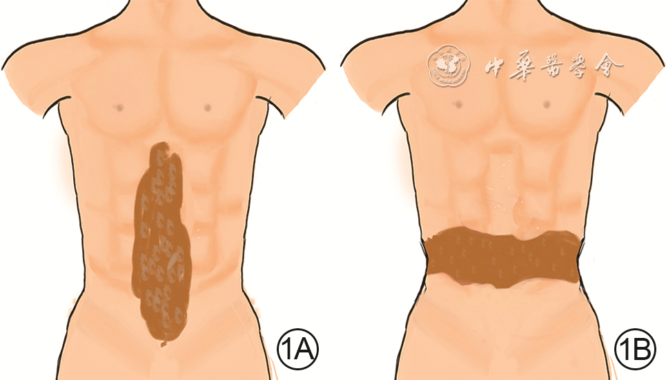
  Objective  To observe the expansion rule of directional skin and soft tissue expander (hereinafter referred to as expander) in abdominal scar reconstruction.  Methods  A prospective self-controlled study was conducted. Twenty patients with abdominal scar who met the inclusion criteria and admitted to Zhengzhou First People's Hospital from January 2018 to December 2020 were selected by random number table method, including 5 males and 15 females, aged 12-51 (31±12) years, with 12 patients of type Ⅰ scar and 8 patients of type Ⅱ scar. In the first stage, two or three expanders with rated capacity of 300-600 mL were placed on both sides of the scar, of which at least one expander had rated capacity of 500 mL (as the follow-up observation object). After the sutures were removed, water injection treatment was started, with the expansion time of 4 to 6 months. After the water injection volume reached 2.0 times of the rated capacity of expander, abdominal scar excision+expander removal+local expanded flap transfer repair was performed in the second stage. The skin surface area at the expansion site was measured respectively when the water injection volume reached 1.0, 1.2, 1.5, 1.8, and 2.0 times of the rated capacity of expander, and the skin expansion rate of the expansion site at corresponding multiples of expansion (1.0, 1.2, 1.5, 1.8, and 2.0 times) and adjacent multiple intervals (1.0-1.2, 1.2-1.5, 1.5-1.8, and 1.8-2.0 times) were calculated. The skin surface area of the repaired site at 0 (immediately), 1, 2, 3, 4, 5, and 6 months after operation, and the skin shrinkage rate of the repaired site at different time points (1, 2, 3, 4, 5, and 6 months after operation) and different time periods (0-1, 1-2, 2-3, 3-4, 4-5, and 5-6 months after operation) were calculated. Data were statistically analyzed with analysis of variance for repeated measurement and least significant difference-t test.  Results  Compared with the expansion of 1.0 time ((287.6±2.2) cm2 and (47.0±0.7)%), the skin surface area and expansion rate of the expansion site of patients ((315.8±2.1), (356.1±2.8), (384.9±1.6), and (386.2±1.5) cm2, (51.7±0.6)%, (57.2±0.6)%, (60.4±0.6)%, and (60.5±0.6)%) were significantly increased when the expansion reached 1.2, 1.5, 1.8, and 2.0 times (with t values of 46.04, 90.38, 150.14, 159.55, 45.11, 87.83, 135.82, and 118.48, respectively, P<0.05). Compared with the expansion of 1.2 times, the skin surface area and expansion rate of the expansion site of patients were significantly increased when the expansion reached 1.5, 1.8, and 2.0 times (with t values of 49.82, 109.64, 122.14, 144.19, 49.51, and 105.85, respectively, P<0.05). Compared with the expansion of 1.5 times, the skin surface area and expansion rate of the expansion site of patients were significantly increased when the expansion reached 1.8 times (with t values of 38.93 and 39.22, respectively, P<0.05) and 2.0 times (with t values of 38.37 and 38.78, respectively, P<0.05). Compared with the expansion of 1.8 times, the skin surface area and expansion rate of the expansion site of patients both had no statistically significant differences when the expansion reached 2.0 times (with t values of 4.71 and 4.72, respectively, P>0.05). Compared with the expansion of 1.0-1.2 times, the skin expansion rate of the expansion site of patient was significantly increased when the expansion reached 1.2-1.5 times (t=6.95, P<0.05), while the skin expansion rate of the expansion site of patient was significantly decreased when the expansion reached 1.5-1.8 and 1.8-2.0 times (with t values of 5.89 and 40.75, respectively, P<0.05). Compared with the expansion of 1.2-1.5 times, the skin expansion rate of the expansion site of patient was significantly decreased when the expansion reached 1.5-1.8 and 1.8-2.0 times (with t values of 10.50 and 41.92, respectively, P<0.05). Compared with the expansion of 1.5-1.8 times, the skin expansion rate of the expansion site of patient was significantly decreased when the expansion reached 1.8-2.0 times (t=32.60, P<0.05). Compared with 0 month after operation, the skin surface area of the repaired site of patient at 1, 2, 3, 4, 5, and 6 months after operation was significantly decreased (with t values of 61.66, 82.70, 96.44, 102.81, 104.51, and 102.21, respectively, P<0.05). Compared with 1 month after operation, the skin surface area of the repaired site of patient was significantly decreased at 2, 3, 4, 5, and 6 months after operation (with t values of 37.37, 64.64, 69.40, 72.46, and 72.62, respectively, P<0.05), while the skin shrinkage rate was significantly increased (with t values of 32.29, 50.00, 52.67, 54.76, and 54.62, respectively, P<0.05). Compared with 2 months after operation, the skin surface area of the repaired site of patient was significantly decreased at 3, 4, 5, and 6 months after operation (with t values of 52.41, 60.41, 70.30, and 65.32, respectively, P<0.05), while the skin shrinkage rate was significantly increased (with t values of 52.97, 59.29, 69.68, and 64.50, respectively, P<0.05). Compared with 3 months after operation, the skin surface area of the repaired site of patient was significantly decreased at 4, 5, and 6 months after operation (with t values of 5.53, 38.00, and 38.52, respectively, P<0.05), while the skin shrinkage rate was significantly increased (with t values of 25.36, 38.59, and 37.47, respectively, P<0.05). Compared with 4 months after operation, the skin surface area (with t values of 41.10 and 50.50, respectively, P>0.05) and skin shrinkage rate (with t values of 48.09 and 50.00, respectively, P>0.05) of the repaired site of patients at 5 and 6 months after operation showed no statistically significant differences. Compared with 5 months after operation, the skin surface area and skin shrinkage rate of the repaired site of patient at 6 months after operation showed no statistically significant differences (with t values of 9.40 and 9.59, respectively, P>0.05). Compared with 0-1 month after operation, the skin shrinkage rate of the repaired site of patient at 1-2, 2-3, 3-4, 4-5, and 5-6 months after operation was significantly decreased (with t values of 13.56, 40.00, 49.21, 53.97, and 57.68, respectively, P<0.05). Compared with 1-2 months after operation, the skin shrinkage rate of the repaired site of patients at 2-3, 3-4, 4-5, and 5-6 months after operation was significantly decreased (with t values of 12.37, 27.72, 30.16, and 31.67, respectively, P<0.05). Compared with 2-3 months after operation, the skin shrinkage rate of the repaired site of patients at 3-4, 4-5, and 5-6 months after operation was significantly decreased (with t values of 33.73, 41.31, and 54.10, respectively, P<0.05). Compared with 3-4 months after operation, the skin shrinkage rate of the repaired site of patient at 4-5 and 5-6 months after operation showed no statistically significant differences (with t values of 10.90 and 23.60, respectively, P>0.05). Compared with 4-5 months after operation, the skin shrinkage rate of the repaired site of patient at 5-6 months after operation showed no statistically significant difference (t=20.90, P>0.05).  Conclusions  The expander can effectively expand the abdominal skin, thus repairing the abdominal scar deformity. Maintained expansion for one month after the water injection expansion reaches 1.8 times of the rated capacity of the expander can be set as a phase Ⅱ operation node.
A prospective study on the development and application verification of the quantitative evaluation software for three-dimensional morphology of pathological scars based on photo modeling technology
Jiang Weiqian, Pan Feng, Chai Mi, Wulan Hasi, Yu Xuedong, Guo Lingli
2023, 39(2): 158-164. doi: 10.3760/cma.j.cn501225-20220513-00184
Abstract:
  Objective  To develop a quantitative evaluation software for three-dimensional morphology of pathological scars based on photo modeling technology, and to verify its accuracy and feasibility in clinical application.  Methods  The method of prospective observational study was adopted. From April 2019 to January 2022, 59 patients with pathological scars (totally 107 scars) who met the inclusion criteria were admitted to the First Medical Center of Chinese PLA General Hospital, including 27 males and 32 females, aged 33 (26, 44) years. Based on photo modeling technology, a software for measuring three-dimensional morphological parameters of pathological scars was developed with functions of collecting patients' basic information, and scar photography, three-dimensional reconstruction, browsing the models, and generating reports. This software and the clinical routine methods (vernier calipers, color Doppler ultrasonic diagnostic equipment, and elastomeric impression water injection method measurement) were used to measure the longest length, maximum thickness, and volume of scars, respectively. For scars with successful modelling, the number, distribution of scars, number of patients, and the longest length, maximum thickness, and volume of scars measured by both the software and clinical routine methods were collected. For scars with failed modelling, the number, distribution, type of scars, and the number of patients were collected. The correlation and consistency of the software and clinical routine methods in measuring the longest length, maximum thickness, and volume of scars were analyzed by unital linear regression analysis and the Bland-Altman method, respectively, and the intraclass correlation coefficients (ICCs), mean absolute error (MAE), and mean absolute percentage error (MAPE) were calculated.  Results  A total of 102 scars from 54 patients were successfully modeled, which located in the chest (43 scars), in the shoulder and back (27 scars), in the limb (12 scars), in the face and neck (9 scars), in the auricle (6 scars), and in the abdomen (5 scars). The longest length, maximum thickness, and volume measured by the software and clinical routine methods were 3.61 (2.13, 5.19) and 3.53 (2.02, 5.11) cm, 0.45 (0.28, 0.70) and 0.43 (0.24, 0.72) cm, 1.17 (0.43, 3.57) and 0.96 (0.36, 3.26) mL. The 5 hypertrophic scars and auricular keloids from 5 patients were unsuccessfully modeled. The longest length, maximum thickness, and volume measured by the software and clinical routine methods showed obvious linear correlation (with r values of 0.985, 0.917, and 0.998, P<0.05). The ICCs of the longest length, maximum thickness, and volume of scars measured by the software and clinical routine methods were 0.993, 0.958, and 0.999 (with 95% confidence intervals of 0.989-0.995, 0.938-0.971, and 0.998-0.999, respectively). The longest length, maximum thickness, and volume of scars measured by the software and clinical routine methods had good consistency. The Bland-Altman method showed that 3.92% (4/102), 7.84% (8/102), and 8.82% (9/102) of the scars with the longest length, maximum thickness, and volume respectively were outside the 95% consistency limit. Within the 95% consistency limit, 2.04% (2/98) scars had the longest length error of more than 0.5 cm, 1.06% (1/94) scars had the maximum thickness error of more than 0.2 cm, and 2.15% (2/93) scars had the volume error of more than 0.5 mL. The MAE and MAPE of the longest length, maximum thickness, and volume of scars measured by the software and clinical routine methods were 0.21 cm, 0.10 cm, 0.24 mL, and 5.75%, 21.21%, 24.80%, respectively.  Conclusions  The quantitative evaluation software for three-dimensional morphology of pathological scars based on photo modeling technology can realize the three-dimensional modeling and measurement of morphological parameters of most pathological scars. Its measurement results were in good consistency with those of clinical routine methods, and the errors were acceptable in clinic. This software can be used as an auxiliary method for clinical diagnosis and treatment of pathological scars.
Effects of three-dimensional bioprinting antibacterial hydrogel on full-thickness skin defect wounds in rats
Jin Ronghua, Zhang Zhenzhen, Xu Pengqin, Xia Sizhan, Weng Tingting, Zhu Zhikang, Wang Xingang, You Chuangang, Han Chunmao
2023, 39(2): 165-174. doi: 10.3760/cma.j.cn501120-20210809-00274
Abstract:
  Objective  To explore the effects of three-dimensional (3D) bioprinting gelatin methacrylamide (GelMA) hydrogel loaded with nano silver on full-thickness skin defect wounds in rats.  Methods  The experimental research method was adopted. The morphology, particle diameter, and distribution of silver nanoparticles in nano silver solution with different mass concentrations and the pore structure of silver-containing GelMA hydrogel with different final mass fractions of GelMA were observed by scanning electron microscope and the pore size was calculated. On treatment day 1, 3, 7, and 14, the concentration of nano silver released from the hydrogel containing GelMA with final mass fraction of 15% and nano silver with final mass concentration of 10 mg/L was detected by mass spectrometer. At 24 h of culture, the diameters of inhibition zone of GelMA hydrogel containing final mass concentration of 0 (no nano silver), 25, 50, and 100 mg/L nano silver against Staphylococcus aureus and Escherichia coli were detected. Fibroblasts (Fbs) and adipose stem cells (ASCs) were isolated respectively by enzymatic digestion using the discarded prepuce after circumcision from a 5-year-old healthy boy who was treated in the Department of Urology of the Second Affiliated Hospital of Zhejiang University School of Medicine in July 2020, and the discarded fat tissue after liposuction from a 23-year-old healthy woman who was treated in the Department of Plastic Surgery of the Hospital in July 2020. The Fbs were divided into blank control group (culture medium only), 2 mg/L nano sliver group, 5 mg/L nano sliver group, 10 mg/L nano sliver group, 25 mg/L nano sliver group, and 50 mg/L nano sliver group, which were added with the corresponding final mass concentrations of nano sliver solution, respectively. At 48 h of culture, the Fb proliferation viability was detected by cell counting kit 8 method. The Fbs were divided into 0 mg/L silver-containing GelMA hydrogel group, 10 mg/L silver-containing GelMA hydrogel group, 50 mg/L silver-containing GelMA hydrogel group, and 100 mg/L silver-containing GelMA hydrogel group and then were correspondingly treated. On culture day 1, 3, and 7, the Fb proliferation viability was detected as before. The ASCs were mixed into GelMA hydrogel and divided into 3D bioprinting group and non-printing group. On culture day 1, 3, and 7, the ASC proliferation viability was detected as before and cell growth was observed by live/dead cell fluorescence staining. The sample numbers in the above experiments were all 3. Four full-thickness skin defect wounds were produced on the back of 18 male Sprague-Dawley rats aged 4 to 6 weeks. The wounds were divided into hydrogel alone group, hydrogel/nano sliver group, hydrogel scaffold/nano sliver group, and hydrogel scaffold/nano sliver/ASC group, and transplanted with the corresponding scaffolds, respectively. On post injury day (PID) 4, 7, 14, and 21, the wound healing was observed and the wound healing rate was calculated (n=6). On PID 7 and 14, histopathological changes of wounds were observed by hematoxylin eosin staining (n=6). On PID 21, collagen deposition of wounds was observed by Masson staining (n=3). Data were statistically analyzed with one-way analysis of variance, analysis of variance for repeated measurement, Bonferroni correction, and independent sample t test.  Results  The sliver nano particles in nano silver solution with different mass concentrations were all round, in scattered distribution and uniform in size. The silver-containing GelMA hydrogels with different final mass fractions of GelMA all showed pore structures of different sizes and interconnections. The pore size of silver-containing GelMA hydrogel with 10% final mass fraction was significantly larger than that of silver-containing GelMA hydrogels with 15% and 20% final mass fractions (with P values both below 0.05). On treatment day 1, 3, and 7, the concentration of nano silver released from silver-containing GelMA hydrogel in vitro showed a relatively flat trend. On treatment day 14, the concentration of released nano silver in vitro increased rapidly. At 24 h of culture, the diameters of inhibition zone of GelMA hydrogel containing 0, 25, 50, and 100 mg/L nano silver against Staphylococcus aureus and Escherichia coli were 0, 0, 0.7, and 2.1 mm and 0, 1.4, 3.2, and 3.3 mm, respectively. At 48 h of culture, the proliferation activity of Fbs in 2 mg/L nano silver group and 5 mg/L nano silver group was both significantly higher than that in blank control group (P<0.05), and the proliferation activity of Fbs in 10 mg/L nano silver group, 25 mg/L nano silver group, and 50 mg/L nano silver group was all significantly lower than that in blank control group (P<0.05). Compared with the that of Fbs in 0 mg/L silver-containing GelMA hydrogel group, the proliferation activity of Fbs in 50 mg/L silver-containing GelMA hydrogel group and 100 mg/L silver-containing GelMA hydrogel group was all significantly decreased on culture day 1 (P<0.05); the proliferation activity of Fbs in 50 mg/L silver-containing GelMA hydrogel group was significantly increased (P<0.05), while the proliferation activity of Fbs in 100 mg/L silver-containing GelMA hydrogel group was significantly decreased on culture day 3 (P<0.05); the proliferation activity of Fbs in 100 mg/L silver-containing GelMA hydrogel group was significantly decreased on culture day 7 (P<0.05). The proliferation activity of ASCs in 3D bioprinting group show no statistically significant differences to that in non-printing group on culture day 1 (P>0.05). The proliferation activity of ASCs in 3D bioprinting group was significantly higher than that in non-printing group on culture day 3 and 7 (with t values of 21.50 and 12.95, respectively, P<0.05). On culture day 1, the number of dead ASCs in 3D bioprinting group was slightly more than that in non-printing group. On culture day 3 and 5, the majority of ASCs in 3D bioprinting group and non-printing group were living cells. On PID 4, the wounds of rats in hydrogel alone group and hydrogel/nano sliver group had more exudation, and the wounds of rats in hydrogel scaffold/nano sliver group and hydrogel scaffold/nano sliver/ASC group were dry without obvious signs of infection. On PID 7, there was still a small amount of exudation on the wounds of rats in hydrogel alone group and hydrogel/nano sliver group, while the wounds of rats in hydrogel scaffold/nano sliver group and hydrogel scaffold/nano sliver/ASC group were dry and scabbed. On PID 14, the hydrogels on the wound surface of rats in the four groups all fell off. On PID 21, a small area of wounds remained unhealed in hydrogel alone group. On PID 4 and 7, the wound healing rates of rats in hydrogel scaffold/nano sliver/ASC group were significantly higher than those of the other three groups (P<0.05). On PID 14, the wound healing rate of rats in hydrogel scaffold/nano sliver/ASC group was significantly higher than the wound healing rates in hydrogel alone group and hydrogel/nano sliver group (all P<0.05). On PID 21, the wound healing rate of rats in hydrogel alone group was significantly lower than that in hydrogel scaffold/nano sliver/ASC group (P<0.05). On PID 7, the hydrogels on the wound surface of rats in the four groups remained in place; on PID 14, the hydrogel in hydrogel alone group was separated from the wounds of rats, while some hydrogels still existed in the new tissue of the wounds of rats in the other three groups. On PID 21, the collagen arrangement in the wounds of rats in hydrogel alone group was out of order, while the collagen arrangement in the wounds of rats in hydrogel/nano sliver group, and hydrogel scaffold/nano sliver/ASC group was relatively orderly.  Conclusions  Silver-containing GelMA hydrogel has good biocompatibility and antibacterial properties. Its three-dimensional bioprinted double-layer structure can better integrate with new formed tissue in the full-thickness skin defect wounds in rats and promote wound healing.
Meta-analysis of the effects of xenogeneic acellular dermal matrix dressings in the treatment of wounds in burn patients
You Aijia, Li Wenjie, Zhou Junli, Li Chun
2023, 39(2): 175-183. doi: 10.3760/cma.j.cn501120-20220106-00008
Abstract:
  Objective  To evaluate the efficacy and safety of xenogeneic acellular dermal matrix (ADM) dressings for the treatment of wounds in burn patients.  Methods  The meta-analysis method was adopted. Databases including Chinese Journal Full-text Database, Wanfang Database, VIP Database, and Chinese Biomedical Database were retrieved with the search terms in Chinese version of "异种脱细胞真皮基质, 敷料, 烧伤创面, 烧伤" and PubMed, Embase, Web of Science, and Cochrane Library were retrieved with the search terms in English version of "xenogeneic acellular dermal matrix, dressing, burn wound, burn" to obtain the publicly published randomized controlled trials on the efficacy of xenogeneic ADM dressings for the treatment of wounds in burn patients from the establishment of each database to December 2021. The outcome indexes included wound healing time, ratio of scar hyperplasia, Vancouver scar scale (VSS) score, ratio of complications, ratio of skin grafting, and ratio of bacteria detection. Rev Man 5.3 and Stata 14.0 statistical softwares were used to conduct a meta-analysis of eligible studies.  Results  A total of 1 596 burn patients from 16 studies were included, including 835 patients in experimental group who received xenogeneic ADM dressings therapy and 761 patients in control group who received other methods therapy. The bias risk of all the 16 included studies was uncertain. Compared with those in control group, patients in experimental group had significantly shorter wound healing time, lower VSS scores (with standardized mean differences of -2.50 and -3.10, 95% confidence intervals of -3.02--1.98 and -4.87--1.34, respectively, P values both <0.05), and lower ratios of scar hyperplasia, complications, skin grafting, and bacteria detection (with relative risks of 0.58, 0.23, 0.32, and 0.27, 95% confidence intervals of 0.43-0.80, 0.14-0.37, 0.15-0.67, and 0.11-0.69, respectively, P<0.05). Subgroup analysis showed that the difference of intervention measures in control group might be the source of heterogeneity in wound healing time. There was no publication bias in ratio of scar hyperplasia (P≥0.05), while there was publication bias in wound healing time, VSS score, and ratio of complications (P<0.05).  Conclusions  Xenogeneic ADM dressings can shorten the wound healing time of burn patients, reduce the VSS score and the ratios of scar hyperplasia, complications, skin grafting, and bacteria detection.
Reviews · Wound Healing and Its Regulation Mechanisms
Research advances on the mechanism of non-coding RNA regulated diabetic wound healing
Li Xiaoliang, Xie Jiangfan, Ye Xiangyang, Li Yanguang, Liu Dewu
2023, 39(2): 184-189. doi: 10.3760/cma.j.cn501225-20221101-00477
Abstract:
Diabetic wounds are a common complication of diabetic patients, and the incidence has been increasing in recent years. In addition, its poor clinical prognosis seriously affects the quality of life of patients, which has become the focus and difficulty of diabetes treatment. As the RNA regulating gene expression, non-coding RNA can regulate the pathophysiological process of diseases, and play an important role in the healing process of diabetic wounds. In this paper, we reviewed the regulatory role, diagnostic value, and therapeutic potential of three common non-coding RNA in diabetic wounds, in order to provide a new solution for the diagnosis and treatment of diabetic wounds at the genetic and molecular level.
Research advances on the mechanism of Wnt/β-catenin signaling pathway in body surface wound healing
Wu Qian, Tan Xiaoyu, Wang Yijia, Cheng Shaowen, Cui Hongwang, Yao Jiangling
2023, 39(2): 190-195. doi: 10.3760/cma.j.cn501225-20220816-00348
Abstract:
Wound healing is a slow and complex biological process, including inflammatory reaction, cell proliferation, cell differentiation, cell migration, angiogenesis, extracellular matrix deposition, tissue remodeling, and so on. Wnt signaling pathway can be divided into classical pathway and non-classical pathway. Wnt classical pathway, also known as Wnt/β-catenin signaling pathway, plays an important role in cell differentiation, cell migration, and maintenance of tissue homeostasis. Many inflammatory factors and growth factors are involved in the upstream regulation of this pathway. The activation of Wnt/β-catenin signaling pathway plays an important role in the occurrence, development, regeneration, repair and related treatment of skin wounds. This article review the relationship between Wnt/β-catenin signaling pathway and wound healing, meanwhile summarizes its effects on important processes of wound healing, such as inflammation, cell proliferation, angiogenesis, hair follicle regeneration, and skin fibrosis, as well as the role of inhibitors of Wnt signaling pathway in wound healing.
Review
Research advances on the role and mechanism of microRNA in hypertrophic scar
Tian Wenrong, Zuo Jun, Ai Jiang, Qi Yusong, Bu Panpan, Zhao Jiaojun, Yu Yang, Ma Shaolin
2023, 39(2): 196-200. doi: 10.3760/cma.j.cn501225-20220508-00179
Abstract:
Hypertrophic scar (HS) affects the function and beauty of patients, and brings a heavy psychological burden to patients. However, the specific pathogenesis mechanism of HS in molecular biology level is not yet clear, and this disease is still one of the clinical diseases difficult to prevent and cure. MicroRNA (miR) is a family of single-stranded endogenous noncoding RNAs that can regulate gene expression. The abnormal transcription of miR in hypertrophic scar fibroblasts can affect the transduction and expression of downstream signal pathway or protein, and the exploration of miR and its downstream signal pathway and protein helps deeply understand the occurrence and development mechanism of scar hyperplasia. This article summarized and analyzed how miR and multiple signal pathways involve in the formation and development of HS in recent years, and further outlined the interaction between miR and target genes in HS.