2024 Vol. 40, No. 7

Feature
Holistic integrative medicine declaration
China Institute for Development Strategy of Holistic Integrative Medicine
2024, 40(7): 601-603. doi: 10.3760/cma.j.cn501225-20240627-00253
Abstract:
Holistic integrative medicine(HIM), has been officially proposed since 2012. Its theoretical system has been continuously improved, and its practical methods have become increasingly diverse, becoming an inevitable choice and path for the medical development in the new era. This article demonstrates ten major propositions for HIM, elaborating on the connotation and extension of HIM from the perspectives of epistemology and methodology, in order to achieve the transformation and adaptive evolution of modern medicine.
Guideline and Consensus
Practical guidelines for the diagnosis, prevention and treatment of invasive fungal infection post burn injury in China: 2024 edition
2024, 40(7): 604-617. doi: 10.3760/cma.j.cn501225-20240103-00003
Abstract:
Burn patients are at high risk for invasive fungal infections. The incidence of invasive fungal infection for burn victims is increasing dramatically during recent years and becoming one of the major causes of death in patients with severe burns. This updated guideline was initiated and organized by Burn Medicine Branch of China International Exchange and Promotion Association for Medical and Healthcare, and Editorial Committee of Chinese Journal of Burns and Wounds. Based on the current evidence-based medical evidence, clinical practice, and authoritative guidelines from other disciplines, we updated and revised the Guidelines for the diagnosis, prevention and treatment of invasive fungal infections post burn injury (2012 edition), aiming to further standardize the diagnosis, prevention and treatment of invasive fungal infection post burns.
Expert Forum
Neutrophil and burn sepsis
Sun Bingwei, Wang Yifan, Yang Yunxi
2024, 40(7): 618-624. doi: 10.3760/cma.j.cn501225-20240329-00109
Abstract:
Sepsis is the most common complication of severe burns and a primary risk factor for endangering the lives of critically severe burn patients. The mortality rate of burn sepsis patients was up to 75%. Neutrophils are one of the innate immune cells and are the earliest and most recruited immune cells at the site of infection, which play a key role in the removal of local infection and the repair of damaged tissue. Many studies have deeply revealed the pathogenesis and progression mechanism of burn sepsis, in which the role of neutrophils has gradually become clear. This paper elaborated on the key mechanisms of neutrophils in the occurrence and evolution of burn sepsis, explored the value of neutrophils in the early warning and prognosis judgement, and the potential therapeutic methods of burn sepsis based on the unique biological behavior of neutrophils in burn sepsis.
Original Article·Burn Metabolism and Nutrition
Changes in entero-insular axis function and its role in mice with severe burns
Liu Xinzhu, Li Dawei, Jiang Min, Li Zhisheng, Feng Baigong, Shen Chuan'an
2024, 40(7): 625-633. doi: 10.3760/cma.j.cn501225-20240520-00189
Abstract:
  Objective  To explore the changes in entero-insular axis function and its role in mice with severe burns.  Methods  This study was an experimental study. Ninety C57BL/6J male mice aged 8-10 weeks were divided into sham injury group and burn group (with 45 mice in each group) according to the random number table. A full-thickness scald (hereinafter referred to as burn) wound of 30% of the total body surface area was created on the back of mice in burn group, and the mice in sham injury group were simulated to cause a sham injury. Twenty-four hours after injury, the fasting blood glucose was measured (n=12), followed by intraperitoneal glucose tolerance test and oral glucose tolerance test; the curve of blood glucose concentration changes over time was plotted, and the area under the curve was calculated (n=6); the blood was taken from the heart before intraperitoneal injection or gavage of glucose solution and at 30, 60, and 120 minutes after intraperitoneal injection or gavage of glucose solution for measuring the plasma insulin and glucagon like peptide-1 (GLP-1) levels using enzyme-linked immunosorbent assay (ELISA), with a sample number of 3; the ileal tissue was taken from 3 mice in each group for detecting the GLP-1 expression and apoptosis levels of intestinal L cells by immunofluorescence staining and TdT-mediated dUTP nick-end labeling staining; the pancreatic islets were collected from 6 mice in each group for glucose-stimulated insulin secretion experiments. After incubation with low glucose (2.8 mmol/L glucose) and high glucose (16.7 mmol/L glucose), the supernatant was taken and the insulin level was detected using ELISA. Thirty-six C57BL/6J male mice aged 8-10 weeks were divided into sham injury group, burn group, and burn+exendin-4 (Ex-4) group (with 12 mice in each group) according to the random number table. The mice in sham injury group and burn group were subjected to the same corresponding treatment as before. The mice in burn+Ex-4 group were injured in the same way as the burn group mice followed by treatment with GLP-1 receptor agonist Ex-4. Twenty-four hours after injury, mouse pancreatic islets were collected, the protein expressions of heavy-chain binding protein (BIP), protein kinase R-like endoplasmic reticulum kinase (PERK), phosphorylated PERK (p-PERK), eukaryotic translation initiation factor 2α (eIF2α), phosphorylated eIF2α (p-eIF2α), and CCAAT/enhancer-binding protein homologous protein (CHOP) were detected using Western blotting, and the p-PERK/PERK and p-eIF2α/eIF2α ratios were calculated (n=3), the apoptosis rate of pancreatic islet cells was detected using flow cytometry (n=3), the glucose stimulated insulin secretion experiment was conducted as before to detect insulin levels in the supernatant (n=6).  Results  Twenty-four hours after injury, the fasting blood glucose of mice in burn group was (7.3±1.0) mmol/L, which was significantly higher than (5.1±0.6) mmol/L in sham injury group (t=6.36, P<0.05). Twenty-four hours after injury, in the intraperitoneal glucose tolerance test and oral glucose tolerance test, the areas under the curve of blood glucose concentration changes over time of mice in burn group were significantly larger than those in sham injury group (with t values of 4.32 and 6.03, respectively, P<0.05); compared with those in sham injury group, the plasma insulin levels of mice before intraperitoneal injection of glucose solution and the plasma GLP-1 levels of mice before intraperitoneal injection or gavage of glucose solution in burn group were significantly decreased (P<0.05), and the plasma levels of insulin of mice at 30, 60, and 120 minutes after intraperitoneal injection or gavage of glucose solution, as well as the plasma levels of GLP-1 of mice at 30 and 60 minutes after gavage of glucose solution were significantly decreased in burn group (P<0.05). Twenty-four hours after injury, compared with those in sham injury group, the GLP-1 expression level of intestinal L cells of mice in burn group was significantly decreased (t=7.74, P<0.05), and the apoptosis level was significantly increased (t=14.28, P<0.05). Twenty-four hours after injury, the insulin level in the supernatant of mice pancreatic islet incubated with high glucose in burn group was (8.5±0.4) ng/mg, which was significantly lower than (15.7±0.3) ng/mg in sham injury group (t=18.68, P<0.05). Twenty-four hours after injury, compared with those in sham injury group, the protein expression levels of BIP, p-PERK/PERK, p-eIF2α/eIF2α, and CHOP in the pancreatic islets of mice in burn group were significantly increased (P<0.05); compared with those in burn group, the protein expression levels of BIP, p-PERK/PERK, p-eIF2α/eIF2α, and CHOP in the pancreatic islets of mice in burn+Ex-4 group were significantly decreased (P<0.05). Twenty-four hours after injury, the apoptosis rate of pancreatic islet cells of mice in burn group was (32.0±3.0)%, which was significantly higher than (10.3±2.5)% in sham injury group (P<0.05); the apoptosis rate of pancreatic islet cells of mice in burn+Ex-4 group was (20.0±3.6)%, which was significantly lower than that in burn group (P<0.05). Twenty-four hours after injury, the insulin level in the supernatant of mice pancreatic islet incubated with high glucose in burn group was significantly lower than that in sham injury group (P<0.05), while the insulin level in the supernatant of mice pancreatic islet incubated with high glucose in burn+Ex-4 group was significantly higher than that in burn group (P<0.05).  Conclusions  After severe burns, the mice display dysfunction of the entero-insular axis, increased apoptosis of intestinal L cells, decreased synthesis and secretion of GLP-1, endoplasmic reticulum stress and increased apoptosis in pancreatic islet cells and a decrease in glucose-stimulated insulin secretion. The GLP-1 receptor agonist Ex-4 can protect the function of pancreatic islet cells of mice with severe burns, reducing the apoptosis level of pancreatic islet cells and promoting insulin secretion possibly via the alleviation of endoplasmic reticulum stress.
Selection and analysis of calculation formulas for resting energy expenditure in patients with severe burns based on different metabolic stages
Zou Wen, Han Chunmao, Jin Ronghua, Shen Tao
2024, 40(7): 634-642. doi: 10.3760/cma.j.cn501225-20240229-00080
Abstract:
  Objective  To explore the changes in resting energy expenditure (REE) values in patients with severe burns under different metabolic stages and the selection of the optimal calculation formula.  Methods  This study was a retrospective and observational study. From April 2020 to December 2023, 40 patients (32 males and 8 females, aged (54±17) years) with severe burns meeting inclusion criteria were treated in the Second Affiliated Hospital of Zhejiang University School of Medicine. After admission, the patients were given routine clinical treatments such as sedation and analgesia, debridement, and skin grafting. At 3, 5, 7, 9, 11, 14 days after injury and every 7 days thereafter, the REE values (i.e., REE measured values) were measured by indirect calorimetry in patients with severe burns who met the measurement conditions till the patients recovered or died. On the day the patient's REE was measured, Milner, Hangang, the Third Military Medical University, Carlson, and Peng Xi team's linear formula were used respectively to calculate the REE value (i.e., REE formula values). The post-injury time to measure REE in patients was calculated, and the clinical characteristics of patients in acute inhibition, hypermetabolic, metabolic balance, and metabolic remodeling phases were compared. The REE measured values and the difference between the REE formula values and the REE measured values of patients under the 4 different metabolic phases were calculated.Compared with the REE measured values, the 10% accuracy rate and 20% accuracy rate were calculated to evaluate the accuracy of the REE formula values. The absolute percentage error (APE) of the REE formula values were calculated to evaluate the deviation. The metabolic formula (i.e., the optimal calculation formula) that was closest to the measured REE values was screened out, and further exploration was conducted to identify the key factors that affected the accuracy of the optimal calculation formula under different metabolic phases.  Results  The post-injury time to measure REE in patients with severe burns was (40±19) days. Comparisons showed that under the 4 different metabolic phases, patients in the metabolic remodeling phase had the highest age, height, weight, body mass index, total body surface area. Age in the metabolic remodeling phase was significantly higher than that in the acute inhibition and hypermetabolic phases (with t values of -3.02 and -4.20, respectively, with all P values <0.05), weight was significantly higher than that in the hypermetabolic and metabolic balance phases (with t values of -1.97 and -2.61, respectively, with all P values <0.05), body mass index was significantly higher than that in the hypermetabolic phase (t=-2.90, P<0.05), and total body surface area was significantly larger than that in the hypermetabolic and metabolic balance phases (with t values of -2.02 and -2.27, respectively, with all P values <0.05). There was no significant change in patients' REE measured values under the 4 different metabolic stages (P>0.05). Except for the Peng Xi team's linear formula (P>0.05), the difference between REE measured values and REE formula values calculated by using Milner, Hangang, the Third Military Medical University, and Carlson formulas respectively was statistically significant under different metabolic stages (with H values of 14.50, 27.15, and 37.26, respectively, F=11.80, P<0.05). Comprehensive analysis of 10% accuracy, 20% accuracy, and APE showed that in the acute inhibition phase, the REE formula values calculated by Peng Xi team's linear formula was closest to REE measured values, and the APE of the REE formula values calculated by Peng Xi team's linear formula was significantly lower than those calculated by Milner formula, Hangang formula, the Third Military Medical University formula, and Carlson formula (with t values of 9.00, -2.10, 5.95, and 6.68, respectively, with all P values <0.05). In the hypermetabolic phase, the REE formula values calculated by Hangang formula were closest to REE measured values, with significantly lower APE of the REE formula values calculated by Hangang formula than those calculated by using Milner formula, the Third Military Medical University formula, Carlson formula, and Peng Xi team's linear formula (with t values of 10.20, 10.33, 10.65, and 5.87, respectively, with all P values <0.05). In the metabolic balance phase, the REE formula values calculated by Hangang formula were again closest to REE measured values, with significantly lower APE of the REE formula values calculated by Hangang formula than those calculated by Milner formula, the Third Military Medical University formula, and Carlson formula (with t values of 7.11, 8.52, and 8.60, respectively, with all P values <0.05). In the metabolic remodeling phase, the REE formula values calculated by the Third Military Medical University were closest to REE measured values, with significantly lower APE of the REE formula values calculated by the Third Military Medical University formula than those calculated by Milner formula, Hangang formula, and Carlson formula (with t values of 5.12, 2.45, and 6.26, respectively, with all P values <0.05). No significant key factors affected the accuracy of the Peng Xi team's linear formula in the acute inhibition phase (P>0.05). In the hypermetabolic phase, total burn area was a key factor affecting the accuracy of Hangang formula (with odds ratio of 1.00, with 95% confidence interval of 1.00-1.10, P<0.05). In the metabolic balance phase, post-injury days was a key factor affecting the accuracy of Hangang formula (with odds ratio of 1.30, with 95% confidence interval of 1.10-1.40, P<0.05). In the metabolic remodeling phase, no significant key factors affected the accuracy of the Third Military Medical University formula (P>0.05).  Conclusions  When calculating REE values in patients with severe burns, it is recommended to use the Peng Xi team's linear formula during the acute inhibition phase, the Hangang formula during the hypermetabolic and metabolic balance phases, and the Third Military Medical University formula during the metabolic remodeling phase. Additionally, it is crucial to ensure the accuracy of key factors affecting the optimal calculation formula in the hypermetabolic and metabolic balance phases.
Original Article
Clinical effects of chimeric perforator flaps in repairing wounds with bone or internal fixation exposure and wounds with osteomyelitis
Cheng Lin, Liu Xianqi, Du Weili, Dai Qiang, Che Kexin, Shen Yuming
2024, 40(7): 643-649. doi: 10.3760/cma.j.cn501225-20231120-00198
Abstract:
  Objective  To explore the clinical effects of chimeric perforator flaps in repairing wounds with bone or internal fixation exposure and wounds with osteomyelitis.  Methods  This study was a retrospective observational study. From January 2018 to December 2022, 20 patients with wounds with bone or internal fixation exposure and wounds with osteomyelitis who met the inclusion criteria were admitted to Beijing Jishuitan Hospital Affiliated to Capital Medical University, including 19 males and 1 female, aged from 21 to 73 years. Among the 21 wounds, there were 5 wounds with bone exposure, 12 wounds with osteomyelitis, and 4 wounds with internal fixation exposure. After the debridement in the first stage, the wound area was 6 cm×3 cm to 22 cm×10 cm. Then vacuum sealing drainage was carried out for 5 to 7 days. In the second stage, the wounds were covered with pedicled chimeric medial sural artery perforator flap, pedicled chimeric posterior tibialis artery perforator flap, free chimeric perforator flap pedicled with descending branch of lateral circumflex femoral artery, free chimeric medial sural artery perforator flap or free chimeric deep circumflex iliac artery perforator flap with incision area of 7 cm×5 cm to 25 cm×12 cm. The chimeric muscle flap was used to fill and cover irregular deep cavities. The wounds in the flap donor sites were sutured directly or repaired with medium-thickness skin grafts from the thigh. The survival of flap and the healing of wound in flap donor site were observed after operation. The recurrence of infection was followed up.  Results  Among the 18 free chimeric perforator flaps, 16 flaps survived successfully; one flap experienced a venous crisis on the day of surgery and survived completely after emergency exploration and re-anastomosis; another one flap had partial distal necrosis, which healed after dressing changes. All the wounds in the flap donor sites healed evenly. All 3 pedicled chimeric perforator flaps survived; one of them developed sub-flap infection but healed after debridement and bone cement placement. The wound in the donor site of 1 flap developed incision dehiscence, which healed successfully after redebridement and suturing. The donor site wounds of the rest 2 flaps healed well. During 3 to 12 months of follow-up, the patients with wounds with bone or internal fixation exposure showed no signs of abnormal exudation or infection, and no infection recurrence was observed in patients with wounds with osteomyelitis.  Conclusions  The application of chimeric perforator flaps is effective in covering wounds, filling dead spaces, and controlling infection in wounds with bone or internal fixation exposure and wounds with osteomyelitis. Moreover, this method minimizes the damage to the donor site.
Clinical effects of combined tissue flap transplantation for repairinggiant chest wall defects
Yu Junyi, Song Dajiang, Liu Xu, Wang Zhiyuan, Li Zan, Zhang Yixin, Zhou Bo, Lyu Chunliu, Tang Yuanyuan, Yi Liang, Luo Zhenhua, Yang Liyi
2024, 40(7): 650-656. doi: 10.3760/cma.j.cn501225-20231120-00199
Abstract:
  Objective  To investigate the clinical effects of combined tissue flap transplantation in repairing giant chest wall defects.  Methods  This study was a retrospective observational study. From August 2013 to December 2020, 31 patients with chest wall tumor or radiation ulcer after radical resection of chest wall tumor and conformed to the inclusion criteria were admitted to the Department of Breast Oncoplastic Surgery of Hunan Cancer Hospital, including 12 males and 19 females, aged 25-71 years. After resection of tumor or ulcer and wound debridement, the area of secondary chest wall defect was 300-600 cm2 with length of 16-35 cm and width of 16-32 cm. According to the actual situation of the patients and the preoperative design, the chest wall defects were repaired with the flexible combination of perforator flaps and myocutaneous flaps from different donor sites, and the area of the combined tissue flap was 260-540 cm2 with length of 20-30 cm and width of 13-20 cm. Free posteromedial thigh perforator flap+free anterolateral thigh myocutaneous flap were used in 2 patients, free deep inferior epigastric artery perforator flap+free anterolateral thigh myocutaneous flap were used in 5 patients, free deep inferior epigastric artery perforator flap+pedicled rectus abdominis myocutaneous flap+free anterolateral thigh myocutaneous flap were used in 7 patients, free deep inferior epigastric artery perforator flap+pedicled rectus abdominis myocutaneous flap+pedicled latissimus dorsi myocutaneous flap were used in 2 patients, and bilateral free anterolateral thigh myocutaneous flaps were used in 15 patients. For the remaining small area of superficial tissue defect after being repaired by combined tissue flaps, skin graft was used to repair or delayed local flap transfering was performed after the tissue flaps survived and edema subsided. The appropriate blood vessels in the donor and recipient sites were selected for anastomosis to reconstruct the blood supply of tissue flaps. The wounds in the donor sites of tissue flaps that can be directly sutured were sutured directly; for those that cannot be sutured directly, the skin grafting or delayed suture was performed. The anastomosis of blood vessels in the recipient sites, operation length, and postoperative hospital stay were recorded. The survivals of tissue flaps and skin grafts, the shape and texture of reconstructed chest wall, the wound healing, scar formation, and function of donor sites of tissue flaps, and the scar formation of the donor sites of skin grafts were observed after operation. Tumor recurrence and death of recurrent patients were followed up after operation.  Results  The blood vessels in the recipient sites were anastomosed as follows: proximal internal thoracic vessels for 24 times, distal internal thoracic vessels for 12 times, trunk of thoracodorsal vessels for 4 times, anterior serratus branches of thoracodorsal vessels for 8 times, and thoracoacromial vessels for 12 times. The operation length was 6.0 to 8.5 hours, and the postoperative hospital stay was 9 to 21 days. Necrosis at the edge of partial tissue flaps occurred in 4 patients after operation, which healed after dressing change, and the tissue flaps and skin grafts of the other patients survived completely. The shape and texture of the reconstructed chest wall were good. Four patients had poor wound healing in the donor sites of abdominal tissue flaps, which healed after dressing change and local drainage. Only linear scar was left in the donor sites of all tissue flaps, and there was no obvious dysfunction in the donor sites of tissue flaps. Mild hypertrophic scar was left in the donor sites of skin grafts. During follow-up of 9 to 36 months after operation, 6 patients had tumor recurrence, and the recurrence time was 5 to 20 months after operation. After comprehensive treatment for patients with tumor recurrence, 3 patients died.  Conclusions  Transplantation of combined tissue flaps in repairing the giant chest wall defects can shorten the time of total operation and hospital stay, and avoid multiple operations. After operation, patients had good chest wall appearance, with reduced tumor recurrence in patients with chest wall tumor.
Methods and clinical effects of reconstructing facial and cervical scars with expanded flaps based on the "MLT" principle
Tao Mingqiu, Huang Mitao, Liang Pengfei, Zhang Minghua, Zhang Pihong, He Zhiyou, Zeng Jizhang, Zhou Jie, Cui Xu, Guo Le, Zhou Situo, Yang Yan, Xie Tinghong, Huang Xiaoyuan
2024, 40(7): 657-664. doi: 10.3760/cma.j.cn501225-20231215-00244
Abstract:
  Objective  To explore the methods and clinical effects of reconstructing facial and cervical scars with expanded flaps based on the "MLT" principle.  Methods  The study was a retrospective observational study. From January 2019 to May 2022, 74 patients with facial and cervical scars after burn or trauma injuries who met the inclusion criteria were admitted to Xiangya Hospital of Central South University, including 38 males and 36 females, aged from 5 to 58 years, including 24 patients with simple facial involvement, 24 patients with simple cervical involvement, and 26 patients with both facial and cervical involvement, with scar area ranging from 12 to 145 cm². By following the "MLT" principle (color and texture similar to the face; flap area large enough to reconstruct the entire defect; skin tissue thin enough to transmit the expression, so as to facilitate the shape of the face and five features); in the stage Ⅰ surgery, the skin and soft tissue expanders (hereinafter referred to as the expanders) were implanted, and in the stage Ⅱ surgery, the expander removal+scar resection+flap transplantation to repair the secondary wound was performed, and the wound in the donor area of flap was directly sutured. After operation, silicone gel preparation and laser therapy were used to prevent scar hyperplasia. The expansion ratio and time period of expanders, the occurrence of complications of skin and soft tissue expansion surgery, the type of flap used, and the survival of flap after the stage Ⅱ surgery were observed and recorded. The long-term effect of facial and cervical reconstruction and the recovery of donor area and recipient area of flap were evaluated during the postoperative follow-up after surgery.  Results  The expansion ratio of 135 expanders ranged from 1.36 to 3.00 times, and the expansion time period ranged from 6 to 14 months. During skin and soft tissue expansion surgery, 8 patients had poor healing of incisions after expander placement, 7 patients had expander rupture, 5 patients had infection in incisions after expander placement, 3 patients had expander exposure, 2 patients had difficult filling the injection pot, and 1 patient had water leakage from the injection pot. Dorsal shoulder expanded flaps with double blood supply of transverse cervical artery and circumflex scapular artery were used in 8 patients, the expanded flaps of anterior transverse carotid artery perforator were used in 11 patients, the expanded flaps of internal thoracic artery perforator were used in 12 patients, tandem expanded flaps of upper chest and neck were used in 16 patients, dorsal thoracic artery perforator expanded flaps were used in 5 patients, and adjacent rotary propulsive expanded flaps were used in 22 patients. After the stage Ⅱ surgery, the flaps of 71 patients were completely survived. One patient had blood circulation disorder in the flap, and the flap survived after hyperbaric oxygen treatment. Necrosis occurred at the end of the flaps in 2 patients, which healed after dressing change. After the surgery, 42 patients were followed up for 3 to 24 months. The color, texture, and thickness of flaps were good and similar to the surrounding normal skin tissue in the recipient area, the appearance and function of the face and neck were significantly improved, and the wound location in the donor and recipient areas of flaps was concealed with slight scar formation.  Conclusions  In the reconstruction of facial and neck scars, by following the "MLT" principle, the expanded flap was carefully designed before surgery, the local aesthetic features within the subunit are reconstructed during the stage Ⅱ surgery, and standard anti-scar treatment measures are actively adopted after surgery. After reconstruction, the color, texture, and thickness of flaps were close to the normal skin in face and neck, and the appearance and function of face and neck are significantly improved, with less linear scars left. It is beneficial to improve the therapeutic effect.
Clinical effects of parallel combined flow-through perforator flaps in the treatment of circular hot crush injuries in limbs with blood supply disorder
Zhou Shuping, Li Shimin, Shi Yingguang, Zheng Liwu, Chang Chaonan, Chen Junjie, Wang Huanpeng, Sun Ke, Wang Xiuhuan, Liu Linbo
2024, 40(7): 665-672. doi: 10.3760/cma.j.cn501225-20231201-00222
Abstract:
  Objective  To explore the clinical effects of parallel combined flow-through perforator flaps in the treatment of circular hot crush injuries in limbs with blood supply disorder.  Methods  The study was a retrospective observational study. From April 2016 to December 2022, 4 cases with circular hot crush injuries in limbs with blood supply disorder were admitted to the Department of Burns and Plastic Surgery of the 988th Hospital of Joint Logistics Support Force of PLA, including 3 males and 1 female, aged from 24 to 48 years. Among them, 2 cases were injured in the calf and 2 cases were injured in the forearm. After emergency debridement, the area of skin and soft tissue defects was from 20 cm×20 cm to 44 cm×20 cm. The patients had defects in tibialis anterior and posterior tibial arteries with a length of 13 to 18 cm, and in ulnar and radial arteries with a length of 9 to 12 cm. Flaps were designed and cut, including a flow-through anterolateral thigh perforator flap with area of 20 cm×9 cm to 24 cm×21 cm carrying the descending branch of the lateral circumflex femoral artery and the accompanying veins of 8 to 18 cm in length; and a flow-through posterior tibial artery perforator flap with area of 21 cm×13 cm and 20 cm×14 cm carrying the posterior tibial artery, the accompanying veins with a length of 14 and 17 cm respectively, and the great saphenous vein with a length of 22 and 21 cm. The circular hot crush injury wounds in the calf with blood supply disorder were repaired by a parallel combination of flow-through posterior tibial artery perforator flap and flow-through anterolateral thigh perforator flap, and the circular hot crush injury wounds in the forearm with blood supply disorder were repaired by a parallel combination of bilateral flow-through anterolateral thigh perforator flap, and the injured main vessels were reconstructed. The donor site wounds of flap were closed directly or treated with split-thickness skin grafts from abdomen. After surgery, the blood supply and survival of the flap and distal affected limb, the healing of wounds in the donor and recipient sites, the survival of the skin graft in the flap donor site were observed. During follow-up, the condition of flaps, the appearance, blood supply, and function of affected limbs were observed. At the last follow-up, the foot and ankle functions were evaluated according to the scoring standards of American Orthopedic Foot and Ankle Association, and the wrist and hand function was evaluated according to the trial standard of replantation of amputated upper limb function assessment of the Hand Surgery of Chinese Medical Association.  Results  The flaps and distal affected limbs of 4 patients had good blood circulation and successfully survived after surgery. The wounds of 3 cases successfully healed, while one patient with circular hot crush injury in the forearm experienced exudation in the recipient site in the later stage, and the wound healed after re-expansion and suturing. The donor site wounds healed smoothly, and the skin grafts successfully survived. During follow-up of 12 to 24 months after surgery, the flaps were slightly swollen, the limbs had good appearance, normal blood circulation, and fine functional recovery. At the last follow-up, the foot and ankle function of 2 patients with circular hot crush injuries in the calf was evaluated as good in 1 case and commonly in 1 case; the wrist and hand function of 2 patients with circular hot crush injuries in the forearm was evaluated as excellent in 1 case and good in 1 case.  Conclusions  The parallel combined flow-through perforator flap can reconstruct the damaged main blood vessels and repair the wound at the same time. It can not only effectively save the limb, but also restore part of the function of the affected limb. It is one of the effective methods to treat the circular hot crush injuries in limbs with blood supply disorder.
Diagnosis and treatment methods of nontuberculous mycobacteria infections related to cosmetic surgery
Guo Jingdong, Chen Bo, Peng Liang, Zhang Dongmei, Hou Haixin, Li Wantong, Zhu Xiongxiang
2024, 40(7): 673-678. doi: 10.3760/cma.j.cn501225-20231031-00160
Abstract:
  Objective  To investigate diagnosis and treatment methods of nontuberculous mycobacteria (NTM) infections related to cosmetic surgery.  Methods  This study was a retrospective observational study. From January 2015 to December 2023, 24 patients with NTM infections related to cosmetic surgery meeting the inclusion criteria were admitted to Shenzhen Hospital of Southern Medical University and Northern Theater Command General Hospital, all of whom were female and aged 29-51 years. Among them, 20 cases underwent lipolysis needle injection surgery, 2 cases underwent facial contouring surgery, 1 case underwent foreign body injection, and 1 case underwent wrinkle removal surgery. The clinical manifestations of infections caused by NTM included scattered dark purplish subcutaneous nodules at the infection site with unclear boundaries, thinning of the skin on the surface of the nodules and ulceration, and powdery-white fish meat like tissue and a small amount of light yellow exudation being seen at the ulceration. In order to improve the detection rate of NTM, bacterial culture of wound exudation combined with solid tissue samples, multiple bacterial cultures, and extending culture time were performed. All patients were treated with damage control debridement on the infected site and comprehensive treatment including the use of antibiotics based on the results of drug sensitivity tests. The types of NTM infections, drug sensitivity test results, wound healing time, number of bacterial cultures, and surgical debridement times were recorded. The wound healing was observed during follow-up.  Results  The bacterial culture results showed that there were 22 cases of infections with Mycobacterium abscessus and 2 cases with Mycobacterium fortuitum. The drug sensitivity test results showed that the NTM was sensitive to antibiotics such as clarithromycin, amikacin, and doxycycline. The average healing time of the wound was 83.6 days with an average of 2.85 times bacterial cultures on the specimen, and an average of 2.52 times of debridement surgery. Follow up for 6 months to 5 years, all wounds of the patients were healed. Among them, 22 patients had a basically normal appearance with slight scars, while 2 patients had obvious scars and depressions.  Conclusions  It is difficult to treat NTM infections related to cosmetic surgery. Damage control debridement for the infected site can ensure wound healing and reduce the damage and postoperative deformities in facial and other cosmetic areas.
Effects of trehalose gel on full-thickness skin defect wounds in rats and scar hyperplasia in rabbit ears
Jin Jian, Chu Yungao
2024, 40(7): 679-688. doi: 10.3760/cma.j.cn501225-20240118-00020
Abstract:
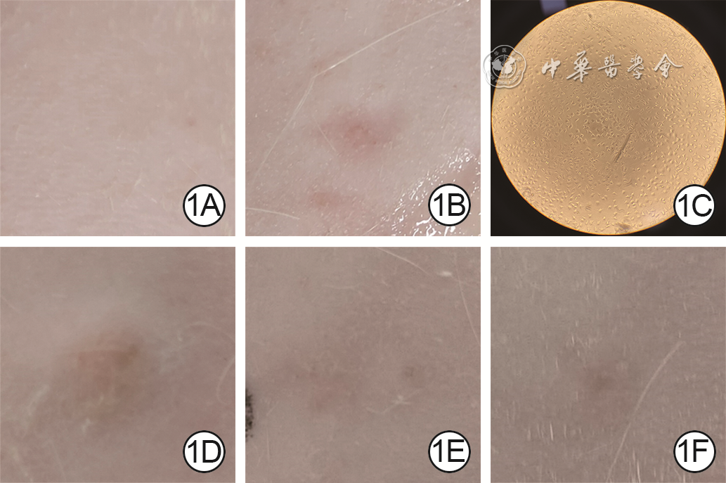
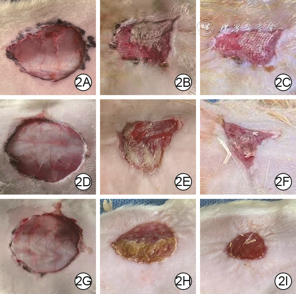
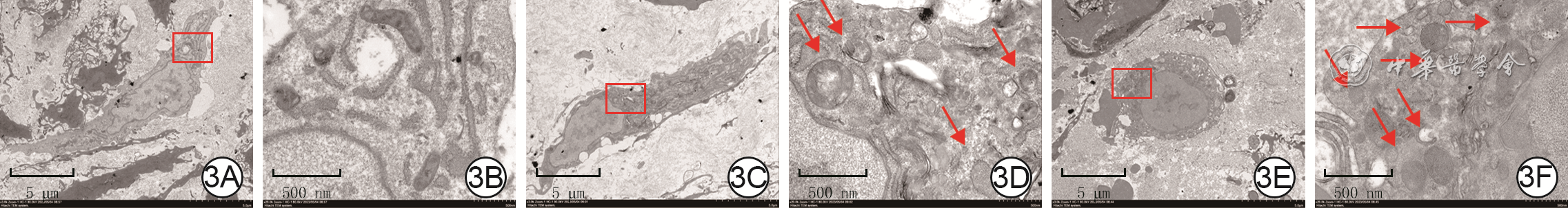
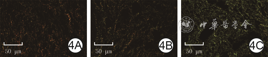
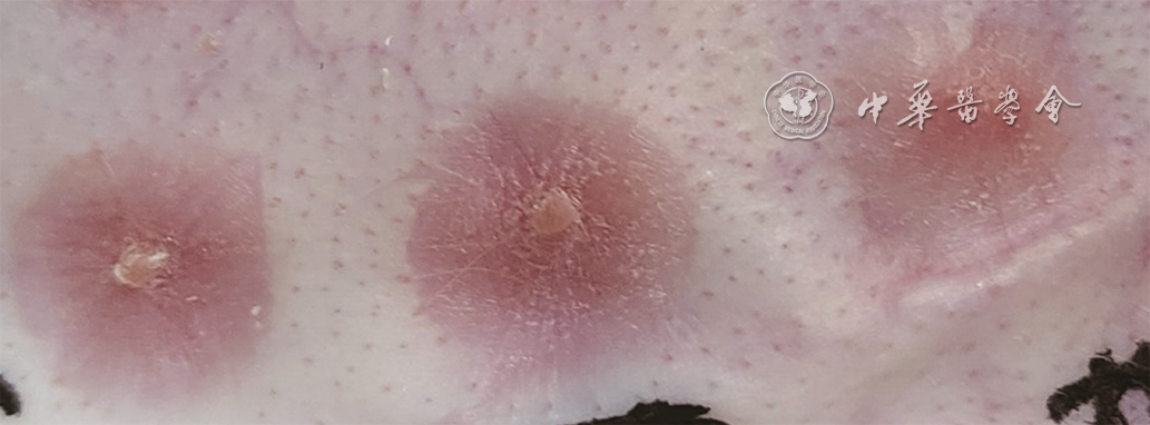
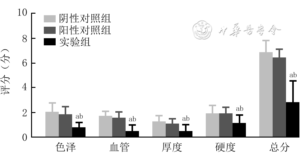
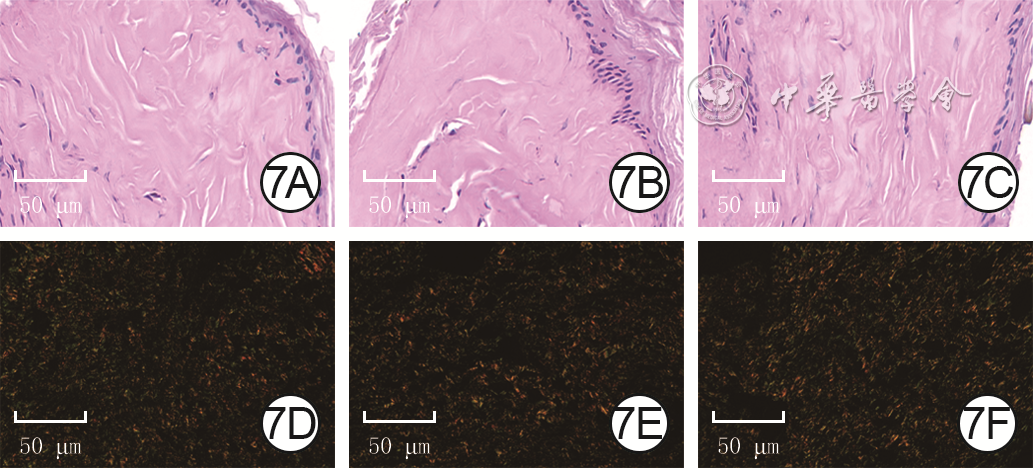
  Objective  To investigate the effects of trehalose gel on full-thickness skin defect wounds in rats and scar hyperplasia in rabbit ears.  Methods  The study was an experimental study. The trehalose gel and carbomer gel were prepared, their appearance after irradiation sterilization, and their physicochemical characterization such as viscosity, film forming rate, bacteria resistance rate, heavy metal content, moisture retention rate, water vapor permeability, sterility, and biocompatibility such as cytotoxicity, intradermal irritation, and sensitization were observed. Thirty male Sprague-Dawley rats aged 8-10 weeks were divided into experimental group, positive control group, and negative control group using a random number table method, with 10 rats in each group. The full-thickness skin defect wound models were prepared on the back of rats in negative control group, positive control group, and experimental group, and were treated with routine dressing change, carbomer gel dressing change, and trehalose gel dressing change, respectively. The wound healing rates on 6 and 12 days after injury and the wound healing time were recorded. On 6 days after injury, the number of autophagosomes and autophagolysosomes in rat wound tissue was detected using transmission electron microscopy. The content of microtubule associated protein light chain 3Ⅰ (LC3Ⅰ) and LC3Ⅱ in rat wound tissue was detected using enzyme-linked immunosorbent assay method and their ratio was calculated. The proportion of type Ⅰ and Ⅲ collagens and their ratio, as well as the total collagen proportion in rat wound tissue were detected using sirius red picric acid staining method. The number of samples in the aforementioned experiments was all 5. Three male New Zealand albino model rabbits aged 3-4 months were taken, and 3 wounds deep to the perichondrium were created on each of the rabbit ears, with six wounds in each group and being grouped and treated as mentioned above. On 30 days after wound healing, the scar tissue of the rabbit ear was observed and evaluated using the Vancouver scar scale. The thickness of the epidermis and dermis in the scar tissue of the rabbit ear was measured using hematoxylin eosin staining, and the proportion of type Ⅰ and Ⅲ collagens and their ratio, as well as the total collagen proportion and arrangement in the scar tissue of the rabbit ear were measured using sirius red picric acid staining method. The number of samples was 6.  Results  The irradiated trehalose gel and carbomer gel were light yellow and transparent, without odor and impurities. The viscosity, film forming rate, bacteria resistance rate, and moisture retention rate of trehalose gel were significantly better than that of carbomer gel (with t values of 4.13, 3.50, 4.03, and 5.80, respectively, P<0.05), but the water vapor permeability was significantly lower than that of carbomer gel (t=-4.14, P<0.05). No heavy metals or bacteria were detected in any gel. Both of the two gel had no cytotoxicity, and the intradermal irritation and sensitization were negative. On 6 and 12 days after injury, the wound healing rates of rats in positive control group were significantly higher than that in negative control group (with t values of -6.82 and -4.58, respectively, P<0.05); the wound healing rate of rats in experimental group was significantly higher than those in positive control group (with t values of -8.90 and -4.25, respectively, P<0.05) and negative control group (with t values of -8.78 and -4.25, respectively, P<0.05). The wound healing time ((20.4±2.5), (23.4±2.5) d) of rats in positive control group and experimental group was significantly shorter than (27.0±2.1) d in negative control group (with t values of 2.45 and -4.49, respectively, P<0.05). On 6 days after injury, the number of autophagosomes and autophagolysosomes in wound tissue in experimental group of rats were significantly higher than those in positive control group (with t values of 7.37 and 9.33, respectively, P<0.05) and negative control group (with t values of -7.06 and -8.54, respectively, P<0.05). On 6 days after injury, the content of LC3 Ⅱ and LC3 Ⅱ/LC3 Ⅰ in wound tissue in positive control group of rats were significantly higher than that in negative control group (with t values of -4.48 and -2.47, respectively, P<0.05); the content of LC3Ⅰ and LC3 Ⅱ/LC3 Ⅰ in wound tissue in experimental group of rats were significantly higher than those in negative control group (with t values of 11.98 and 6.04, respectively, P<0.05) and positive control group (with t values of -6.64 and -4.17, respectively, P<0.05), the content of LC3Ⅰ was significantly lower than that in negative control group (t=2.33, P<0.05). On 6 days after injury, the proportions of total collagen and type Ⅰ collagen in wound tissue of rats in the three groups were similar, P>0.05. On 6 days after injury, the proportion of type Ⅲ collagen in wound tissue of rats in positive control group was significantly higher than that in negative control group (t=-3.19, P<0.05), and the type Ⅰ collagen/type Ⅲ collagen was significantly lower than that in negative control group (t=2.18, P<0.05); the proportion of type Ⅲ collagen in the wound tissue of rats in experimental group was significantly higher than those in negative control group and positive control group (with t values of -2.38 and 5.91, respectively, P<0.05), and type Ⅰ collagen/type Ⅲ collagen was significantly lower than those in negative control group and positive control group (with t values of 3.08 and -4.35, respectively, P<0.05). On 30 days after wound healing, it was observed that the rabbit ear scar proliferation in positive control group was similar to that in negative control group, while the rabbit ear scar proliferation in experimental group was significantly reduced. On 30 days after wound healing, the color, blood vessels, thickness, hardness score, and total score of rabbit ear scars in experimental group were significantly lower than those in positive control group (with t values of 3.80, 3.80, 2.39, 2.71, and 4.84, respectively, P<0.05) and negative control group (with t values of -3.81, -4.78, 0.04, -2.71, and -5.14, respectively, P<0.05). On 30 days after wound healing, there was no significant difference in the epidermal thickness of rabbit ear scar tissue among experimental group, negative control group, and positive control group (P>0.05); the dermal thickness of rabbit ear scar tissue in positive control group was significantly smaller than that in negative control group (t=5.42, P<0.05), while the dermal thickness of rabbit ear scar tissue in experimental group was significantly smaller than those in negative control group and positive control group (with t values of 11.91 and 8.49, respectively, P<0.05). On 30 days after wound healing, the collagen protein arrangement of scar tissue of rabbits in the three groups was disordered, and the total collagen proportion was similar (P>0.05). The proportion of type Ⅰ collagen of scar tissue in experimental group was significantly lower than that in positive control group (t=3.00, P<0.05), the content of type Ⅲ collagen was significantly higher than those in negative control group and positive control group (with t values of -4.46 and 4.05, respectively, P<0.05), and the type Ⅰcollagen/type Ⅲ collagen was significantly lower than those in negative control group and positive control group (with t values of 8.50 and -5.25, respectively, P<0.05).  Conclusions  Compared with carbomer gel, trehalose gel has a more suitable physicochemical characterization for wound healing, and has good biocompatibility. It can promote the wound healing of full-thickness defects in rats and reduce scar hyperplasia in rabbit ears based on autophagy activation.
Review
Research advances on the mechanism of circadian rhythm genes in wound healing
Teng Ying, Qi Fang, Xu Guangchao, Wang Dali
2024, 40(7): 689-693. doi: 10.3760/cma.j.cn501225-20230831-00071
Abstract:
The healing after skin injury is a dynamic process of the interaction of various cells, cytokines, and extracellular matrix, and the abnormality of any link will affect the speed and quality of wound healing. Circadian rhythm is a spontaneous periodic oscillation cycle in organisms, which plays an important role in maintaining homeostasis and regulating physiological activities. Studies have shown that circadian rhythm genes play a key role in wound healing. This review summarizes the effects of circadian rhythm genes on different stages of wound healing and its possible mechanism of action, in order to provide a new perspective for wound healing and provide theoretical basis for formulating more effective treatment strategies for refractory wounds.
Research advances on in-situ cell electrospinning and its application in wound repair
Liu Huazhen, Zhang Yi, Gao Chuang, Lu Chunxiang, Guo Zilong, Sun Wenbin, Xiao Shichu, Liu Yuanyuan
2024, 40(7): 694-698. doi: 10.3760/cma.j.cn501225-20231123-00204
Abstract:
Currently, there are limited strategies for convenient and rapid wound repair in clinical practice. In recent years, in-situ cell electrospinning (IS-CE) technology, developed from in-situ electrospinning (IS-E) technology, has emerged. IS-CE technology involves encapsulating living cells within micro-nanofibers to construct living fibrous tissue scaffolds in situ, making some progress in wound repair applications. However, this technology still faces limitations such as low cell survival rate and poor fiber stability. This article provides a comprehensive review on the current status of both IS-E and IS-CE technologies, as well as the application of IS-CE technology in wound repair. In addition, the advantages, limitations, and improvement methods of IS-CE technology applied in wound treatment are emphatically discussed, aiming to provide insights for its application in tissue engineering and wound repair.
Academic Information
Summary of the 19th Chinese Symposium of Burns and Wounds
Lyu Yanling, Mo Yu, Liang Guangping, Luo Gaoxing, Peng Yizhi, Sun Dan, Qiu Kaizhen, Wu Luyao, Li Tingting, Liu Zhixin
2024, 40(7): 699-700. doi: 10.3760/cma.j.cn501225-20240626-00251
Abstract:
The 19th Chinese Symposium of Burns and Wounds was successfully held in Foshan of Guangdong Province from June 20th to 22nd in 2024. There were more than 700 delegates attending the academic event. The theme of the congress was expansion, integration and standardization, which could promote academic exchanges, multi-disciplinary fusion, and standardization of clinical treatment of burns and wounds. A total of nearly 200 famous experts and scholars had their speeches on the two-day keynote forum and special academic seminars including critical care, wound repair, scar prevention and treatment, rehabilitation nursing, and disciplinary integration sessions. The congress ended successfully with abundant fruits and friendship.