2025 Vol. 41, No. 1

Academician Forum
Biomaterial is becoming a promising research field for trauma, burns, and wound repair
Fu Xiaobing
2025, 41(1): 1-4. doi: 10.3760/cma.j.cn501225-20241125-00461
Abstract:
I learned from some specialists in fields of trauma, burns, and tissue repair in different conferences that the new research fields in trauma, burns, and tissue repair combined with biomaterials have been established in recent years. I feel that the direction of scientific research in the subject field characterized by the treatment of "injury" is undergoing new changes, expanding into new fields related to "injury" treatment such as biomaterials and biomedical engineering, especially the close integration with biomaterials. These reflect the characteristics of cross-industry, cross-field, and multi-disciplinary intersection and integration. These new developments could lead to revolutionary breakthroughs in future research and clinical treatment of trauma, burns, and tissue repair. In this paper, I would like to summarize the fruitful achievements in these fields and put forward some suggestions for the next development of the discipline.
Expert Forum
Mastering the technique of chimeric perforator flap to further improve the reconstructive level of compound tissue defects
Tang Juyu
2025, 41(1): 5-10. doi: 10.3760/cma.j.cn501225-20241106-00435
Abstract:
The precise repair of compound tissue defects remains a significant challenge in the field of microsurgical reconstruction. In recent years, the integrated advent of perforator flap and chimeric flap techniques has led to the development of the chimeric perforator flap technique. In chimeric perforator flaps, the perforator flap is connected to bone, muscle, or fascial flaps solely through the vascular pedicle, allowing each tissue component considerable freedom of movement. Bone flaps can effectively reconstruct bone defects, muscle flaps can accurately restore functional muscle deficits and fill deep dead spaces, fascial flaps can reconstruct joint capsule or tendon defects, while perforator flaps can freely cover superficial wounds, thus achieving three-dimensional repair of compound tissue defects. Compared with traditional bone flaps and musculocutaneous flaps, chimeric perforator flaps significantly improve the repair outcomes of the recipient area while markedly reducing damage to the donor area. It is recommended to master the technique of chimeric perforator flaps proficiently in order to further enhance the level of reconstruction and repair for compound tissue defects.
Further emphasis on the application of microsurgical techniques in burns
Shen Yuming, Hu Yungang
2025, 41(1): 11-17. doi: 10.3760/cma.j.cn501225-20241012-00380
Abstract:
Over the course of more than 60 years of development, microsurgical techniques have entered the era of intelligence and digitization. This article focuses on the development of microsurgical techniques in the field of tissue repair in China, provides a detailed analysis of the application of microsurgical techniques in the repair of destructive burn wounds and the reconstruction of post-burn scar contracture deformity, and systematically introduces the flap repair strategies in various tissue forms. With advances of science and technology, and breakthroughs in emerging fields such as tissue engineering, regenerative medicine and gene editing, microsurgical techniques have demonstrated tremendous potential in the area of tissue repair, with broad prospects for future applications.
Wound Repair by Microsurgery
Clinical effects of various special forms of the descending branch of the lateral circumflex femoral artery perforator flaps in repairing high-voltage electrical burn wounds on the wrist
Du Weili, Xiong Feng, Che Kexin, Cheng Lin, Dai Qiang, Shen Yuming
2025, 41(1): 18-27. doi: 10.3760/cma.j.cn501225-20240930-00365
Abstract:
  Objective  To explore the clinical effects of various special forms of the descending branch of the lateral circumflex femoral artery (DLCFA) perforator flaps in repairing high-voltage electrical burn wounds on the wrist.  Methods  This study was a retrospective observational study. From September 2014 to June 2024, 79 male patients with high-voltage electrical burns on the wrist, aged 20 to 62 years and met the inclusion criteria, were admitted to Beijing Jishuitan Hospital Affiliated to Capital Medical University, with wrist high-voltage electrical burn wound (hereinafter referred to as wrist wound) types being classified as type Ⅱ or type Ⅲ. In the early stage after injury, debridement was performed on the patients' wrists. Based on the wound condition and flap indications, the flow-through, lobed, chimeric, flow-through-lobed, lobed-chimeric, flow-through-chimeric, or flow-through-lobed-chimeric DLCFA perforator flap was employed individually, and the flow-through-chimeric DLCFA perforator flap and tensor fascia lata myocutaneous flap were employed in combination to repair the wounds. The donor site wounds were repaired using direct sutures or skin grafting. The number of various DLCFA perforator flaps resected during surgery and the number of various types of wrist wounds repaired were recorded, as well as the closure status of the donor site wound. The postoperative flap survival, occurrence of vascular crisis, wound or suture site healing, and patency of the reconstructed artery in flow-through flaps were recorded. During follow-up, the appearance of the flap, scar formation, and the presence of thigh muscle herniation were observed.  Results  Intraoperatively, 11 flow-through DLCFA perforator flaps were resected to repair 11 type Ⅱ wrist wounds, 13 lobed DLCFA perforator flaps were resected to repair 9 type Ⅱ and 4 type Ⅲ wrist wounds, 16 chimeric DLCFA perforator flaps were resected to repair 16 type Ⅱ wrist wounds, 11 flow-through-lobed DLCFA perforator flaps were resected to repair 5 type Ⅱ and 6 type Ⅲ wrist wounds, 10 lobed-chimeric DLCFA perforator flaps were resected to repair 5 type Ⅱ and 5 type Ⅲ wrist wounds, 6 flow-through-chimeric DLCFA perforator flaps were resected to repair 6 type Ⅱ wrist wounds, 7 flow-through-lobed-chimeric DLCFA perforator flaps were resected to repair 7 type Ⅲ wrist wounds, and 5 flow-through-chimeric DLCFA perforator flaps combined with tensor fascia lata myocutaneous flaps were resected to repair 5 type Ⅲ wrist wounds. Seventy-four patients had their donor site wounds closed by direct suturing, while 5 patients had their donor site wounds closed by skin grafting. Postoperatively, the flaps in 3 patients developed vascular crisis, including 1 case of arterial crisis and 2 cases of venous crises but survived after emergency vascular exploration and other treatments; the remaining flaps survived completely. Postoperatively, 3 patients had seepage beneath their flaps, which were closed after dressing changes; the remaining patients' wounds or suture sites all healed. Anteriography showed that all reconstructed arteries in 35 patients who underwent flow-through flap transplantation were patent postoperatively. During the follow-up period of 3 months to 1 year, 20 patients had bloated flap, while the rest had good flap appearance; linear scars were left in the donor sites that underwent direct wound closure, and the skin-grafted areas of the donor site wounds showed no significant patchy hypertrophic scarring; no thigh muscle herniation occurred.  Conclusions  Taking the full advantage of perforator flaps, various special forms of the DLCFA perforator flaps are used to repair the three-dimensionally injury wounds caused by high-voltage electrical burns on the wrist, which not only minimizes the damage to the donor site but also allow the recipient site to be well repaired, showing good appearance in the recent follow-up.
Clinical effects of the anterolateral thigh perforator flaps transplantation in repairing the wounds after radical resection of skin malignant tumors in the head, face, and neck
Sun Xiangdong, Li Chengzhi, Wulamu Aili, Maijimi Simayi, Da Chengli, Zhang Xingqin, Aihemaitijiang Aihetaier, Abudusaimijiang Aximu, Wu Yuanquan
2025, 41(1): 28-35. doi: 10.3760/cma.j.cn501225-20240920-00341
Abstract:
  Objective  To investigate the clinical effects of the anterolateral thigh perforator flaps transplantation in repairing the wounds after radical resection of skin malignant tumors in the head, face, and neck.  Methods  This study was a retrospective observational study. From May 2020 to December 2023, 27 patients with skin malignant tumors in the head, face, and neck were admitted to the Department of Burns and Plastic Surgery of the First People's Hospital of Kashi Prefecture, including 19 males and 8 females, aged 53 to 89 years. There were 21 cases with squamous cell carcinoma and 6 cases with basal cell carcinoma. The wound area after radical resection of tumors was 9.0 cm×7.0 cm to 27.0 cm×21.5 cm. The wounds were repaired with the lobulated, combined, or various forms of combination of the anterolateral thigh perforator flaps, and the harvesting flap area in single donor site was 10.0 cm×8.0 cm to 27.0 cm×11.0 cm. The wounds in the flap donor sites were closed by suturing in 26 patients, while the wound in the flap donor site in 1 patient was repaired with medium-thickness skin graft in the lower leg. The survival of the flap, and the occurrence of vascular crisis and infection were observed after surgery. During follow-up after surgery, the tumor recurrence, shape and texture of the flaps, and the function and scar formation of the limbs where the donor site was located were observed.  Results  Only one patient developed venous crisis of the flap 27 hours after surgery, and the flap survived after vascular exploration and reanastomosis of the vein; the flaps in the other patients survived after surgery. One patient had an infected effusion under the flap after surgery, which healed after dressing change. After 6-36 months of postoperative follow-up, no tumor recurrence was observed; the flap had good appearance, texture, and elasticity; the limb where the donor site was located functioned normally, with only linear scars left.  Conclusions  For complex wounds after radical resection of skin malignant tumors in the head, face, and neck, different forms of anterolateral thigh perforator flaps can be used to repair the wounds according to the condition of the wounds, and the wounds in the recipient sites heal well after surgery, with minimal damage to the donor site.
Clinical application effects of free transplantation of lobulated inguinal flaps
Zhang Wei, Zhang Weidong, Xu Junhui, Chen Lan, Gong Xiang, Liu Feng, Zhou Jinxiu, Yang Fei, Xie Weiguo
2025, 41(1): 36-44. doi: 10.3760/cma.j.cn501225-20241014-00384
Abstract:
  Objective  To investigate the clinical application effects of free transplantation of lobulated inguinal flaps.  Methods  This study was a retrospective observational study. From July 2019 to April 2024, 34 patients with skin defect wounds whose wounds in one part met the inclusion criteria were admitted to Tongren Hospital of Wuhan University & Wuhan Third Hospital, including 28 males and 6 females, aged 26 to 59 years. The wound area in the recipient area ranged from 3.0 cm×2.0 cm to 25.0 cm×20.0 cm. The lobulated inguinal flap pedicled with the branch of the superficial circumflex iliac artery were obtained in 19 patients, and the lobulated inguinal flap pedicled with the main artery of the superficial circumflex iliac artery and the superficial inferior epigastric artery were obtained in 15 patients. The total area of the flaps ranged from 6.0 cm×2.2 cm to 27.0 cm×23.0 cm. The flaps were divided into 2 to 4 lobes, and the area of each lobe ranged from 2.0 cm×1.0 cm to 17.0 cm×12.0 cm. Each lobe of the flaps was reassembled, spliced, or directly transplanted onto the wounds, and the donor wounds were sutured in layers. The survival of each lobe of the flaps and wound healing in the recipient and donor areas were observed, and the wound recovery in the recipient and donor areas were followed up. At the last follow-up, the patient's satisfaction with the efficacy was assessed by 5-grade Likert scale.  Results  A small amount of necrosis appeared in the tip of one lobe of the flaps in 4 patients after surgery, which healed after trimming. The flaps of the remaining 30 patients survived. The wounds in the recipient areas healed smoothly. There was a small amount of necrosis at the suture edge of the donor areas in 3 patients, which healed after local trimming and dressing change. The donor wounds healed well in the remaining 31 patients. During the follow-up of 6 to 42 months, all the recipient wounds were well repaired, and the shape of the donor areas was good. At the last follow-up, 15 patients were very satisfied with the efficacy, 15 were relatively satisfied, and 4 were generally satisfied.  Conclusions  Through preoperative ultrasonic examination and positioning, the inguinal flap is designed according to the course of blood vessels and lobulated with the branch of the superficial circumflex iliac artery or the main artery of the superficial circumflex iliac artery and the superficial inferior epigastric artery as the pedicles. The anatomical process is reliable and the blood flow of the flap after being lobulated is rich, which can meet the repair needs of various skin defect wounds. The repair effect is good, and the damage in the donor area is small, which is worthy of promotion.
Clinical efficacy of layered thinning superficial circumflex iliac artery perforator flap based on color Doppler ultrasound positioning
Zhang Wentong, Yang Yong, Li Feng, Li Bin, Wang Dandan, Chen Tao, Li Jianfeng
2025, 41(1): 45-52. doi: 10.3760/cma.j.cn501225-20240927-00358
Abstract:
  Objective  To explore the clinical efficacy of layered thinning superficial circumflex iliac artery perforator (SCIP) flap based on color Doppler ultrasound (CDU) positioning.  Methods  The study was a retrospective observational study. From February 2023 to February 2024, 14 patients who met the inclusion criteria were admitted to the Department of Hand Surgery of Beijing Jishuitan Hospital Affiliated to Capital Medical University, including 11 males and 3 females, aged 15 to 60 years. The wound area was from 7 cm×4 cm to 14 cm×11 cm. Before the flap transplantation surgery, CDU was used to accurately locate the deep fascial exit point of the superficial branch of the superficial circumflex iliac artery. During the surgery, the SCIP flap was thinned in layers to repair the hand and forearm wounds of 11 patients and foot wounds of 3 patients. The flap incision area ranged from 8 cm×5 cm to 15 cm×12 cm. The donor area wounds of flaps were sutured directly. During the surgery, the deep fascial exit point of the superficial branch of the superficial circumflex iliac artery in the flap donor area was observed and compared with the result of CDU positioning before the surgery, and the flap thickness was measured. The flap survival and occurrence of adverse reactions were observed after the surgery. During follow-up, the appearance and texture of flaps, and the wound healing in the donor area was observed. At the last follow-up, the function of the wrist and hand in the affected limbs was evaluated according to the trial standards for evaluation of partial function of upper extremity by the Hand Surgery Society of Chinese Medical Association, and the function of the foot and ankle in the affected limbs was evaluated according to the scoring standard of American Orthopaedic Foot and Ankle Society.  Results  During the surgery, the deep fascial exit point of the superficial branch of the superficial circumflex iliac artery in the flap donor area was consistent with the result of CDU positioning before the surgery, and the distance between them was less than 10 mm. The flap thickness was 5 to 8 mm with an average of 6.2 mm. All the flaps survived after surgery. During the follow-up of 5 to 12 months, the flaps had good appearance and texture, all the wounds in the donor areas healed with only linear scar left. At the last follow-up, the function of the wrist and hand in the affected limbs was evaluated as excellent in 9 cases, good in one case, and fair in one case; the function of the foot and ankle in the affected limbs was evaluated as excellent in one case and good in two cases.  Conclusions  CDU examination can provide precise preoperative perforator positioning for layered thinning of SCIP flap and contribute to the optimization of flap design, so as to avoid the problem of flap necrosis caused by improper preoperative design to some extent and improve the safety of surgery. In addition, the layered thinning SCIP flap results in less damage to the donor area and is beneficial for the recovery of the affected limb function, which is worthy of clinical promotion.
Clinical effect of antibiotic-loaded bone cement implantation combined with free chimeric tissue flap transplantation in the sequential treatment of severe gouty wounds
Xiao Shun'e, Li Hai, Zhang Tianhua, Wu Xiangkui, Wu Bihua, Wei Zairong, Deng Chengliang
2025, 41(1): 53-60. doi: 10.3760/cma.j.cn501225-20240919-00340
Abstract:
  Objective  To evaluate the clinical effect of antibiotic-loaded bone cement implantation combined with free chimeric tissue flap transplantation in the sequential treatment of severe gouty wounds.  Methods  This study was a retrospective observational study. From July 2019 to July 2022, 11 male patients with severe gouty wounds who were aged 33 to 71 years and met the inclusion criteria were admitted and treated at the Affiliated Hospital of Zunyi Medical University. The wounds were located on the hands in 2 cases, the ankles in 5 cases, and the feet in 4 cases. After debridement, the wound area ranged from 5.0 cm×4.0 cm to 22.0 cm×6.0 cm. All wounds were sequentially repaired with antibiotic-loaded bone cement implantation combined with free chimeric tissue flaps transplantation. Two cases were repaired by free perforating branch of superficial circumflex iliac artery with chimeric osseous flaps, with the areas of harvested skin flaps being 5.5 cm×4.0 cm and 8.0 cm×6.0 cm, respectively, and the volumes of iliac bone flaps being 2.0 cm×2.0 cm×1.5 cm and 3.5 cm×2.0 cm×2.0 cm, respectively. Two cases were repaired by free perforating branch of deep circumflex iliac artery with chimeric osseous flaps, with the areas of harvested skin flaps being 6.0 cm×4.0 cm and 7.5 cm×5.0 cm, respectively, and the volumes of iliac bone flaps being 2.0 cm×1.5 cm×1.5 cm and 2.5 cm×2.0 cm×1.5 cm, respectively. Seven cases were repaired by free chimeric myocutaneous flaps based on the descending branch of the lateral circumflex femoral artery. The areas of harvested skin flaps ranged from 9.5 cm×6.0 cm to 25.0 cm×6.5 cm, and the volumes of muscle flaps ranged from 4.0 cm×3.0 cm×2.0 cm to 6.0 cm×5.0 cm×2.5 cm. The donor site wounds were directly sutured. The chimeric tissue flap was freely transplanted to the recipient wound site, of which the iliac bone graft was used to fill the bone defect, the muscle flap was utilized to fill the wound cavity, and the skin flap was employed to cover the wound surface; the arteries and veins in the vascular pedicle were anastomosed with those in the recipient area. At admission and 3 days post antibiotic-loaded bone cement implantation, the changes in white blood cell count, neutrophil and hypersensitive C-reactive protein level, as well as the bacterial culture of wound secretions specimen, and the growth of granulation tissue were observed. After stage Ⅱ surgery, the survival of transplanted chimeric tissue flaps, the occurrence of vascular crisis, and the healing of wounds in donor and recipient sites were observed. During follow-up, the blood supply, appearance, and texture of the transplanted tissue flaps in the recipient sites, the function and appearance of the affected limbs and fingers, and the complications in the donor and recipient sites were observed.  Results  Three days post antibiotic-loaded bone cement implantation, white blood cell count, hypersensitive C-reactive protein level, and neutrophil significantly decreased compared with those at admission (with Z values of -2.93 and -2.93 respectively, t=8.63, P<0.05). At admission, all patients exhibited bacterial infections with redness and swelling around the wounds. Three days post antibiotic-loaded bone cement implantation, bacterial cultures of wound secretions specimen were negative, local redness resolved, and granulation tissue showed good growth. After stage Ⅱ surgery, all chimeric flaps survived without vascular crises. The wound healing in the recipient site of the dorsum of the foot in one patient was poor and delayed but healed after dressing changes; all the other recipient sites in remaining patients healed successfully. The donor incision healed well in all patients. During 6 to 24 months of follow-up, the flaps in the recipient area demonstrated good blood circulation, texture, and appearance. Bone healing was achieved in 4 patients with iliac grafts. Nine patients with lower limb wounds were able to bear weight, and the functions including gripping, palm alignment, and finger alignment were significantly improved in 2 patients with hand wounds. No significant complications were observed in donor or recipient sites.  Conclusions  In treating patients with severe gouty wounds, the sequential strategy of stage Ⅰ debridement with antibiotic-loaded bone cement implantation followed by stage Ⅱ free chimeric osseous flaps or myocutaneous flaps repair can achieve effectively control of postoperative wound infection, promote wound healing, and well restore the functions of affected finger or limb with no obvious complications, which is worthy of promotion for clinical application.
Clinical effects of free bilateral turbocharged anterolateral thigh flaps in tandem in repairing extensive wounds in the foot and ankle
Yang Liang, Zhou Rong, Ju Jihui, Niu Zefeng, Liu Zhongzheng, Guo Liping, Liu Zhijin, Jin Qianheng, Ge Chengwei, Wang Guiyang, Yang Lin, Cheng Junnan
2025, 41(1): 61-69. doi: 10.3760/cma.j.cn501225-20240508-00164
Abstract:
  Objective  To explore the clinical effects of free bilateral turbocharged anterolateral thigh flaps in tandem in repairing extensive wounds in the foot and ankle.  Methods  The study was a retrospective observational study. From April 2020 to June 2023, 12 patients with extensive wounds in the foot and ankle who met the inclusion criteria were admitted to the Department of Wound Repair Surgery of Suzhou Ruihua Orthopedic Hospital, including 8 males and 4 females, aged 21 to 65 years. The wound area after debridement ranged from 27 cm×14 cm to 37 cm×20 cm. The bilateral perforator flaps pedicled with either oblique or descending branches of the lateral circumflex femoral artery were designed and harvested based on the size and shape of the wounds. The individual flap incision area ranged from 16 cm×9 cm to 34 cm×12 cm. The non-homologous perforator of the flap on the one side was turbocharged by anastomosing it with the gross muscular branch or main vessel of the oblique or descending branch of the lateral circumflex femoral artery from the flap. Subsequently, the proximal end of the oblique or descending branch of the lateral circumflex femoral artery and its accompanying vein from the flap on the one side were connected end-to-end with either the anterior tibial artery and vein, posterior tibial artery and vein, or dorsal foot artery and vein in the recipient area, the distal end of the oblique or descending branch of the lateral circumflex femoral artery and its accompanying vein from the flap on the one side were anastomosed end-to-end with a source vessel originating from flap on the other side. The wounds in the flap donor areas were sutured directly. The number and source of perforators carried by the flaps and the duration of the flap repair surgery were recorded. The survival of the flap, the occurrence of vascular crisis, and the wound healing at both donor and recipient areas were observed after surgery. The flap condition, appearance and function of the affected limb were observed during follow-up. At the last follow-up, the sensory function of the flap was assessed using the British Medical Research Council's sensory rating standard, the foot and ankle function of the affected limb was evaluated according to the American Orthopedic Foot and Ankle Society scoring standard.  Results  A total 24 flaps were successfully harvested, carrying 60 perforators, including 34 perforators from the oblique branch of the lateral circumflex femoral artery, 24 perforators from the descending branch of the lateral circumflex femoral artery, one perforator from the transverse branch of the lateral circumflex femoral artery, and one perforator from the direct branch of the femoral artery. The duration of the flap repair surgery ranged from 4.2 to 9.0 hours. The flaps of 12 patients exhibited complete survival after surgery. A total of two flaps of two patients experienced venous crisis after surgery but survived through emergency exploration. One patient encountered undesirable wound healing at the donor area of flap on the one side after surgery, which healed after dressing change, debridement, and suturing. The remaining patients' donor area wounds healed. Two patients displayed impaired wound healing in the recipient area, which improved after dressing change and resection of residual sequestrum, and the wounds in the recipient area of other patients healed successfully. During the follow-up of 4-26 months, the flaps demonstrated favorable color and texture, slight edematous appearance, and partial sensory recovery, as well as good aesthetic and functional restoration of the affected limbs. At the last follow-up, the sensory function of the flap was assessed as grade S2 in 9 cases and grade S3 in 3 cases; the foot and ankle function of the affected limb was evaluated as excellent in two cases, good in 9 cases, and fair in one case.  Conclusions  The bilateral turbocharged anterolateral thigh flaps have numerous sources of perforators. By implementing supercharging of non-homologous perforators within the flap, the vascular supply to the flap is turbocharged, thereby mitigating the risk of extensive flap necrosis. The flap is an effective approach for repairing extensive wounds in the foot and ankle, resulting in improved function of the affected limb after repair.
Clinical efficacy of the first dorsal metatarsal artery pedicled lateral toe bilobed flap in repairing the finger pulp defects of two adjacent fingers
Li Shimin, Zhou Shuping, Chen Junjie, Li Sen, Shi Yingguang, Zheng Liwu, Chang Chaonan, Wang Huanpeng, Sun Ke, Yin Daqing
2025, 41(1): 70-76. doi: 10.3760/cma.j.cn501225-20240407-00121
Abstract:
  Objective  To explore the clinical efficacy of the first dorsal metatarsal artery pedicled lateral toe bilobed flap in repairing the finger pulp defects of two adjacent fingers.  Methods  This study was a retrospective observational study. From January 2018 to December 2022, 9 patients with finger pulp defects in two adjacent fingers who met the inclusion criteria were admitted to the Department of Burns and Plastic Surgery of the 988th Hospital of Joint Logistics Support Force of PLA, including 6 males and 3 females, aged 26 to 48 years. The injured fingers were the index finger and middle finger (5 cases) or the middle finger and ring finger (4 cases). After debridement, the wound area of a single finger ranged from 1.2 cm×0.8 cm to 3.2 cm×2.8 cm. The finger pulp defects of two adjacent fingers were repaired with the first dorsal metatarsal artery pedicled lateral toe bilobed flap, and the two adjacent fingers were sutured together. The area of single flap ranged from 1.5 cm×1.0 cm to 3.5 cm×3.0 cm. The wound in the flap donor site was sutured directly or repaired with full-thickness skin graft from the groin region. The finger separation surgery was performed 3 weeks after surgery. The survival and blood supply of flaps, and survival of skin grafts and wound healing of the donor sites were observed after surgery. During follow-up, the texture, sliding, and shape of the flap, movement function of the finger, and the shape and function of the foot donor site were observed. At the last follow-up, the sensory of the flap was evaluated according to the sensory evaluation standard of the British Medical Research Council, and the hand function was evaluated according to the functional evaluation trial standard for severed finger replantation of the Hand Surgery Society of the Chinese Medical Association.  Results  After surgery, all the flaps of 9 patients survived without vascular crisis. The flaps were soft in texture and good in shape. One patient had partial necrosis at the edge of the skin graft in the toe, and the wound healed after dressing change; the skin grafts in the toe in the other 8 patients survived, and the wounds healed well. During follow-up of 12 to 18 months after surgery, the flaps had soft texture, good elasticity, low sliding, and good shape. The finger movement function was normal. The wound in foot donor site recovered well without ulceration and deformity, and walking was not affected. At the last follow-up, the sensation of the flaps was sensitive, of which 8 flaps reached S3 and 10 flaps reached S3+ in sensation, and the two-point discrimination distance of the flaps was 9-13 mm. The functional scores of the affected fingers were 85 to 95, all of which were excellent.  Conclusions  The first dorsal metatarsal artery pedicled lateral toe bilobed flap can repair finger pulp defects of two adjacent fingers at the same time, and the appearance, sensation, and function of the affected fingers recovered well after surgery, with less damage to the foot donor site. It is one of good methods to repair finger pulp defects of two adjacent fingers in clinic.
Original Article
Clinical application effects of tissue flaps prepared from the discarded limbs after amputation in patients with destructive wounds in the lower limbs
Huang Mitao, He Zhiyou, Zhang Pihong, Zhang Minghua, Cui Xu, Guo Le, Huang Xiaoyuan, Liang Pengfei
2025, 41(1): 77-83. doi: 10.3760/cma.j.cn501225-20240605-00212
Abstract:
  Objective  To explore the clinical application effects of tissue flaps prepared from the discarded limbs after amputation in patients with destructive wounds in the lower limbs.  Methods  The study was a retrospective observational study. From March 2019 to March 2024, 7 male patients with destructive wounds in the lower limbs who met the inclusion criteria were admitted to the Department of Burns and Plastic Surgery of Xiangya Hospital of Central South University, aged 35 to 57 years. After clinical treatment, the damaged limbs were still difficult to preserve. Five patients with destructive wounds in the lower limbs were repaired with pedicled tissue flaps prepared from the discarded limbs after amputation, with wound areas of 15 cm×10 cm to 25 cm×15 cm and tissue flap incision areas of 15 cm×10 cm to 20 cm×15 cm. Two patients with destructive wounds in the lower limbs were repaired with free tissue flaps prepared from the discarded limbs after amputation, with wound areas of 22 cm×18 cm and 25 cm×15 cm and tissue flap incision areas of 23 cm×20 cm and 25 cm×18 cm. The survival of the tissue flap, the healing and appearance of wounds in the recipient site were followed up. At the last follow-up, the recovery status of the recipient site was evaluated according to the comprehensive flap evaluation scale, and the status of corrective prosthesis fitting was recorded.  Results  During the follow-up of 6 to 24 months, all the tissue flaps successfully survived, with good healing and appearance of wounds in the recipient site. At the last follow-up, the scores of the recovery status of the recipient site ranged from 36 to 39 (with an average of 37.2). All amputated limbs were able to accommodate corrective prostheses with no distal ulceration.  Conclusions  Utilizing tissue flaps prepared from the discarded limbs after amputation in patients with destructive wounds in the lower limbs to repair wounds effectively uses the discarded tissue and avoid creating new donor sites, which is a method to be considered for repairing multiple destructive wounds.
Two-sample Mendelian randomization analysis of the causal relationship between human immune cell phenotypes and keloids
Gan Wenjun, Wang Jingru, He Jia, Chen Xiaodong
2025, 41(1): 84-93. doi: 10.3760/cma.j.cn501225-20231130-00219
Abstract:
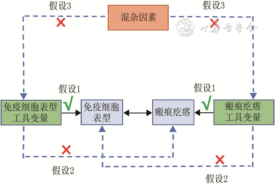
  Objective  To explore the causal relationship between human immune cell phenotypes and keloids.  Methods  This study was based on a two-sample Mendelian randomization (MR) analysis. Human immune cell phenotypes were considered as the exposure factors, and keloid was the outcome. Data on immune cell phenotypes (3 757 samples) and keloids (668 samples) were obtained from the genome-wide association study database. Using single nucleotide polymorphisms (SNPs) significantly associated with immune cell phenotypes as instrumental variables with the influence of weak instrumental variables being excluded, two-sample MR analysis was employed to evaluate the causal relationship between 731 human immune cell phenotypes and keloids. The inverse variance weighted (IVW) method was used to infer causal relationships, and the MR-Egger, weighted median, and weighted mode methods were used for validation. For SNPs of immune cell phenotypes meeting the hypothesis, the Cochran Q test was used to assess heterogeneity, and the MR-Egger regression and MR-PRESSO outlier tests were used to evaluate horizontal pleiotropy.  Results  A total of 18 204 SNPs meeting the significant threshold (P<1×10⁻⁵) were selected as instrumental variables for 731 immune cell phenotypes, and none of these SNPs were weak instrumental variables (with F values all >10). According to the IVW method, 21 immune cell phenotypes were identified with potential causal relationships to keloids, among which the CD62L- monocyte absolute count, CD19 on naive-mature B cell, CD19 on IgD+ B cell, CD27 on plasma blast-plasma cell, CD86 on CD62L+ myeloid dendritic cell, CD45 on natural killer T cell, CD25 on CD39+ CD4+ regulatory T cell, CD45 on monocytic myeloid-derived suppressor cells, CD8 on effector memory CD8+ T cell, and CD45RA on resting CD4+ regulatory T cell showed significant positive correlations with keloids (with odds ratios of 1.12, 1.09, 1.08, 1.21, 1.13, 1.12, 1.17, 1.11, 1.10, and 1.07, respectively, 95% confidence intervals of 1.03-1.23, 1.02-1.16, 1.01-1.15, 1.06-1.38, 1.02-1.25, 1.01-1.24, 1.03-1.33, 1.00-1.23, 1.00-1.20, and 1.01-1.13, respectively, P<0.05), while the activated and secreted CD4+ regulatory T cell absolute count, CD25 on unswitched memory B cell, plasmacytoid dendritic cell absolute count, CD14 on monocytic myeloid-derived suppressor cells, CD8 on natural killer T cell, CD20 on IgD+ CD38+ B cell, CD11c+ CD62L- monocyte absolute count, CD66b++ myeloid cell absolute count, CD11c on granulocytes, CD14 on CD14+ CD16+ monocyte, and CD3 on central memory CD8+ T cell showed significant negative correlations with keloids (with odds ratios of 0.95, 0.93, 0.93, 0.93, 0.91, 0.89, 0.89, 0.88, 0.87, 0.86, and 0.85, respectively, 95% confidence intervals of 0.90-1.00, 0.87-0.99, 0.88-0.99, 0.87-0.99, 0.84-1.00, 0.81-0.98, 0.81-0.98, 0.79-0.99, 0.78-0.96, 0.75-0.99, and 0.74-0.96, respectively, P<0.05). MR-Egger method confirmed the potential causal relationship existing respectively between CD25 on CD39+ CD4+ regulatory T cell, CD86 on CD62L+ myeloid dendritic cell, CD19 on IgD+ B cell, CD45RA on resting CD4+ regulatory T cell, CD3 on central memory CD8+ T cell and keloids (with odds ratios of 1.32, 1.22, 1.11, 1.09, and 0.73, respectively, 95% confidence intervals of 1.03-1.70, 1.04-1.44, 1.02-1.21, 1.01-1.19, and 0.55-0.95, respectively, P<0.05). The weighted median method confirmed the potential causal relationship existing respectively between CD45 on natural killer T cell, activated and secreted CD4+ regulatory T cells absolute count, CD20 on IgD+ CD38+ B cell, CD66b++ myeloid cell absolute count and keloids (with odds ratios of 1.15, 0.93, 0.87, and 0.83, respectively, 95% confidence intervals of 1.01-1.31, 0.86-1.00, 0.77-0.98, and 0.71-0.96, respectively, P<0.05). Among them, the potential causal relationship between CD20 on IgD+ CD38+ B cell and keloids was further verified by the weighted mode method (with odds ratio of 0.86, 95% confidence interval of 0.77-0.97, P<0.05). According to the aforementioned IVW method analysis results, the SNPs associated with the 21 immune cell phenotypes that had a significant causal relationship with keloids showed no significant heterogeneity (P>0.05) or significant horizontal pleiotropy (P>0.05).  Conclusions  From a genetic perspective, the potential causal relationships between 21 human immune cell phenotypes and keloids have been revealed, of which 10 immune cell phenotypes may be risk factors for keloids, while 11 immune cell phenotypes may act as protective factors for keloids.
Effects of nutritional therapy based on indirect calorimetry in patients with extremely severe burns during hypermetabolic stage
Jiang Bin, Du Yong, Xia Yilan, Chen Huaqing, Lu Fuchang
2025, 41(1): 94-100. doi: 10.3760/cma.j.cn501225-20240320-00103
Abstract:
  Objective  To explore the effects of nutritional therapy based on indirect calorimetry in patients with extremely severe burns during hypermetabolic stage.  Methods  This study was a retrospective cohort study. From March 25, 2022 to March 30, 2024, 20 extremely severe burn patients who met the inclusion criteria were admitted to the Department of Burn and Wound Repair of the Second Affiliated Hospital of Zhejiang University School of Medicine. There were 15 males and 5 females, aged (46±12) years. The patients were divided into death group (6 cases) and survival group (14 cases) according to their treatment outcomes. At the 3rd, 7th, 14th, and 21st d after injury, the resting energy expenditure (REE) of patients in the two groups was measured by indirect calorimetry once a day, and the nutritional treatment scheme was formulated according to the results of REE. The REE value, levels of albumin and interleukin-6 (IL-6), actual energy intake, and enteral nutrition energy intake of patients in the two groups at the 3rd, 7th, 14th, and 21st d after injury were collected (the last two indicators were respectively expressed as the mean values of the 3rd to 6th, 7th to 10th, 14th to 17th, and 21st to 24th d after injury). The actual energy intake/REE value and enteral nutrition energy intake/actual energy intake were calculated.  Results  The REE values of patients in survival group and death group were (8 143±2 328), (9 843±2 610), (10 149±2 248), (9 608±2 838) kJ and (6 816±2 057), (10 691±2 515), (11 031±2 850), (8 990±2 018) kJ, respectively at the 3rd , 7th, 14th, and 21st d after injury. The REE value and enteral nutrition energy intake of patients in the two groups showed trends of increase first and decrease then from the 3rd to 21st d after injury, while the differences in the above two indicators at each time point between the two groups of patients were not statistically significant (P>0.05). The actual energy intake of patients in survival group at the 21st d after injury was significantly higher than that at the 3rd d after injury (P<0.05), and the enteral nutrition energy intake of patients in death group at the 14th d after injury was significantly higher than that at the 3rd d after injury (P<0.05). There were no statistically significant differences in overall comparison in actual energy intake/REE value and enteral nutrition energy intake/actual energy intake at each time point after injury between and within the two groups of patients (P>0.05). The level of IL-6 of patients in survival group at the 21st d after injury was significantly lower than that at the 3rd and 7th d after injury, respectively (with both P values <0.05), and significantly higher than that at the 14th d after injury (P<0.05); the level of IL-6 of patients in death group at the 21st d after injury was significantly higher than that at the 3rd, 7th, and 14th d after injury (with P values all <0.05). The albumin level of patients in survival group at the 7th d after injury was significantly higher than that at the 3rd d after injury (P<0.05).  Conclusions  The REE value and enteral nutrition energy intake of extremely severe burn patients showed trends of increase first and decrease then, while they have no significant effects on the clinical outcome of the patients. The nutritional treatment scheme based on the indirect calorimetry can basically meet the energy demand of patients with extremely severe burns during hypermetabolic stage. The impacts of the albumin and IL-6 levels on the clinical outcome of extremely severe burn patients still need further research.