2024 Vol. 40, No. 12

Expert Forum
Practice and thinking on the integrated treatment mode for critical burns
Li Xiaojian, Tang Wenbin, Li Shanghua
2024, 40(12): 1101-1106. doi: 10.3760/cma.j.cn501225-20240827-00317
Abstract:
On the basis of further improving the survival rate of patients, the treatment of critical burns should prevent and treat all kinds of serious complications during the rescue process, shorten the time of wound healing, improve the quality of wound healing, and reduce scar hyperplasia and contracture after wound healing, so that patients can return to work and society after being cured. Based on the clinical practice of the Department of Burn and Plastic Surgery of Guangzhou Red Cross Hospital Affiliated to Jinan University in the aspects of integrated treatment modes including pre-hospital first aid and early treatment, early specialist treatment and rehabilitation, multidisciplinary cooperation and doctor-nurse cooperation in the treatment process, and regional treatment system, this paper discusses the composition and application of the integrated treatment mode for critical burns under the condition that a perfect treatment system for critical burns has not yet been formed, for the reference of fellow practitioners.
Discussion on the clinical practice and mode of treating critical burns in a comprehensive intensive care unit led by burn department
Zhang Pihong, Zhang Yikun, Liang Pengfei
2024, 40(12): 1107-1113. doi: 10.3760/cma.j.cn501225-20240906-00329
Abstract:
At present, although many burn centers and treatment units in China have established burn intensive care units (ICU), due to their limited capacity, the sudden nature of burn accidents, and the gradual transformation of professional burn treatment team, more and more critically burned patients are mainly treated in comprehensive ICU. The comprehensive ICU has been gradually adopting a semi-open management mode, with multidisciplinary collaboration led by ICU or burn specialists for critical burn treatment. Based on clinical practice, this paper shares the experience of treating critical burns under a semi-open management mode led by the burn department in comprehensive ICU, briefly introduces the strategies for capacity management and experience of infection prevention and control in the course of critical burns, and explores the mode of comprehensive ICU led by the burn department in treating critical burns, so that the burn surgeon can better treat critical burns by combining burn treatment experience and modern technologies of critical monitoring, diagnosis and treatment.
Original Article · Critical Burns
Establishment and validation of a risk prediction model for pulmonary embolism in severe burn patients
Jiang Shengpan, Gao Xiaoqing, Luan Xiagang, Tan Yiqing
2024, 40(12): 1114-1122. doi: 10.3760/cma.j.cn501225-20240122-00028
Abstract:
  Objective  To screen the risk factors for pulmonary embolism in severe burn patients, based on which, a risk prediction model was established and validated.  Methods  This study was a retrospective case series study. The clinical data of 267 severe burn patients who met the inclusion criteria and were admitted to the Department of Burns of Wuhan Third Hospital from March 2020 to March 2023 were collected, including 159 males and 108 females, aged 18-82 years. The patients were divided into pulmonary embolism group (26 cases) and non-pulmonary embolism group (241 cases) according to whether they were complicated with pulmonary embolism. The following data of patients in the 2 groups were collected and compared, including gender, age, body mass index, bedtime during treatment, cause of burn, albumin level on admission, combination of chronic obstructive pulmonary disease (COPD), combination of diabetes mellitus, combination of hypertension, combination of inhalation injury, and the abbreviated burn severity index (ABSI) on admission. The indicators with statistically significant differences between the two groups were conducted with univariate and multivariate logistic regression analyses to identify the independent risk factors for pulmonary embolism in 267 severe burn patients. Based on these findings, a nomogram prediction model was established. The performance of the prediction model was evaluated by the receiver operating characteristic (ROC) curve, while its validation was conducted through calibration curve and clinical decision curve analysis.  Results  The proportions of beyond 60 years old, bedtime over 7 days during treatment, combination of COPD, and combination of diabetes mellitus (with χ2 values of 7.75, 29.15, 29.86, and 5.94, respectively), and ABSI score on admission (t=6.01) of patients in pulmonary embolism group were significantly higher than those in non-pulmonary embolism group (P<0.05). There were no statistically significant differences in the other indicators between the two groups of patients (P>0.05). The univariate logistic regression analysis showed that age, bedtime during treatment, combination of COPD, combination of diabetes mellitus, and ABSI score on admission were the risk factors for pulmonary embolism in severe burn patients (with odds ratios of 3.40, 14.87, 17.78, 2.80, and 1.88, respectively, 95% confidence intervals of 1.38-8.39, 4.34-50.98, 4.63-68.22, 1.19-6.58, and 1.47-2.41, respectively, P<0.05). The multivariate logistic regression analysis showed that bedtime over 7 days during treatment, combination of COPD, and high ABSI score on admission were the independent risk factors for pulmonary embolism in severe burn patients (with odds ratios of 11.02, 30.82, and 1.86, respectively, 95% confidence intervals of 2.76-43.98, 3.55-267.33, and 1.38-2.50, respectively, P<0.05). Based on the three aforementioned independent risk factors, a nomogram prediction model for the risk of pulmonary embolism in severe burn patients was established. The ROC curve of prediction model showed that the area under the ROC curve was 0.91 (with 95% confidence interval of 0.82-0.99). When the optimal cut-off value of 25% was taken, the sensitivity and specificity of prediction model was 84.6% and 93.4%, respectively. The calibration curve showed that the calibration curve of prediction model was around the ideal curve, with a consistency index of 0.80 in Cox regression (with 95% confidence interval of 0.74-0.87). The clinical decision curve showed that the threshold probability value of the prediction model was in the range of 1% to 98%, with net return rate over 0.  Conclusions  The independent risk factors for pulmonary embolism in severe burn patients include bedtime over 7 days during treatment, combination of COPD, and high ABSI score on admission. The nomogram prediction model established based on this has good predictive value for complicated pulmonary embolism in severe burn patients.
Effects of analgesic and sedation management in critical care medicine on patients with severe burns combined with inhalation injuries and the factors affecting tracheal intubation time
Pan Yanyan, Fan Youfen
2024, 40(12): 1123-1131. doi: 10.3760/cma.j.cn501225-20240625-00248
Abstract:
  Objective  To analyze the effects of analgesic and sedation management in critical care medicine on patients with severe burns combined with inhalation injuries and the factors affecting the tracheal intubation time.  Methods  The study was a historical controlled study. A total of 157 patients with severe burns combined with inhalation injuries who met the inclusion criteria were admitted to the Department of Burns of Ningbo No. 2 Hospital from January 2017 to December 2022, including 126 males and 31 females, aged 14-87 years. Medical records of patients were collected before adopting the analgesic and sedation management in critical care medicine from January 2017 to December 2019 (before intervention, 77 patients) and after adopting the analgesic and sedation management in critical care medicine from January 2020 to December 2022 (after intervention, 80 patients), including the total burn area, degree of inhalation injury, abbreviated burn severity index (ABSI) score, and other general information; the number of patients, dosage, and days of using analgesic drugs (tramadol, fentanyl, and remifentanil), and the total cases and days of analgesia; the number of patients, dosage, days of using sedative drugs (midazolam, dexmedetomidine, and lytic cocktail), and the total cases and days of sedation; the days of tracheal intubation, hospitalization costs, cases and days of mechanical ventilation, and incidence of complications (secondary pneumonia and hypotension). The effects of analgesic and sedation management in critical care medicine on the time of tracheal intubation were analyzed using the interrupted time-series (ITS) analysis and univariate Fine-Gray competing risk model, and the independent factors affecting the time of tracheal intubation were screened using univariate and multivariate Fine-Gray competing risk model.  Results  There were no significant differences in the total burn area, degree of inhalation injury, and ABSI score between patients before and after intervention (P>0.05). The total sedation days and total analgesia days in patients after intervention were 7.0 (2.0, 14.0) and 7.0 (4.0, 14.0) d, respectively, which were significantly more than 3.0 (1.0, 5.0) and 4.0 (3.0, 7.0) d in patients before intervention (with Z values of -2.84 and -2.91, respectively, P<0.05). Compared with those in patients before intervention, the proportions of patients and days of using midazolam and fentanyl were significantly higher (with χ2 values of 5.68 and 6.19, Z values of -3.67 and -2.16, respectively, P<0.05), and the proportion of patients using tramadol was significantly higher (χ2=6.57, P<0.05), while the dosage of dexmedetomidine and the proportion of patients using lytic cocktail were significantly lower after intervention (Z=-2.17, χ2=14.54, P<0.05). The day of tracheal intubation in patients after intervention was 15.0 (9.0, 31.0) d, which was significantly more than 12.0 (9.0, 16.5) d before intervention (Z=-2.57, P<0.05). Compared with those in patients before intervention, the hospitalization costs, the proportion of patients and days of undergoing mechanical ventilation, and the proportion of patients with secondary pneumonia were significantly increased in patients after intervention (Z=-2.62, χ2=8.79, Z=-3.80, χ2=8.67, P<0.05). ITS analysis showed that the days of tracheal intubation in patients decreased by 0.57 d per half year before intervention (P<0.05), and the days of tracheal intubation increased by 0.62 d per half year after intervention (P<0.05). The analysis of univariate Fine-Gray competing risk model showed that after controlling the competing risk event, the day of tracheal intubation in patients in before intervention was significantly shorter than that after intervention (Z=44.81, P<0.05). Multivariate Fine-Gray competing risk model analysis showed that the ABSI score, underlying disease, and days of using midazolam were the independent factors affecting the days of tracheal intubation (with risk ratios of 0.67, 0.34, and 1.93, 95% confidence intervals of 0.66-0.73, 0.16-0.73, and 1.04-3.60, respectively, P<0.05).  Conclusions  After adopting the analgesic and sedation management in critical care medicine in patients with severe burns and inhalation injury, the total days of analgesia and sedation were significantly prolonged, and the days of tracheal intubation, days of mechanical ventilation, and hospitalization costs are increased. The proportions of patients receiving mechanical ventilation and the incidence of secondary pneumonia are also increased. The ABSI score, underlying disease, and days of using midazolam are the independent factors affecting the time of tracheal intubation.
Effects and underlying mechanism of human adipose mesenchymal stem cells-derived exosomes on acute lung injury in septic mice
Bai Xiaozhi, Tao Ke, Liu Yang, Hao Tong, Zhang Hao, Guan Hao
2024, 40(12): 1132-1142. doi: 10.3760/cma.j.cn501225-20240927-00355
Abstract:
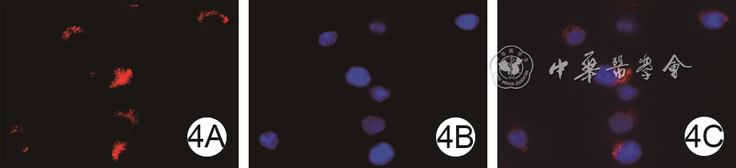
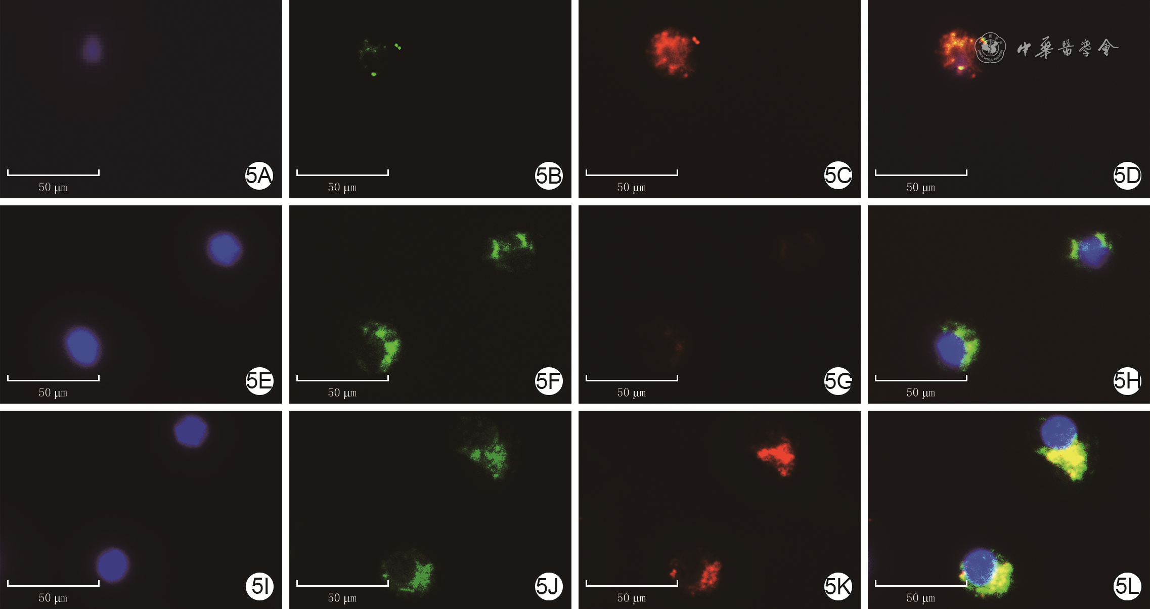
  Objective  To explore the effects and underlying mechanism of human adipose mesenchymal stem cells (ADSC)-derived exosomes on acute lung injury in septic mice.  Methods  The study was an experimental study. Human ADSC of passages 4-5 were selected, and exosomes in their supernatant were isolated and extracted by differential ultracentrifugation. Exosomes were then used after identification. Twenty-four adult male BALB/c mice were selected and divided into normal control group, simple cecal ligation and puncture (CLP) group, and CLP+ADSC-exosome group according to the random number table method (the grouping method was the same below), with 8 mice in each group. The mice in simple CLP group were injected with phosphate buffer after CLP surgery (to establish an animal model of acute lung injury in septic mice), the mice in CLP+ADSC-exosome group were treated according to the corresponding group name, and the mice in normal control group were only injected with phosphate buffer. At 24 hours after surgery, the morphology of lung tissue was observed by hematoxylin-eosin staining, the apoptosis of lung tissue cells was detected by in-situ end-labeling method, the content of tumor necrosis factor-α (TNF-α) and interleukin-1β (IL-1β) in the serum of mice was detected by enzyme-linked immunosorbent assay, the content of malondialdehyde and superoxide dismutase (SOD) in lung tissue was detected by microplate reader, and the expressions of CD86 and CD206 in mouse lung tissue cells was detected by immunofluorescence method. Mouse macrophage RAW264.7 was taken and divided into blank control group, simple lipopolysaccharide (LPS) group, and LPS+ADSC-exosome group. The cells of LPS+ADSC-exosome group and simple LPS group were cultured by adding LPS+ADSC-exosome and LPS, respectively, and cells in blank control group were routinely cultured. Twelve hours after culture, the ATP content, the percentage of mitochondrial reactive oxygen species positive cells, as well as mitochondrial membrane potential in cells were detected by related detection kits. The mRNA expression levels of M1 polarization marker inducible nitric oxide synthase (iNOS), M2 polarization marker arginase-1 (Arg1), and inflammatory factors TNF-α and IL-1β in cells were detected by real-time fluorescence quantitative reverse-transcription polymerase chain reaction method. Three samples were used for mRNA expression detection, and four samples were used for the detection of the other indicators.  Results  At 24 hours after surgery, the structure of mouse lung tissues in normal control group was clear and intact without inflammatory cell infiltration. Compared with that in normal control group, the lung tissue edema as well as the infiltration of inflammatory cells of mice was much more obvious in simple CLP group. However, compared with that in simple CLP group, the lung tissue edema of mice in CLP+ADSC-exosome group was significantly alleviated, the infiltration of inflammatory cells was significantly reduced, and the cell apoptosis and necrosis were significantly improved. Twenty-four hours after surgery, compared with that in normal control group, the levels of TNF-α and IL-1β in the serum of mice in simple CLP group were significantly increased (with t values of 50.82 and 30.81, respectively, P<0.05); compared with that in simple CLP group, the levels of TNF-α and IL-1β in the serum of mice in CLP+ADSC-exosome group were significantly decreased (with t values of 16.36 and 19.25, respectively, P<0.05). Compared with that in normal control group, the content of malondialdehyde in the lung tissue of mice in simple CLP group was significantly increased (t=9.89, P<0.05); and the content of SOD was significantly decreased (t=5.01, P<0.05); compared with that in simple CLP group, the content of malondialdehyde in the lung tissue of mice in CLP+ADSC-exosome group was significantly decreased (t=4.38, P<0.05), and the content of SOD was significantly increased (t=2.97, P<0.05). Twenty-four hours after surgery, compared with that in normal control group, the proportion of CD86 positive cells in the lung tissue of mice in simple CLP group was significantly increased, and the proportion of CD206 positive cells was significantly decreased; compared with that in simple CLP group, the proportion of CD86 positive cells in the lung tissue of mice in CLP+ADSC-exosome group was significantly decreased, and the proportion of CD206 positive cells was significantly increased. After 12 hours of culture, compared with that in blank control group, the ATP content of RAW264.7 cells in simple LPS group was significantly decreased (t=6.28, P<0.05); compared with that in simple LPS group, the ATP content of RAW264.7 cells in LPS+ADSC-exosome group was significantly increased (t=4.01, P<0.05). After 12 hours of culture, compared with (22±4)% in blank control group, (40±6)% of positive cells of mitochondrial reactive oxygen species in RAW264.7 cells in simple LPS group was significantly increased (t=5.04, P<0.05); compared with that in LPS group, (30±5)% of positive cells of mitochondrial reactive oxygen species in RAW264.7 cells in LPS+ADSC-exosome group was significantly decreased (t=2.65, P<0.05). After 12 hours of culture, compared with that in blank control group, the mitochondrial membrane potential of RAW264.7 cells in simple LPS group was significantly decreased; the mitochondrial membrane potential of RAW264.7 cells in LPS+ ADSC-exosome group was between those in blank control group and simple LPS group. After 12 hours of culture, compared with that in blank control group, the mRNA expressions of TNF-α, IL-1β, and iNOS in RAW264.7 cells in simple LPS group were significantly increased (with t values of 16.51, 31.04, and 7.70, respectively, P<0.05), and the decrease in the mRNA expression of Arg1 was not statistically significant (P>0.05); compared with that in simple LPS group, the mRNA expressions of TNF-α, IL-1β, and iNOS in RAW264.7 cells in LPS+ADSC-exosome group were significantly decreased (with t values of 11.38, 22.58, and 5.28, respectively, P<0.05), and the mRNA expression of Arg1 was significantly increased (t=7.66, P<0.05).  Conclusions  Human ADSC-exosomes may play a role in improving lung injury in septic mice by improving LPS-induced mitochondrial dysfunction in mice macrophages, inhibiting the polarization of macrophages toward M1, and reducing the inflammatory response.
Original Article
Prospective self-controlled clinical trial on the effects of external sutureless on skin incision healing
Chen Zong'an, Yang Yating, Wang Wenbo, Xia Lingling, Liu Wei, Gao Zhen, Wu Xiaoli
2024, 40(12): 1143-1149. doi: 10.3760/cma.j.cn501225-20240206-00051
Abstract:
  Objective  To compare the impact of whether to apply the external suture after internal suture on skin incision healing.  Methods  This study was a prospective self-controlled clinical trial. From November 2020 to September 2021, 49 patients who underwent skin resection surgery and met the inclusion criteria were admitted to Shanghai Ninth People's Hospital of Shanghai Jiao Tong University School of Medicine, including 39 females and 10 males, aged 18 to 55 years. The surgical sites included the upper limbs (17 cases), lower limbs (2 cases), shoulders (3 cases), neck (8 cases), abdomen (8 cases), chest (10 cases), and back (1 case). The surgical incision of each patient was divided equally into the external suture segment (with internal suture and external suture) and the external sutureless segment (with internal suture only) using the random sequence method. On the day of suture removal and at the 1st month of follow-up after surgery, the incision dehiscence was observed. At the 12th month of follow-up after surgery, the scar hyperplasia was observed and the scar hyperplasia rate of the incision was calculated. At the 6th and 12th months of follow-up after surgery, the scar width of incision was measured. At the 1st, 6th, and 12th months of follow-up after surgery, the Vancouver scar scale (VSS) was used to assess scar condition of the incision in terms of color, thickness, vascularization, and pliability, and the total score was calculated. The number of patients corresponding to the VSS score in the 1st and 12th months of follow-up after surgery was 41 and 46, respectively, while the number of patients corresponding to other indicators was 49.  Results  On the day of suture removal and at the 1st month of follow-up after surgery, no dehiscence was observed in either the external suture segment or external sutureless segment of any patient's surgical incision. At the 12th month of follow-up after surgery, the scar hyperplasia rate in the external suture segment was 2.04% (1/49), which was not significantly different from 4.08% (2/49) in the external sutureless segment (P>0.05). At the 6th and 12th months of follow-up after surgery, the scar widths in the external sutureless segment of incision were 1.48 (1.01, 1.91) and 1.41 (1.13, 1.93) mm, respectively, which were not significantly different from 1.38 (1.00, 1.94) and 1.45 (1.17, 1.84) mm in the external suture segment (with Z values of 191.00 and 152.00, respectively, P>0.05). At the 1st, 6th, and 12th months of follow-up after surgery, there were no statistically significant differences either in scores of scar color, thickness, vascularization, pliability, or in the total VSS score of the incision scar between the external suture segment and the external sutureless segment (P>0.05).  Conclusions  After skin incision is finely sutured internally with tension reduction, the presence or absence of external suture has no significant effect on the healing of the incision and the appearance of postoperative scar.
Clinical effects of Meek skin grafting combined with platelet-rich plasma in repairing extensive deep burn wounds
Zhang Ting, Liu Jiaqi, Yang Yunshu, Han Juntao, Hu Dahai, Zheng Zhao
2024, 40(12): 1150-1157. doi: 10.3760/cma.j.cn501225-20231124-00206
Abstract:
  Objective  To investigate the clinical effects of Meek skin grafting combined with platelet-rich plasma (PRP) in repairing of extensive deep burn wounds.  Methods  This study was a retrospective observational study. From August 2018 to August 2023, 44 patients who met the inclusion criteria were admitted to the First Affiliated Hospital of Air Force Medical University, including 23 males and 21 females. Their age ranged from 22 to 62 years and the total burn area was 30%-80% total body surface area, the burns on limbs or torso were deep partial-thickness to full-thickness. According to the treatment process of deep burns on the limbs or torso, the patients were divided into observation group (21 cases) and control group (23 cases). patients in observation group were treated with Meek skin grafting combined with PRP at the same time, while patients in control group were treated with Meek skin grafting alone. The survival of Meek skin grafts was observed on the 10th day after operation. The wound healing rate in the operation area of patients was observed on the 14th day after operation. The postoperative wound healing time and positive rate of bacterial culture of wound secretion specimens were recorded.  Results  On the 10th day after operation, the skin grafting area of patients in observation group were dry, and the skin grafts adhered closely to the wound base with ruddy color, with a survival rate of (89±4)%; whereas in control group, some of Meek skin grafts fell off in the skin graft area, and the residual wounds were scattered in irregular map shape, accompanied by purulent secretions. The survival rate of skin grafts was (79±6)%, which was significantly lower than that in observation group (t=6.72, P<0.05). On the 14th day after operation, 19 patients in observation group had complete wound healing in the operation area, and the other 2 patients had small residual wounds, which healed after 1 week delay through dressing changes; in control group, the wounds in 12 patients healed completely, the wounds in 6 patients healed after supplementary stamp skin grafting, and the wounds in 5 patients healed with delay after routine dressing changes. The wound healing rate was significantly lower than that in observation group (P<0.05). After operation, the wound healing time of the operation area of patients in observation group was (13.3±1.6) days, which was significantly shorter than (16.4±3.5) days in control group (t=3.72, P<0.05); there was no statistically significant difference in the positive rate of bacterial culture of wound secretion specimens between observation group and control group after operation (P>0.05).  Conclusions  Compared with Meek skin grafting alone, Meek skin grafting combined with autologous PRP can promote the survival of skin grafts, accelerate the expansion and fusion of skin grafts, and shorten the wound healing time, thereby improving the therapeutic effect in the repairing of extensive deep burn wounds.
Clinical curative effects of two types of pedicled flaps in repairing the full-thickness electric burn wounds deep to tendon and bone in the knee
Xue Jidong, Liang Yan, Di Haiping, Xing Peipeng, Dong Guoyun, Liang Zhanling, Xia Chengde
2024, 40(12): 1158-1165. doi: 10.3760/cma.j.cn501225-20240419-00143
Abstract:
  Objective  To compare the clinical curative effects of saphenous artery flap and retrograde anterolateral femoral perforator flap in repairing full-thickness electric burn wounds deep to tendon and bone in the knee.  Methods  This study was a retrospective observational study. From July 2018 to February 2022, 34 patients with full-thickness electric burn wounds deep to tendon and bone in the knee and conformed to the inclusion criteria were admitted to Zhengzhou First People's Hospital, including 26 males and 8 females, aged 18 to 54 years. According to the repair method of the electric burn wounds in the knee, the patients were divided into saphenous artery flap group (18 cases) repaired with saphenous artery flap and anterolateral femoral flap group (16 cases) repaired with retrograde anterolateral femoral perforator flap. The exposed area of bone and/or tendon after debridement was 5 cm×4 cm to 12 cm×7 cm, 5 patients were combined with open joint, and the resected area of the flap was 9 cm×6 cm to 25 cm×12 cm in saphenous artery flap group; the exposed area of bone and/or tendon after debridement was 7 cm×5 cm to 15 cm×7 cm, 6 patients were combined with open joint, and the resected area of the flap was 15 cm×10 cm to 39 cm×25 cm in anterolateral femoral flap group. According to the resected width of the flap, the wounds in the flap donor areas were repaired by direct suture or medium thickness skin graft in the trunk. The survival of the flap was observed after surgery. At the last follow-up, the color and texture of the flap were observed, and the two-point discrimination distance of the flap was detected. The sensory recovery of the flap was evaluated using the trial standards for evaluation of partial function of upper extremity by the Hand Surgery Society of Chinese Medical Association, and the excellent ratio of sensory recovery was calculated. The function of knee joint was assessed using the Knee Subjective Score Scale of International Knee Documentation Committee. The repairing effect of the flap was evaluated using comprehensive evaluation scale of flap, and the excellent ratio was calculated.  Results  Most of the flaps in patients in the 2 groups survived well after surgery, only 2 patients in the anterolateral femoral flap group had distal flap necrosis of 3 to 5 cm2, which healed after skin grafting or local suture. At the last follow-up of 12 to 18 months after surgery, the color and texture of the flap in patients in the two groups were similar to those of the skin tissue in the knee. The excellent ratio of sensory recovery of the flap was 18/18 in patients in saphenous artery flap group, which was significantly higher than 5/16 in anterolateral femoral flap group (P<0.05). The two-point discrimination distance of the flap was (11.7±1.5) mm in patients in saphenous artery flap group, which was significantly shorter than (21.5±1.7) mm in anterolateral femoral flap group (t=-1.84, P<0.05). The score of the knee joint function and the excellent ratio of the repairing effect of the flap had no statistically significant differences in patients in the two groups (P>0.05).  Conclusions  The full-thickness electric burn wounds deep to tendon and bone in the knee can be repaired with saphenous artery flap and retrograde anterolateral femoral perforator flap. After being repaired with those two types of flaps, the function of the knee joint recovers well, while the sensory recovery is better after being repaired by the saphenous artery flap.
Analysis of biofilm genes and quorum sensing genes of carbapenem resistant Acinetobacter baumannii in the wounds of diabetic foot patients
Xu Jun, Han Xiaocui, He Lu, Feng Shuhong, Sun Dongjian, Cao Chen, Liu Xijiao, Zhang Yanyan, Ding Baixing, Chang Bai
2024, 40(12): 1166-1175. doi: 10.3760/cma.j.cn501225-20240715-00269
Abstract:
  Objective  To investigate the biofilm genes and quorum sensing genes of carbapenem resistant Acinetobacter baumannii (CRAB) in the wounds of diabetic foot patients.  Methods  This study was a retrospective observational study. The 233 strains of Acinetobacter baumannii were cultured from 177 inpatients (128 males and 49 females, aged (56±10) years) with diabetic foot admitted to the Department of Diabetic Foot of Tianjin Medical University Chu Hsien-I Memorial Hospital from October 2020 to September 2023. Two hundred and thirty-three Acinetobacter baumannii strains were detected by bacterial culture from the diabetic foot wounds of the aforementioned patients. All Acinetobacter baumannii strains were identified using matrix-assisted laser desorption/ionization time-of-flight mass spectrometry, followed by analysis of their resistance rate using kinetic turbidimetric assay by a fully automated microbiological analysis system. Ten CRAB strains (from 10 patients, 9 males and 1 female, aged (63±13) years) and 10 carbapenem sensitive Acinetobacter baumannii (CSAB; from 10 patients, 8 males and 2 females, aged (63±9) years) strains were randomly selected, and the whole DNA genome was extracted and sequenced. The antibiotic resistance genes were annotated using a comprehensive antibiotic resistance gene database, and a phylogenetic tree was drawn to analyze the homologous relationship between CRAB and CSAB. The 7 housekeeping genes of Acinetobacter baumannii was entered into the PubMLST website to analyze the multi-locus sequence typing of CRAB and CSAB. All the measured genes were put into the PubMLST website to search for the biofilm genes bap, csuA, csuB, csuA/B, csuC, csuD, csuE, pgaA, pgaB, pgaC, pgaD, bfmR, bfmS, ompA carried by each Acinetobacter baumannii, as well as the quorum sensing genes abaI and abaR, and flagellar gene pilA. The differences in carrying these genes between CRAB and CSAB were compared. The biofilm genes and quorum sensing genes carried by CRAB and CSAB carrying oxacillinase (OXA) resistance gene blaOXA were analyzed. Gross observation was performed to check if there was gel-like membrane-like substance in the diabetic foot wounds infected with CRAB and CSAB, and if so, the microstructure was observed by scanning electron microscope.  Results  Among the detected Acinetobacter baumannii, the positive detection rates of CSAB, CRAB, multi-drug resistant Acinetobacter baumannii, and pan-drug resistant Acinetobacter baumannii were 16.7% (39/233), 83.3% (194/233), 95.3% (222/233), and 34.3% (80/233), respectively, and no fully drug-resistant Acinetobacter baumannii was detected. Among 233 strains of Acinetobacter baumannii, the resistance rate to carbapenem antibiotics exceeded 80%; the resistance rate of cefoperazone/sulbactam was relatively low, at 37%; the resistance rates to the other cephalosporin antibiotics (cefotaxime, ceftazimide, cefotetan, and cefuroxime) were more than 80%; the resistance rates to all penicillin antibiotics were greater than 80%; the resistance rates to quinolone antibiotics were all over 60%; the resistance rate to minocycline was only 12%; the resistance rates to tigecycline and colistin did not exceed 1%. The phylogenetic tree showed that 10 CRAB strains were highly homologous, while 10 CSAB strains had low homology. The analysis of multi-locus sequence typing showed that 10 CRAB strains were all the same type; among the 10 CSAB strains, except 1 strain without typing, the remaining 9 CSAB strains had 7 types. Eight of 10 CRAB strains contained complete biofilm genes and quorum sensing genes. The biofilm genes from the strains of CSAB were incomplete and none carried the bap gene. Neither CRAB nor CSAB carried the flagellar gene pilA. Compared with that carried by CRAB, biofilm genes bap, csuA, csuC, and csuD and quorum sensing genes abaI and abaR carried by CSAB were significantly decreased (P<0.05). The main blaOXA categories carried by CRAB were blaOXA-23-like (specifically BlaOXA-167) and blaOXA-51-like (specifically blaOXA-66), both of which had carbapenase activity. Eight of 10 CRAB strains carried both blaOXA-66 and blaOXA-167, and all of them had relatively complete quorum sensing genes and biofilm genes. The main blaOXA categories carried by CSAB were blaOXA-51-like and blaOXA-213-like. Although they had carbapenemase activity, clinical drug sensitivity test showed that they were all sensitive to carbapenem antibiotics. Gel-like and membrane-like substance could be seen in wounds infected with CRAB, which were biofilm; no gel-like and membrane-like substance was found in the wound infected with CSAB.  Conclusions  CRAB and CSAB in diabetic foot wounds are significantly different in terms of multi-locus sequence typing, carrying biofilm genes, quorum sensing genes, and blaOXA gene, leading to differences in antibiotic resistance between the two.
Effect of rat platelet-rich plasma gel on autologous adipose-derived mesenchymal stem cells overexpressing glia-derived neurotrophic factor
Cai Weixia, Zheng Zhao, Liu Jiaqi, Liu Yang, Zhang Ting, Ji Peng, Tian Chenyang
2024, 40(12): 1176-1183. doi: 10.3760/cma.j.cn501225-20240408-00126
Abstract:
  Objective  To investigate the effect of rat platelet-rich plasma (PRP) gel on autologous adipose-derived mesenchymal stem cells (ADSCs) overexpressing glial-derived neurotrophic factor (GDNF).  Methods  This study was an experimental study. Five adult male Sprague-Dawley rats were used, and the primary ADSCs were obtained by collagenase digestion, and then the cells were identified successfully. The 3rd passage of ADSCs were obtained and divided into negative control group infected with unloaded adenovirus and overexpressing GDNF group infected with overexpressing GDNF adenovirus, according to random number table method (the grouping method was the same below). After 48 hours of culture, the infection of cells was observed. Five adult male Sprague-Dawley rats were used, and the PRP was obtained after collecting blood by differential centrifugation. PRP was prepared into a gel and its microstructure was observed by scanning electron microscope. The ADSCs of 3rd passage were added into the PRP solution mixture and cultured for 48 hours after gelation. The cell growth was observed by hematoxylin-eosin staining and calcein/propyl iodide staining. ADSCs infected with unloaded adenovirus and ADSCs infected with overexpressing GDNF adenovirus were routinely cultured in PRP gel. After 48 hours of culture, the cell growth was detected by calcein/propyl iodide staining. After culture for 24, 48, 72 hours and 1, 2, 3, 4 weeks, the supernatant of cell culture medium was collected, the absorbance value was determined by microplate analyzer, and the GDNF content was calculated, with the sample number of 3. After 48 hours of culture, the expression of S100 protein (a specific marker of Schwann cells) was detected by immunofluorescence assay.  Results  After 48 hours of culture, the proportions of cells infected with adenovirus in negative control group and overexpressing GDNF group were close to 90%, and the cell growth was good. The cells in negative control group grew normally. The morphology of the cells in overexpressing GDNF group changed significantly with 80%-90% of the cells having two or more protrusions, and the protrusions were interwoven to form a network wherever the cells gathered. PRP gel formed a three-dimensional network structure with different pore sizes. After 48 hours of culture, ADSCs could be well attached to PRP gel, and 98% of the cells were alive. After 48 hours of culture, ADSCs infected with unloaded adenovirus grew well and showed typical ADSC-like spindle-shaped growth. ADSCs infected with overexpressing GDNF adenovirus grew well, and most of the cells had two or more protrusions, and the protrusions were interwoven into a network. After culture for 24, 48, 72 hours and 1, 2, 3, 4 weeks, the content of GDNF in the supernatant of ADSCs infected with overexpressing GDNF adenovirus was (90±10), (133±15), (150±10), (137±15), (132±18), (120±10), and (127±16) pg/mL, which was significantly higher than (42±7), (44±7), (43±6), (47±6), (49±5), (49±6), and (51±4) pg/mL of ADSCs infected with unloaded adenovirus (with t values of 6.20, 8.08, 15.18, 9.12, 7.99, 9.61, and 7.86, respectively, P<0.05). After 48 hours of culture, the fluorescence intensity of S100 protein expression of ADSCs infected with overexpressing GDNF adenovirus was significantly stronger than that of ADSCs infected with unloaded adenovirus.  Conclusions  The prepared autologous three-dimensional PRP gel has good biocompatibility and can carry rat GDNF-overexpressing ADSCs and release GDNF slowly, inducing ADSCs to differentiate into Schwann cells that express high level of S100 protein.
Long term efficacy of absorbable sutures coated with silver nanoparticles on the suture track of skin incisions in mice
He Feng, Du Juan, Song Yanbiao, Ye Mao, Wei Yandong, Liu Xuelai
2024, 40(12): 1184-1192. doi: 10.3760/cma.j.cn501225-20231007-00104
Abstract:
    Objective   To explore the long term efficacy of absorbable sutures coated with silver nanoparticles on the suture track of skin incisions in mice.    Methods   This study was an experimental research. Eighteen half male and half female BALB/c mice aged 10-12 weeks were divided into normal Vicryl suture group, antibiotic Vicryl suture group, and silver nanoparticle Vicryl suture group according to the random number table, with 6 mice in each group, half male and half female. After the skin incisions were created on the back of all mice, the skin incisions of mice in normal Vicryl suture group, antibiotic Vicryl suture group, and silver nanoparticle Vicryl suture group were performed with continuous intradermal suture by absorbable Vicryl sutures, absorbable Vicryl sutures coated with triclosan antimicrobial, and absorbable Vicryl sutures coated with silver nanoparticles, respectively. On the 25th day after surgery, the skin tissue in the suture track of incisions was collected, immunohistochemical staining was used to detect the percentages of F4/80, interleukin-6 (IL-6), and tumor necrosis factor α (TNF-α) positive areas in the suture track of incisions of mice, the TdT-mediated dUTP nick-end labeling (TUNEL) staining was used to detect the percentage of TUNEL staining positive area in the suture track of incisions of mice, Masson staining was used to observe the collagen fiber formation in the suture track of incisions of mice, and immunohistochemical staining was used to detect the percentage of tissue cathepsin K positive area in the suture track of incisions of mice. The number of the above experimental samples was 6.    Results   On the 25th day after surgery, the percentages of F4/80, IL-6, and TNF-α positive areas in the suture track of incisions of mice in silver nanoparticle Vicryl suture group ((19.2±1.6)%, (20.2±1.7)%, and (16.0±1.6)%) were significantly lower than those in normal Vicryl suture group ((100±6.4)%, (100±7.6)%, and (100±9.6)%) and antibiotic Vicryl suture group ((47.2±3.2)%, (53.8±5.0)%, and 43.2%, P<0.05). The percentages of F4/80, IL-6, and TNF-α positive areas in the suture track of incisions of mice in antibiotic Vicryl suture group were significantly lower than those in normal Vicryl suture group (P<0.05). On the 25th day after surgery, the percentage of TUNEL staining positive area in the suture track of incisions of mice in silver nanoparticle Vicryl suture group was significantly lower than those in normal Vicryl suture group and antibiotic Vicryl suture group (with both P values<0.05). The percentage of TUNEL staining positive area in the suture track of incisions of mice in antibiotic Vicryl suture group was significantly higher than that in normal Vicryl suture group (P<0.05). On the 25th day after surgery, the collagen fiber deposition was not obvious in the suture track of incisions of mice in normal Vicryl suture group, no obvious collagen fiber formation was observed in the suture track of incisions of mice in antibiotic Vicryl suture group, while the collagen fibers were mostly densely arranged and well-ordered in the suture track of incisions of mice in silver nanoparticle Vicryl suture group. On the 25th day after surgery, the percentage of tissue cathepsin K positive area in the suture track of incisions of mice in silver nanoparticle Vicryl suture group was significantly lower than those in normal Vicryl suture group and antibiotic Vicryl suture group (with P values both<0.05), the percentage of tissue cathepsin K positive area in the suture track of incisions of mice in antibiotic Vicryl suture group was significantly lower than that in normal Vicryl suture group (P<0.05).    Conclusions   In the suture track of incisions of mice, silver nanoparticle Vicryl sutures exhibit superior long-term anti-inflammatory effects compared with absorbable Vicryl sutures coated with triclosan antimicrobial and normal absorbable Vicryl sutures. Additionally, cell apoptosis is reduced, collagen fiber deposition is obvious, and delayed-type hypersensitivity reactions is the weakest in the suture track of incisions of mice sutured by silver nanoparticle Vicryl sutures. The silver nanoparticle Vicryl sutures have potential value for clinical application.
Review
Research advances on extracellular vesicles in promoting diabetic wound healing through the regulation of immune microenvironment
Zhou Ziyue, Xu Guangchao, Wang Dali
2024, 40(12): 1193-1198. doi: 10.3760/cma.j.cn501225-20231215-00243
Abstract:
Peripheral neuropathy and poor blood circulation caused by long-term hyperglycemia in patients with diabetes often leads to foot wounds, ulcers, and gangrene, which accompanied by an imbalance in the wound immune microenvironment. Extracellular vesicle (EV) therapy is an emerging biologic treatment method. EV possesses advantages such as good biocompatibility, high efficacy, low immunogenicity, and low tissue toxicity. By modulating the immune responses and cellular functions in the microenvironment, EV can reduce inflammation levels in wounds, showing great potential in accelerating diabetic wound healing. This review summarizes the mechanism by which EV participates in regulating wound healing through the modulation of various inflammatory and non-inflammatory cells in the microenvironment of diabetic wounds, discusses the advantages and limitations of various approaches of the clinical translation of EV, proposes the concept of combining EV therapy with the disease microenvironment, thereby providing new insights for further researches on the clinical application of EV.
Research advances on the role of plant-derived extracellular vesicles in wound repair
Wang Weiqi, Yang Yueting, Su Zhihong, Li Jin, Hu Wenhui, Zhang Peihua
2024, 40(12): 1199-1204. doi: 10.3760/cma.j.cn501225-20240226-00072
Abstract:
Plant-derived extracellular vesicles (EVs) are nanoscale vesicles secreted by plant cells, containing various proteins, lipids, and RNA molecules, which can regulate the information exchange and material transfer between cells, thus are important carriers of cellular communication. Currently, animal-derived EVs have been widely used in interspecies communication, cellular exchange, and drug carrier research; while plant-derived EVs have received widespread attention due to their superior biocompatibility and targeting and low immunogenicity. At the same time, research and analysis techniques of plant-derived EVs have made great progress. This review focuses on the isolation techniques, characterization and identification of plant-derived EVs and their role in wound repair, aiming to provide new ideas and avenues for future research on plant-derived EVs and to provide reference for their clinical applications.