Current Issue

2026 Vol. 42, No. 10

Guideline and Consensus
Expert consensus on the surgical management of pressure injuries (2026 edition)
2026, 42(10): 903-918. doi: 10.3760/cma.j.cn501225-20250213-00058
Abstract:
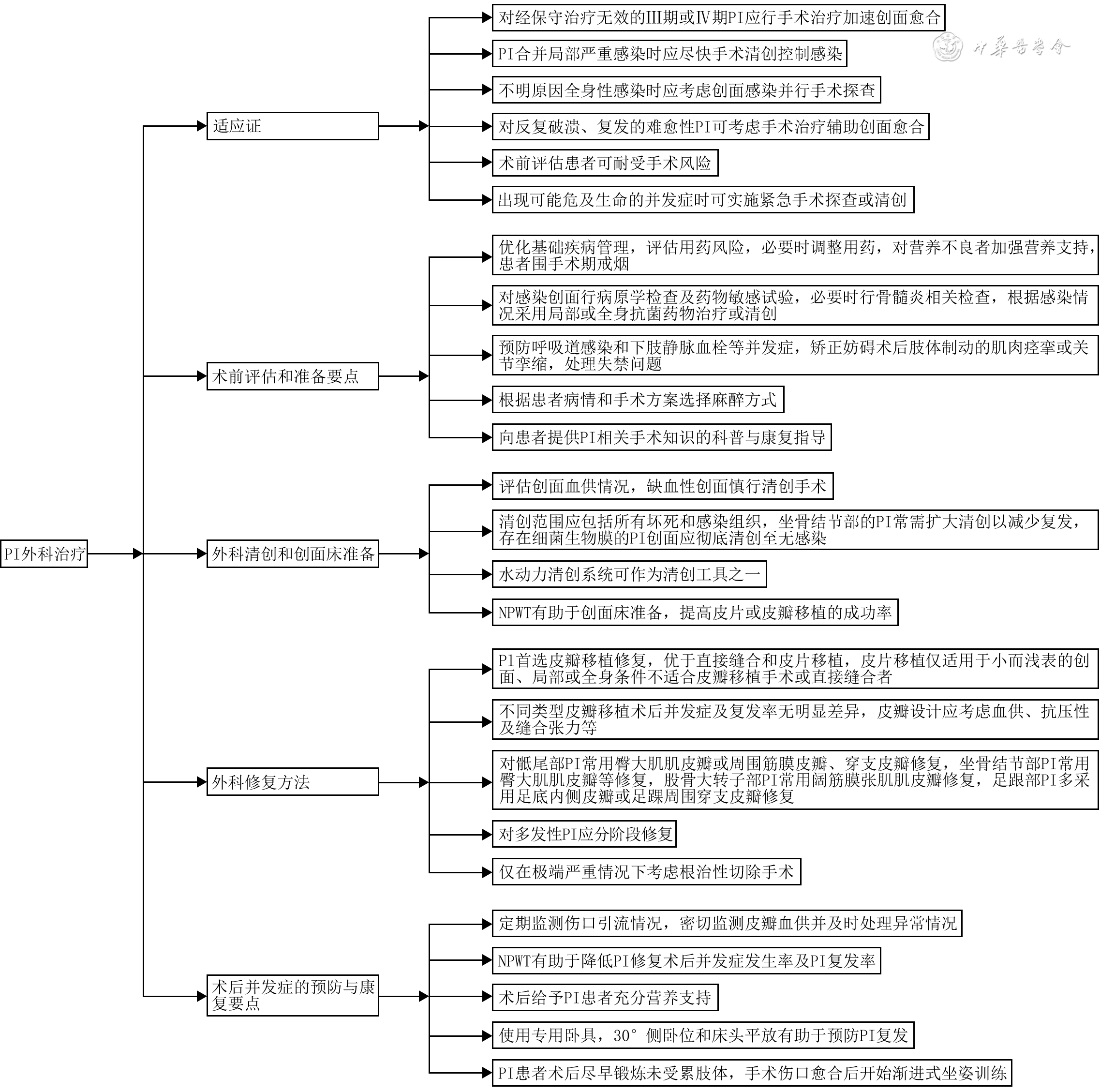
Pressure injuries are common chronic hard-to-heal wounds. With the acceleration of population aging and the increasing prevalence of chronic diseases, the incidence of pressure injuries has increased year by year, accompanied by a growing demand for surgical management. Despite continuous advances in relevant clinical techniques, a unified standardized pathway covering the entire process of surgical management of pressure injuries remains lacking, which adversely affects patient outcomes. To provide further guidance for clinical practice, the Wound Repair Professional Committee of the Chinese Medical Doctor Association organized a multidisciplinary panel of experts from China to develop the Expert consensus on the surgical management of pressure injuries (2026 edition). The consensus covered indications for surgical management of pressure injuries, key considerations for preoperative assessment and preparation, surgical debridement and wound-bed preparation, methods of surgical repair, and key considerations for the prevention of postoperative complications and rehabilitation. It aimed to standardize clinical practice, improve treatment outcomes, and promote consistent improvement in the capacity for surgical management of pressure injuries across healthcare institutions at all levels.
Expert Forum
Being attentive to sarcopenia in elderly patients with refractory wounds
Zhu Feng, Guo Guanghua
2026, 42(10): 919-925. doi: 10.3760/cma.j.cn501225-20260529-00220
Abstract:
Sarcopenia is a major threat to the health of elderly patients. Elderly patients with refractory wounds are at increased risk of sarcopenia and have poorer outcomes, however, its clinical recognition and management remain inadequate. Based on an understanding of the bidirectional deterioration between sarcopenia and refractory wounds, healthcare professionals should draw on relevant consensus and guidelines, incorporate sarcopenia into routine screening, comprehensive assessment, and dynamic re-assessment, and provide integrated management encompassing nutritional support and metabolic regulation, immune and inflammatory modulation, wound care, physical activity and rehabilitation, and psychological support. Future research and clinical practice, supported by technologies such as artificial intelligence and multi-omics, will be conducted to establish consensus on screening, diagnosis, and treatment for sarcopenia in elderly patients with refractory wounds, thereby improving their clinical outcomes and quality of life.
Original Article·Severe Burn Treatment
Analysis of influencing factors for the occurrence of transient hypernatremia in patients with extremely severe burns
Zhang Yue, Zhao Deli, Zhang Wanfu, Han Fei, Guan Hao, Hu Dahai
2026, 42(10): 926-935. doi: 10.3760/cma.j.cn501225-20251219-00534
Abstract:
  Objective  To investigate the influencing factors for the occurrence of transient hypernatremia (TH) in patients with extremely severe burns.  Methods  This study was a bidirectional cohort study. Clinical data of 126 patients with extremely severe burns (97 males and 29 females, aged 18 to 64 years) who met the inclusion criteria and were admitted to the First Affiliated Hospital of Air Force Medical University from August 2021 to December 2025 were retrospectively analyzed. The incidence of hypernatremia was recorded. Among 118 patients who underwent surgical treatment, patients who underwent first escharectomy at ≤5 days after injury were included in early escharectomy group (n=39), and patients who underwent first escharectomy at >5 days after injury were included in late escharectomy group (n=79). The incidence of hypernatremia was compared between the two groups of patients. According to whether they received air-fluidized bed (AFB) therapy, patients were divided into AFB group (n=89) and non-AFB group (n=37). The incidence of hypernatremia was compared between the two groups of patients, and independent risk factors for the occurrence of hypernatremia in patients with extremely severe burns were screened. For patients who underwent pulse contour cardiac output (PiCCO) monitoring during the shock stage, extravascular lung water index (EVLWI) and pulmonary vascular permeability index (PVPI) were compared between the 20 patients with hypernatremia and the 35 patients without hypernatremia. Fifty-five patients with extremely severe burns (35 males and 20 females, aged 20 to 63 years) who met the inclusion criteria and were admitted to this hospital from May to December 2025 were prospectively enrolled. At 3 days after injury, eschar tissue, wound skin tissue, and peripheral venous blood were collected. Using the random number table method, specimens from 5 patients with hypernatremia (set as high Na+ group) and 5 patients without hypernatremia (set as non-high Na+ group) were selected. Sodium content in eschar was determined by inductively coupled plasma mass spectrometry. Serum levels of interleukin-1 (IL-1), IL-6, tumor necrosis factor-α (TNF-α), and hyaluronic acid were detected by enzyme-linked immunosorbent assay. Protein expression level of sodium voltage-gated channel alpha subunit 5 (SCN5A) in wound skin tissue was detected by Western blotting.  Results  In the retrospective cohort study of 126 patients, 50 patients (39.7%) developed hypernatremia 3 to 5 days after injury, among whom 41 had TH, and 9 died. In the 41 patients with TH, the serum sodium began to increase 2 to 3 days after injury, peaked at 5 to 8 days after injury, and gradually recovered by 10 days after injury. The incidence of hypernatremia of patients in late escharectomy group was 32.9% (26/79), which was significantly higher than 7.7% (3/39) in early escharectomy group (χ2=8.96, P<0.05). The incidence of hypernatremia of patients in AFB group was significantly higher than that in non-AFB group (P<0.05). The results of multivariate logistic regression analysis showed that AFB therapy was not an independent risk factor for hypernatremia in patients with extremely severe burns (OR=1.80, with a 95% CI of 0.40 to 8.20, P>0.05), while both total burn area and full-thickness burn area were independent risk factors for hypernatremia in patients with extremely severe burns (with ORs of 3.50 and 2.10, respectively, 95% CIs of 1.90 to 6.70 and 1.20 to 3.80, respectively, P<0.05). Among patients who underwent PiCCO monitoring during the shock stage, EVLWI and PVPI in patients with hypernatremia were significantly higher than those in patients without hypernatremia (with t values of 3.45 and 4.28, respectively, P<0.05). In the prospective cohort study, sodium content in eschar at 3 days after injury in high Na+ group of patients was (128±9) mmol/kg, which was significantly higher than (102±6) mmol/kg in non-high Na+ group (t=5.38, P<0.05). Compared with those in non-high Na+ group, serum levels of IL-1, IL-6, TNF-α, and hyaluronic acid in high Na+ group of patients were significantly increased 3 days after injury (P<0.05), while the protein expression level of SCN5A in wound skin tissue was significantly decreased (P<0.05).  Conclusions  TH after extremely severe burns is not merely an elevation in serum sodium, but involves sodium accumulation in eschar, inflammation-mediated sodium redistribution, and sodium release during the reabsorption phase. Early escharectomy can effectively reduce the risk of occurrence of TH in patients with extremely severe burns, and its mechanism may be associated with the removal of some sodium-containing eschar and a reduction in sodium release.
Analysis of the relationship between albumin balance status during the shock phase and MODS in patients with severe burns and the risk factors for the occurrence of negative albumin balance
Sun Ranran, Lang Jingyuan, Gao Shijie, Heng Xue, Chen Dandan, Li Buying, Li Haisheng, Luo Gaoxing
2026, 42(10): 936-944. doi: 10.3760/cma.j.cn501225-20250616-00268
Abstract:
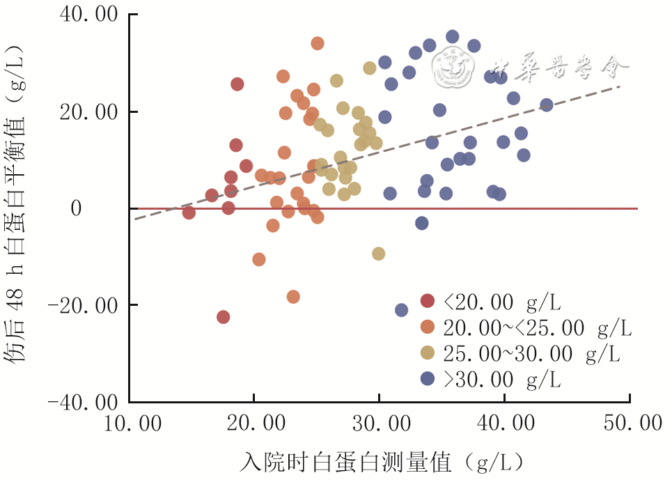
  Objective  To analyze the relationship between albumin balance status during the shock phase and multiple organ dysfunction syndrome (MODS) in patients with severe burns and the risk factors for the occurrence of negative albumin balance (NAB).  Methods  This study was a retrospective case series study. From January 1, 2023 to December 31, 2024, 89 patients with severe burns who met the inclusion criteria were admitted to the First Affiliated Hospital of Army Medical University (the Third Military Medical University), including 73 males and 16 females, aged (48±14) years. The albumin level measured on admission was collected, blood and plasma volumes were estimated based on the Nadler formula, with attenuation correction using the metabolic half-lives of exogenous and endogenous albumin, and the albumin balance value at 48 h post-injury was calculated. The occurrence of NAB was recorded. A correlation analysis was conducted between the albumin level measured on admission and the albumin balance value at 48 h post-injury of patients. The patients with an albumin balance value of ≤0 g/L at 48 h post-injury were included in negative balance group (n=14), while those >0 g/L were included in positive balance group (n=75). The two groups of patients were compared in terms of total burn area and full-thickness burn area, neutrophil percentage-to-albumin ratio (NPAR), aggregate index of systemic inflammation (AISI), systemic inflammation response index (SIRI), systemic immune-inflammation index (SII), neutrophil-to-lymphocyte ratio (NLR), monocyte-to-lymphocyte ratio (MLR), modified sequential organ failure assessment (SOFA) score, and incidence of MODS at 48 h post-injury. The independent risk factors for the occurrence of negative albumin balance at 48 h post-injury in patients with severe burns were screened.  Results  The albumin level measured on admission of patients was (28±7) g/L, and the albumin balance value at 48 h post-injury was 9.01 (3.06, 19.62) g/L, with 15.73% (14/89) of the patients developing NAB. The albumin level measured on admission of patients was significantly positively correlated with the albumin balance value at 48 h post-injury (rs=0.349, P<0.05). The total burn area and full-thickness burn area of patients in negative balance group were significantly larger than those in positive balance group (with Z values of -3.736 and -4.096, respectively, P<0.05). The AISI, SIRI, SII, NPAR, MLR, modified SOFA score, and incidence proportion of MODS at 48 h post-injury in negative balance group were significantly higher than those in positive balance group (with Z values of -2.310, -2.186, -2.130, -3.629, -2.108, and -2.597, respectively, χ2=11.168, P<0.05). Univariate logistic regression analysis results showed that total burn area, full-thickness burn area, and NPAR, AISI, SIRI, SII, NLR, MLR, and modified SOFA score at 48 h post-injury were the risk factors for the occurrence of NAB at 48 h post-injury in patients with severe burns (with ORs of 1.061, 1.053, 3.181, 2.350, 2.483, 2.014, 2.026, 2.316, and 1.283, respectively, 95% CIs of 1.028-1.094, 1.025-1.081, 1.635-6.189, 1.193-4.631, 1.250-4.932, 1.061-3.821, 1.025-4.008, 1.167-4.597, and 1.065-1.546, respectively, P<0.05). Multivariate logistic regression analysis results showed that full-thickness burn area and SII at 48 h post-injury were independent risk factors for the occurrence of NAB at 48 h post-injury in patients with severe burns (with ORs of 1.055 and 2.347, respectively, 95% CIs of 1.026-1.086 and 1.129-4.880, respectively, P<0.05).  Conclusions  NAB during the shock phase may be associated with the incidence of MODS in patients with severe burns, with full-thickness burn area and SII at 48 h post-injury being the independent risk factors for the occurrence of NAB.
Establishment and evaluation of a risk prediction model for the occurrence of early dysfunction of blood coagulation in elderly patients with severe burns
Duan Deqing, Wu Zongyan, Chen Yong, Deng Hong'ao, Hu Shiqiang, Lin Huan, Mao Yuangui, Zhang Hongyan
2026, 42(10): 945-953. doi: 10.3760/cma.j.cn501225-20250507-00209
Abstract:
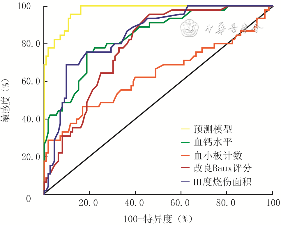
  Objective  To screen risk factors for the occurrence of early dysfunction of blood coagulation in elderly patients with severe burns, and to develop and evaluate a risk prediction model based on these factors.  Methods  This study was a retrospective case-control study. From June 2015 to June 2025, 158 elderly patients with severe burns who met the inclusion criteria were admitted to the First Affiliated Hospital of Nanchang University, including 94 males and 64 females, aged 60 to 97 years. The patients were divided into early dysfunction of blood coagulation group (45 cases) and non-early dysfunction of blood coagulation group (113 cases) according to the presence or absence of early dysfunction of blood coagulation. Gender, age, total burn area, full-thickness burn area, admission time after injury, cause of burn, combination of inhalation injury, combination of underlying diseases, the modified Baux score, acute physiology and chronic health evaluation Ⅱ (APACHEⅡ) score, body temperature, blood pH value, platelet count, base excess level, lactic acid level, hemoglobin level, serum sodium level, serum potassium level, serum calcium level, and plasma fibrinogen (FIB) level on admission, as well as length of hospital stay and clinical prognosis were compared between the two groups of patients. The risk factors and independent risk factors affecting the occurrence of early dysfunction of blood coagulation in elderly patients with severe burns were screened, and the prediction model was constructed according to the independent risk factors. The receiver operating characteristic curve was used to evaluate the predictive value of the prediction model and various independent risk factors for the occurrence of early dysfunction of blood coagulation in aforementioned elderly patients with severe burns.  Results  There were statistically significant differences in the total burn area, full-thickness burn area, the modified Baux score, APACHEⅡ score, platelet count, serum calcium level, blood pH value, and plasma FIB level on admission, as well as in the combination of inhalation injury and clinical prognosis between the two groups of patients (with t values of 2.270, 7.615, 5.975, 2.319, -2.172, and -7.861, respectively, Z values of -2.168 and -2.568, respectively, χ2 values of 20.090 and 10.286, respectively, P<0.05). There were no statistically significant differences in the other indicators between the two groups of patients (P>0.05). The results of univariate logistic regression analysis showed that the total burn area, full-thickness burn area, combination of inhalation injury, the modified Baux score, APACHEⅡ score, platelet count, serum calcium level, and plasma FIB level on admission were all risk factors for the occurrence of early dysfunction of blood coagulation in elderly patients with severe burns (with ORs of 1.022, 1.074, 0.141, 1.045, 1.070, 0.996, <0.001, and 0.568, respectively, 95% CIs of 1.003-1.042, 1.047-1.102, 0.055-0.359, 1.027-1.064, 1.008-1.135, 0.992-0.999, <0.001-0.002, and 0.380-0.849, respectively, P<0.05). The results of multivariate logistic regression analysis showed that the full-thickness burn area, the modified Baux score, platelet count, and serum calcium level on admission were independent risk factors for the occurrence of early dysfunction of blood coagulation in elderly patients with severe burns (with ORs of 1.181, 1.254, 0.991, and <0.001, respectively, 95% CIs of 1.085-1.287, 1.106-1.423, 0.984-0.998, and <0.001-0.009, respectively, P<0.05). In terms of predicting the occurrence of early dysfunction of blood coagulation in elderly patients with severe burns, the area under the receiver operating characteristic curve of the prediction model constructed based on the independent risk factors was 0.977 (with a 95% CI of 0.959-0.994), which was higher than 0.844 (with a 95% CI of 0.781-0.908), 0.782 (with a 95% CI of 0.711-0.853), 0.634 (with a 95% CI of 0.526-0.742), and 0.838 (with a 95% CI of 0.770-0.905) of the full-thickness burn area, the modified Baux score, platelet count, and serum calcium level on admission, respectively.  Conclusions  Full-thickness burn area, the modified Baux score, platelet count, and serum calcium level on admission are independent risk factors for the occurrence of early dysfunction of blood coagulation in elderly patients with severe burns. The prediction model constructed based on these four indicators has a good predictive value for the occurrence of early dysfunction of blood coagulation in elderly patients with severe burns.
Original Article
A prospective clinical study on the application efficacy of SS-DSA combined with CDU in ALTF-based reconstruction of soft tissue defects in extremities
Tian Yongsheng, Zhang Yuji, Wu Junbang, Liu Zeyu, Wang Guiyang, Wang Shi, Dong Shuai, Wu Chenglong, Zhou Rong, Ju Jihui
2026, 42(10): 954-963. doi: 10.3760/cma.j.cn501225-20260317-00110
Abstract:
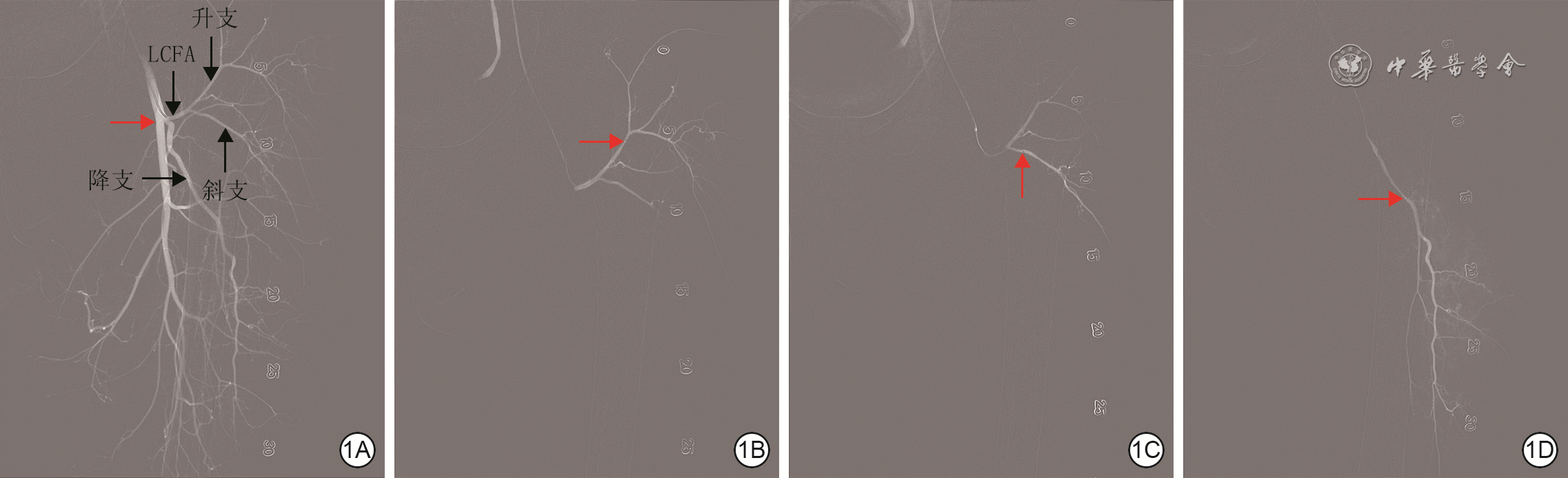
  Objective  To investigate the clinical application efficacy of superselective digital subtraction angiography (SS-DSA) combined with color Doppler ultrasound (CDU) in anterolateral thigh flap (ALTF)-based reconstruction of soft tissue defects in the extremities.  Methods  This was a prospective case series study. From October 2024 to August 2025, 50 patients (37 males and 13 females, aged 23-76 years) with soft tissue defects in the extremities who met the inclusion criteria were treated at Suzhou Ruihua Orthopedic Hospital, and all underwent ALTF reconstruction. Preoperatively, CDU and SS-DSA were used to localize the perforators of the lateral circumflex femoral artery (LCFA) and their source artery branches in the flap donor site. The findings from SS-DSA and CDU were integrated (i.e., the combined method) to identify the dominant perforator and its source artery branch and to develop an individualized flap design. Thorough debridement was performed on the wounds, and the post-debridement defect size ranged from 8 cm×6 cm to 32 cm×20 cm. The flap harvesting area extended approximately 1 cm beyond the outer margins of the recipient site wound. The harvested flaps were transferred to the recipient site wound, and under a microscope, end-to-end or end-to-side anastomosis was performed between the flap vessels and the recipient vessels, followed by wound closure. A total of 51 flaps were harvested from the 50 patients. Donor site wounds were repaired by direct suture or full-thickness skin grafting. The number of LCFA perforators observed intraoperatively and actually used, as well as their source artery branch types, flap thickness, and vascular pedicle length, were recorded. Using intraoperative exploration findings of perforators as the gold standard, the sensitivity and positive predictive value of CDU, SS-DSA, and the combined method for perforator localization were compared. The distances between the perforator localization points of the three methods and the actual intraoperative perforator entry points (i.e., perforator localization error distances) were compared. The accuracy of the combined method in predicting the source artery branches of perforators was calculated. During SS-DSA perforator localization, the total procedure time, dose-area product (DAP), and fluoroscopy time (FT) were recorded, and the estimated glomerular filtration rate (eGFR) of patients before and after angiography was calculated. The puncture site was observed after angiography. Postoperatively, flap survival and donor site wound healing were observed.  Results  A total of 89 LCFA perforators were observed intraoperatively, including 47 originating from the oblique branch, 32 from the descending branch, 5 from the anterior branch, 3 from the transverse branch, and 2 from the ascending branch; 69 LCFA perforators were actually used, including 2 originating from the ascending branch, 5 from the anterior branch, 19 from the descending branch, and 43 from the oblique branch. The flap thickness was (1.4±0.5) cm, and the vascular pedicle length was (13±3) cm. The sensitivities of CDU, SS-DSA, and the combined method for perforator localization were 86.52%, 85.39%, and 95.51%, respectively, and the corresponding positive predictive values were 87.50%, 87.36%, and 96.59%, respectively. The perforator localization error distance of the combined method was 0.31 (0.25, 0.37) cm, which was significantly shorter than those of CDU (0.66 (0.58, 0.74) cm) and SS-DSA (0.67 (0.58, 0.75) cm), with Z values of -7.82 and -7.45, respectively, and both P values <0.05. The combined method achieved 100% (89/89) accuracy in predicting the source artery branches of perforators. The total procedure time of SS-DSA for perforator localization was (32±5) min, the DAP was 102.05 (76.90, 128.50) Gy·cm², and the FT was 6.08 (4.42, 7.20) min. The post-angiography eGFR showed no statistically significant difference compared with the pre-angiography eGFR (P>0.05). No obvious complications were observed at the puncture site after angiography. Postoperatively, all flaps survived uneventfully, and all donor site wounds healed well.  Conclusions  SS-DSA combined with CDU provides high accuracy in perforator localization of the LCFA, optimizes flap design, and demonstrates favorable safety; the ALTF designed with their assistance achieves satisfactory clinical efficacy in the reconstruction of soft tissue defects in the extremities.
Clinical effects of multisegmental lymphatic-venous anastomosis in the treatment of post-traumatic lymphedema
Wang Junjie, Wang Xuebin, Wang Erping, Tang Yin, Guo Zonghui
2026, 42(10): 964-973. doi: 10.3760/cma.j.cn501225-20250219-00073
Abstract:
  Objective  To investigate the clinical effects of multisegmental lymphatic-venous anastomosis (LVA) in the treatment of post-traumatic lymphedema (PTL).  Methods  This study was a retrospective case series study. From January to August 2024, 7 patients with PTL who met the inclusion criteria were admitted to the Department of Microprosthetic Hand Surgery of Ningbo No. 2 Hospital. There were 3 males and 4 females, aged 51-75 years. The lesions involved the upper limb in 3 patients and the lower limb in 4 patients. All patients underwent multisegmental LVA. It involved anastomosing functional collecting lymphatic vessels located in the superficial layer of the subdermal fat distal to the site of lymphatic injury in the limb to adjacent superficial veins with good valvular function and appropriate diameter. At the site of lymphatic injury in the limb, functional collecting lymphatic vessels in the superficial layer of the deep fascia were anastomosed with adjacent superficial veins with good valvular function and appropriate diameter. After surgery, all patients received complete decongestive therapy (CDT). The completion of surgery, occurrence of complications, and recurrence of PTL were recorded for the 7 patients. After excluding one patient with lower-limb involvement whose edema was confined to the knee and below, the circumferences of affected limbs in the remaining patients were compared between before surgery and at 3 days, 1 month, 3 months, and 6 months after surgery. Body weight, muscle balance and bioelectrical impedance of the healthy and affected limbs in 7 patients were measured using a body composition analyzer and compared before surgery and at 14 days after surgery.  Results  All 7 patients completed the surgery successfully. One patient with upper limb involvement developed lymphangitis in the affected limb at 7 days after surgery. After anti-infective treatment followed by CDT, the limb circumference at 1 month after surgery returned to the level at 3 days after surgery. No surgery-related complications occurred in the remaining patients. During postoperative follow-up, no recurrence of PTL was observed in the 7 patients. Before surgery and at 3 days, 1 month, 3 months, and 6 months after surgery, the affected side hand and wrist circumferences in the 3 patients with upper-limb involvement were (20.4±3.6), (20.0±3.0), (19.8±3.2), (19.7±3.0), and (19.5±2.6) cm, respectively; the forearm circumferences were (29±6), (27±5), (26±6), (25±4), and (23±4) cm, respectively; the upper arm circumferences were (35±4), (34±4), (33±5), (32±4), and (31±4) cm, respectively; the affected side foot and ankle circumferences in the 3 patients with lower-limb involvement were (27±4), (25±4), (25±5), (24±4), and (24±4) cm, respectively; the calf circumferences were (38±9), (35±8), (34±8), (33±8), and (33±8) cm, respectively; the thigh circumferences were (49±10), (47±10), (47±10), (46±10), and (46±10) cm, respectively. On the affected side of the patients, the circumferences of forearm, upper arm, foot and ankle, calf, and thigh were all significantly reduced from 3 days to 6 months after surgery compared with those before surgery (P<0.05). The forearm circumference at 1, 3, and 6 months after surgery, the upper arm circumference at 3 and 6 months after surgery, the foot and ankle circumference at 3 months after surgery, the calf circumference at 1, 3, and 6 months after surgery, and the thigh circumference at 3 and 6 months after surgery were all significantly reduced compared with those at 3 days after surgery (P<0.05). The forearm circumference at 3 and 6 months after surgery, the upper arm circumference at 3 and 6 months after surgery, the foot and ankle circumference at 3 months after surgery, and the thigh circumference at 3 and 6 months after surgery were all significantly reduced compared with those at 1 month after surgery (P<0.05). The forearm and upper arm circumferences at 6 months after surgery were significantly reduced compared with those at 3 months after surgery (P<0.05). Compared with those before surgery, the body weight and muscle balance of the affected limb in the 7 patients at 14 days after surgery decreased significantly (with t values of 3.181 and 3.221, respectively, P<0.05), and the bioelectrical impedance of the affected limb increased significantly (t=-6.631, P<0.05).  Conclusions  In the surgical treatment of PTL, the use of multisegmental LVA can reduce the circumference of affected limbs and effectively improve edema symptoms, and combining it with CDT can continuously enhance the therapeutic effect.
Clinical effects of different types of chimeric perforator flap transplantation in the treatment of lower extremity wounds with bone exposure
Wang Jing, Huang Shuqing, Liu Jingyi, Lei Zeyuan, Yang Zhibin
2026, 42(10): 974-982. doi: 10.3760/cma.j.cn501225-20250527-00245
Abstract:
  Objective  To investigate the clinical effects of different types of chimeric perforator flap transplantation in the treatment of lower extremity wounds with bone exposure.  Methods  This study was a retrospective case series study. From January 2019 to August 2024, 15 patients with lower extremity wounds with bone exposure who met the inclusion criteria were admitted to the Department of Plastic Surgery of the Second Affiliated Hospital of Army Medical University (the Third Military Medical University), among whom 12 patients were combined with osteomyelitis. Among the 15 patients, there were 8 males and 7 females, aged 27-66 years, and the wound area after the last debridement ranged from 3.0 cm×2.0 cm to 22.0 cm×4.5 cm. According to the wound area, depth, composition of defective tissue, anatomical site, and vascular conditions of the affected limb, chimeric perforator flaps containing muscle flaps, muscle flaps and iliac bone flaps, or muscle flaps and fascial flaps including circumflex iliac artery chimeric perforator flaps, superficial circumflex iliac artery combined with superficial inferior epigastric artery chimeric perforator flaps, lateral circumflex femoral artery chimeric perforator flaps, and thoracodorsal artery chimeric perforator flaps, were used to repair lower extremity wounds with bone exposure. The area of the chimeric perforator flaps ranged from 4.0 cm×3.0 cm to 25.0 cm×5.0 cm. The donor site wounds of the chimeric perforator flaps were repaired by direct suture, skin grafting, or rotation of adjacent perforator flaps. The survival of the transplanted chimeric perforator flaps in the early postoperative period was observed. Follow-up was conducted to observe the survival and appearance of the transplanted chimeric perforator flaps, wound healing and complications in both donor and recipient sites, scar formation in the donor site, and weight-bearing activity of all patients, and signs of osteomyelitis recurrence in patients combined with osteomyelitis.  Results  In one patient who underwent transplantation of a chimeric perforator flap based on the descending branch of the lateral circumflex femoral artery containing a muscle flap and a fascial flap, venous vascular crisis occurred within 24 hours after surgery, with the flap becoming dark purple; the flap survived after emergency vascular exploration and secondary venous anastomosis and recanalization. In another patient who underwent transplantation of a chimeric perforator flap based on the oblique branch of the lateral circumflex femoral artery containing a muscle flap, partial necrosis of the distal end of the flap occurred 10 days after surgery, and the flap survived after dressing changes and free skin grafting. The transplanted chimeric perforator flaps in the remaining 13 patients survived smoothly. Postoperative follow-up of 3-6 months showed that the transplanted chimeric perforator flaps in 15 patients all survived well, among whom 7 patients had bulky flaps; all the wounds in both donor and recipient sites healed well in 15 patients, with no complications such as infection, sinus tract formation, or persistent pain, and only linear scars remained in the donor sites, but 3 patients had abnormal skin sensation in the donor sites; all 15 patients were able to perform weight-bearing activities; no signs of osteomyelitis recurrence were observed in the 12 patients combined with osteomyelitis.  Conclusions  The use of different types of chimeric perforator flaps to repair lower extremity wounds with bone exposure has the advantages including fewer complications, high flap survival rate, high cure rate of osteomyelitis, and good recovery of lower extremity function, and is a reliable clinical regimen for repairing wounds and reconstructing lower extremity function in lower extremity injuries with bone exposure.
Effects and mechanisms of type Ⅲ collagen on wound healing in diabetic mice with full-thickness skin defect
Gao Chong, Zhang Qiang, Feng Youzhi, Liang Qiu, Song Jie
2026, 42(10): 983-992. doi: 10.3760/cma.j.cn501225-20250618-00271
Abstract:
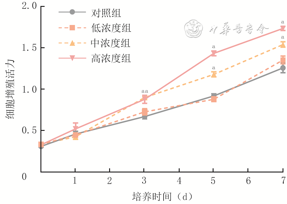
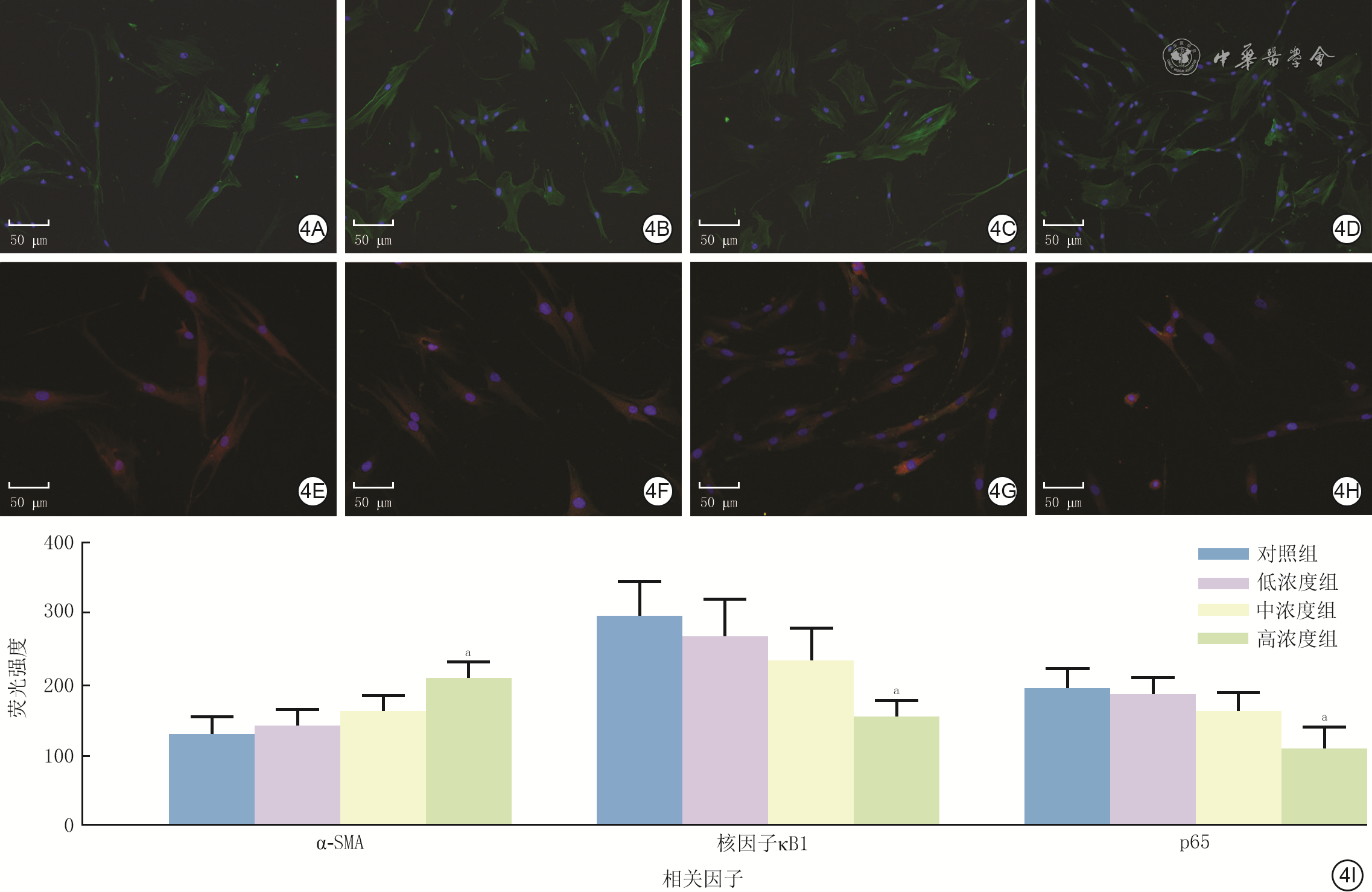
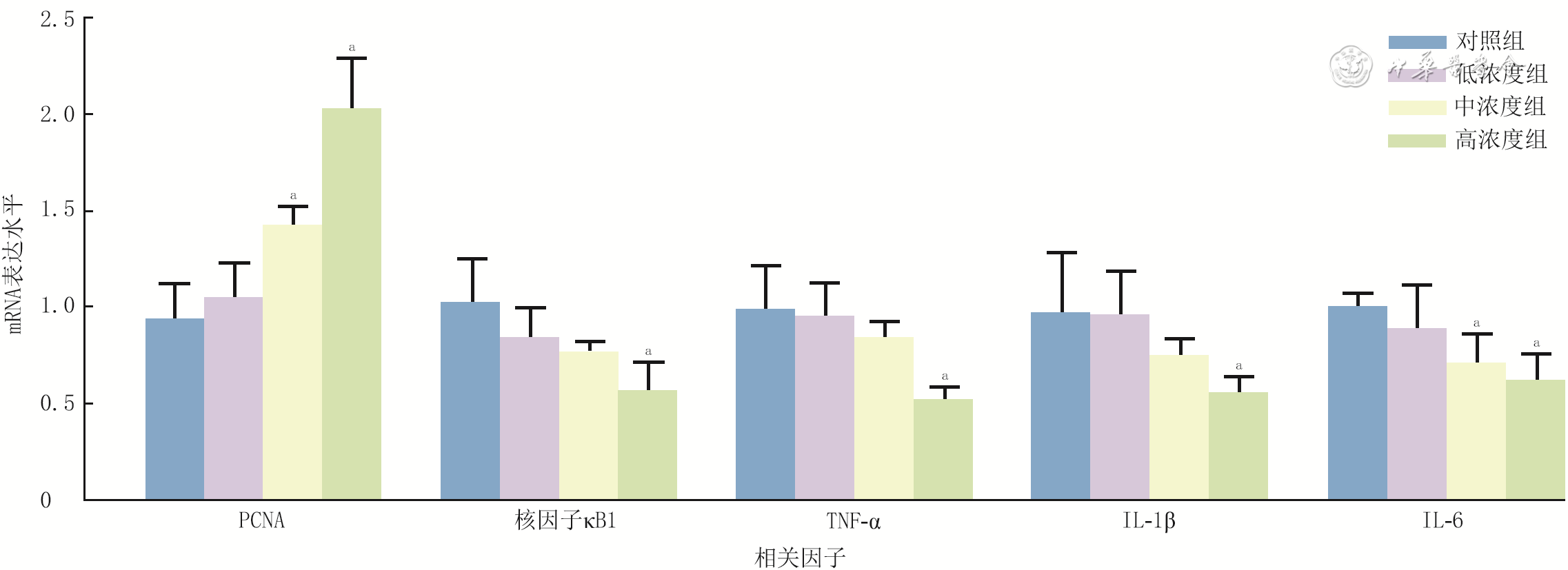
  Objective  To investigate the effects and mechanisms of type Ⅲ collagen (Col Ⅲ) on wound healing in diabetic mice with full-thickness skin defect.  Methods  This study was an experimental study using a grouped design combined with repeated-measures and factorial designs. Skin fibroblasts (Fbs) were isolated from 6 male db/db mice aged 12 weeks by type Ⅱ collagenase digestion and divided into low, medium, and high concentration groups, which were treated respectively with recombinant human Col Ⅲ at final mass concentrations of 0.05, 0.10, and 0.20 mg/mL, and control group that was treated with phosphate-buffered saline (PBS). The scratch healing rate was calculated after cell scratch assay at 48 h after scratch; the number of migrated cells was calculated after Transwell assay at 24 h after culture; the cell proliferative activity was detected by cell counting kit-8 at 3, 5, and 7 d after culture; the expressions of α-smooth muscle actin (α-SMA), nuclear factor-κB1 (NF-κB1), and p65 in cells were detected by immunofluorescence staining at 24 h after culture; the mRNA expressions of proliferating cell nuclear antigen (PCNA), NF-κB1, tumor necrosis factor-α (TNF-α), interleukin-1β (IL-1β), and IL-6 in cells were detected by real-time fluorescence quantitative reverse transcription polymerase chain reaction at 24 h after culture. The sample size was 5 for the above experiments. Full-thickness skin defect wounds were successfully created in 30 male C57BL/6J mice aged 8 weeks with diabetes. The mice were then divided into PBS group and Col Ⅲ group by the random number table method, which were subcutaneously injected with PBS and 0.20 mg/mL Col Ⅲ solution at the wound margin, with 15 mice in each group. The wound healing rates at 7 and 14 d after surgery were calculated. The wound tissue was harvested at 14 d after surgery for Masson staining to quantify the percentage of collagen fiber deposition area, for immunohistochemical staining to detect the expressions of TNF-α and NF-κB1, and for immunofluorescence staining to detect the expressions of CD31 and PCNA.  Results  Compared with those in control group, the scratch healing rates in medium and high concentration groups of cells at 48 h after scratch were significantly increased (P<0.05), the number of migrated cells at 24 h after culture was significantly increased (P<0.05), and the proliferative activities at 3, 5, and 7 d after culture were significantly increased (P<0.05). After 24 h of culture, compared with those in control group, the expression of α-SMA in high concentration group of cells was significantly increased (P<0.05), while the expressions of NF-κB1 and p65 were significantly decreased (P<0.05); the mRNA expression of PCNA in medium concentration group of cells was significantly increased (P<0.05), while that of IL-6 was significantly decreased (P<0.05); the mRNA expression of PCNA in high concentration group of cells was significantly increased (P<0.05), while the mRNA expressions of NF-κB1, TNF-α, IL-1β, and IL-6 were significantly decreased (P<0.05). At 7 and 14 d after surgery, the wound healing rates of mice in Col Ⅲ group were (61.8±4.4)% and (95.1±2.7)%, respectively, which were significantly higher than (52.0±3.9)% and (76.0±7.2)% in PBS group (with t values of 3.68 and 5.53, respectively, P<0.05). At 14 d after surgery, compared with those in PBS group, the percentage of collagen fiber deposition area in the wound tissue of mice in Col Ⅲ group was significantly increased (t=4.41, P<0.05), the expressions of TNF-α and NF-κB1 were significantly decreased (with t values of 4.10 and 3.07, respectively, P<0.05), and the expressions of CD31 and PCNA were significantly increased (with t values of 4.01 and 3.23, respectively, P<0.05).  Conclusions  Col Ⅲ can promote the proliferation and migration of skin Fbs from diabetic mice, inhibit NF-κB-mediated inflammatory response, and promote collagen fiber deposition and angiogenesis in wounds, thereby accelerating the healing of full-thickness skin defect wounds in diabetic mice.
Discipline Construction·Quality and Efficiency in Diagnosis and Treatment, and Disciplinary Development
2026, 42(10): 993-994. doi: 10.3760/cma.j.cn501225-20260830-00319
Abstract:
Discipline Construction
2026, 42(10): 995-996. doi: 10.3760/cma.j.cn501225-20260614-00238
Abstract:
Review
Research advances on the application of artificial intelligence in the warning and management of burn-related delirium
Wang Gui, Qian Hongyan, Yue Xiaomin
2026, 42(10): 997-1002. doi: 10.3760/cma.j.cn501225-20250911-00392
Abstract:
Delirium in burn patients presented a prominent challenge in intensive care, characterized by high incidence, difficult identification, and a narrow intervention window. Artificial intelligence (AI) technology, with its unique advantages of being adept at processing complex high-dimensional data, mining hidden patterns, and real-time continuous monitoring, provided new directions for the warning and management of burn-related delirium. Based on the main driving mechanisms and clinical heterogeneity of burn-related delirium, this article systematically reviewed the research advances of AI in establishment of predictive models for burn-related delirium, multimodal data fusion and interpretable outputs, multi-source data acquisition, intelligent processing and decision support, as well as clinical applications and closed-loop feedback. It analyzed current challenges such as data governance and algorithmic fairness, integration and collaboration in clinical scenarios, and liability attribution and ethical supervision, and prospected future research directions, aiming to provide references and insights for the application of AI in the field of burns.