- Medline/PubMed数据库
- Scopus数据库
- PMC数据库
- CSCD
- 北大核心收录期刊
- 统计源期刊
- 我国高质量科技期刊T1级
- 第6届中国精品科技期刊
| Citation: | Shen X,Sun ZY,Zhang R,et al.Effects of recombinant human metallothionein-Ⅲ combined with wound dressing on wound healing of full-thickness skin defects in mice[J].Chin J Burns Wounds,2024,40(5):425-432.DOI: 10.3760/cma.j.cn501225-20231031-00164.

|

| [1] |
王布雷. 白及多糖水凝胶促皮肤伤口愈合作用研究 [D]. 西安:陕西师范大学, 2022. DOI: 10.27292/d.cnki.gsxfu.2021.001160. |
| [2] |
GuerraA,BelinhaJ,JorgeRN.Modelling skin wound healing angiogenesis: a review[J].J Theor Biol,2018,459:1-17.DOI: 10.1016/j.jtbi.2018.09.020.
|
| [3] |
KagiJH,ValeeBL.Metallothionein: a cadmium- and zinc-containing protein from equine renal cortex[J].J Biol Chem,1960,235:3460-3465.
|
| [4] |
LiuAL,ZhangZM,ZhuBF,et al.Metallothionein protects bone marrow stromal cells against hydrogen peroxide-induced inhibition of osteoblastic differentiation[J].Cell Biol Int,2004,28(12):905-911.DOI: 10.1016/j.cellbi.2004.09.004.
|
| [5] |
CherianMG,JayasuryaA,BayBH.Metallothioneins in human tumors and potential roles in carcinogenesis[J].Mutat Res,2003,533(1/2):201-209.DOI: 10.1016/j.mrfmmm.2003.07.013.
|
| [6] |
CoyleP,PhilcoxJC,CareyLC,et al.Metallothionein: the multipurpose protein[J].Cell Mol Life Sci,2002,59(4):627-647.DOI: 10.1007/s00018-002-8454-2.
|
| [7] |
何婧雯,熊海容,王靖,等.金属硫蛋白的研究现状及进展[J].农产品加工(上半月),2019(9):72-75. DOI: 10.16693/j.cnki.1671-9646(X).2019.09.018.
|
| [8] |
YouHJ,LeeKJ,JeongHG.Overexpression of human metallothionein-III prevents hydrogen peroxide-induced oxidative stress in human fibroblasts[J].FEBS Lett,2002,521(1/2/3):175-179.DOI: 10.1016/s0014-5793(02)02870-3.
|
| [9] |
燕艳,季志会,杜伟,等.金属硫蛋白应用研究进展[J].东北农业大学学报,2010,41(7):150-154.DOI: 10.3969/j.issn.1005-9369.2010.07.030.
|
| [10] |
ZhangW,XieY,LiuW,et al.Role of metallothionein in post-burn inflammation[J].Inflammation,2016,39(2):768-774.DOI: 10.1007/s10753-016-0305-7.
|
| [11] |
LansdownAB.Metallothioneins: potential therapeutic aids for wound healing in the skin[J].Wound Repair Regen,2002,10(3):130-132.DOI: 10.1046/j.1524-475x.2002.20101.x.
|
| [12] |
薛燕.金属硫蛋白在皮肤疾病中的研究进展[J].中国美容医学,2013,22(17):1823-1826.DOI: 10.3969/j.issn.1008-6455.2013.17.031.
|
| [13] |
徐连春,尚剑,孙晔,等.银纳米颗粒及载银抗菌涂层的研究与进展[J].中国组织工程研究,2016,20(25):3793-3800.DOI: 10.3969/j.issn.2095-4344.2016.25.022.
|
| [14] |
AlemdaroğluC,DeğimZ,CelebiN,et al.An investigation on burn wound healing in rats with chitosan gel formulation containing epidermal growth factor[J].Burns,2006,32(3):319-327.DOI: 10.1016/j.burns.2005.10.015.
|
| [15] |
王宏宇, 巴特, 周彪, 等. 不同途径应用人脐带间充质干细胞外泌体治疗小鼠全层皮肤缺损创面的效果[J]. 中华烧伤与创面修复杂志, 2024, 40(4): 314-322. DOI: 10.3760/cma.j.cn501225-20231123-00203.
|
| [16] |
姚梦云,张宁,张庆,等.白细胞介素4修饰的金纳米酶对糖尿病小鼠全层皮肤缺损的作用[J].中华烧伤与创面修复杂志,2023,39(1):15-24.DOI: 10.3760/cma.j.cn501225-20220630-00275.
|
| [17] |
谢军,毛玉洁,王思宇,等.紫草素对大鼠慢性皮肤溃疡创面愈合及新生血管形成的促进作用及其机制[J].解放军医学杂志,2022,47(1):39-45.DOI: 10.11855/j.issn.0577-7402.2022.01.0039.
|
| [18] |
PenkowaM,CarrascoJ,GiraltM,et al.CNS wound healing is severely depressed in metallothionein I- and II-deficient mice[J].J Neurosci,1999,19(7):2535-2545.DOI: 10.1523/JNEUROSCI.19-07-02535.1999.
|
| [19] |
MorelliniNM,GilesNL,ReaS,et al.Exogenous metallothionein-IIA promotes accelerated healing after a burn wound[J].Wound Repair Regen,2008,16(5):682-690.DOI: 10.1111/j.1524-475X.2008.00418.x.
|
| [20] |
AignerGP,PeerV,FiechtnerB,et al.Wound healing and Cadmium detoxification in the earthworm Lumbricus terrestris - a potential case for coelomocytes?[J].Front Immunol,2023,14:1272191.DOI: 10.3389/fimmu.2023.1272191.
|
| [21] |
SunZ,QinJ,YuanH,et al.Recombinant human metallothionein-III alleviates oxidative damage induced by copper and cadmium in Caenorhabditis elegans[J].J Appl Toxicol,2023,43(8):1242-1252.DOI: 10.1002/jat.4460.
|
| [22] |
Cortese-KrottMM,MünchowM,PirevE,et al.Silver ions induce oxidative stress and intracellular zinc release in human skin fibroblasts[J].Free Radic Biol Med,2009,47(11):1570-1577.DOI: 10.1016/j.freeradbiomed.2009.08.023.
|
| [23] |
PavlíkV,SobotkaL,PejchalJ,et al.Silver distribution in chronic wounds and the healing dynamics of chronic wounds treated with dressings containing silver and octenidine[J].FASEB J,2021,35(5):e21580.DOI: 10.1096/fj.202100065R.
|
| [24] |
LinX,JagadapillaiR,CaiJ,et al.Metallothionein induction attenuates the progression of lung injury in mice exposed to long-term intermittent hypoxia[J].Inflamm Res,2020,69(1):15-26.DOI: 10.1007/s00011-019-01287-z.
|
| [25] |
GurtnerGC, WernerS, BarrandonY, et al. Wound repair and regeneration[J]. Nature, 2008,453(7193):314-321. DOI: 10.1038/nature07039.
|
| [26] |
WangZ,ZhaoF,XuC,et al.Metabolic reprogramming in skin wound healing[J/OL].Burns Trauma,2024,12:tkad047[2023-10-31].https://pubmed.ncbi.nlm.nih.gov/38179472/.DOI: 10.1093/burnst/tkad047.
|
| [27] |
ManchandaM,TorresM,InuossaF,et al.Metabolic reprogramming and reliance in human skin wound healing[J].J Invest Dermatol,2023,143(10):2039-2051.e10.DOI: 10.1016/j.jid.2023.02.039.
|
| [28] |
EmingSA,MurrayPJ,PearceEJ.Metabolic orchestration of the wound healing response[J].Cell Metab,2021,33(9):1726-1743.DOI: 10.1016/j.cmet.2021.07.017.
|
| [29] |
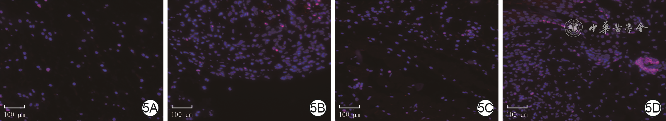
韩莹,时玉峥,魏训东,等.Ki67法检测间充质干细胞对淋巴细胞增殖抑制能力[J].药物评价研究,2022,45(4):673-679.DOI: 10.7501/j.issn.1674-6376.2022.04.009.
|
| [30] |
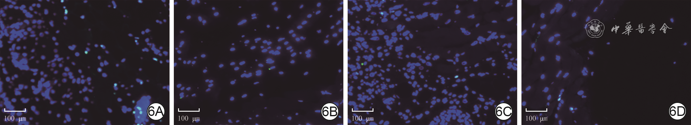
李丽,牛钰清,陈菲,等.介绍一种细胞凋亡TUNEL检测与Ki-67 DAPI三色荧光染色操作流程[J].临床与实验病理学杂志,2019,35(8):980-981.DOI: 10.13315/j.cnki.cjcep.2019.08.026.
|
| [31] |
ChowdhuryD,AlrefaiH,Landero FigueroaJA,et al.Metallothionein 3 controls the phenotype and metabolic programming of alternatively activated macrophages[J].Cell Rep,2019,27(13):3873-3886.e7.DOI: 10.1016/j.celrep.2019.05.093.
|
| [32] |
ÅgrenMS,ChafranskaL,EriksenJO,et al.Spatial expression of metallothionein, matrix metalloproteinase-1 and Ki-67 in human epidermal wounds treated with zinc and determined by quantitative immunohistochemistry: a randomised double-blind trial[J].Eur J Cell Biol,2021,100(3):151147.DOI: 10.1016/j.ejcb.2020.151147.
|
Figures(7) / Tables(1)
Copyright © Chinese Journal of Burns京ICP备07035254号-14
E-mail:shaoshangzazhi@163.com
Supported by:
Beijing Renhe Information Technology Co. Ltd


 DownLoad: