- Medline/PubMed数据库
- Scopus数据库
- PMC数据库
- CSCD
- 北大核心收录期刊
- 统计源期刊
- 我国高质量科技期刊T1级
- 第6届中国精品科技期刊
| Citation: | Lyu KY,Li YS.Mechanisms of surgical incision scar and technological innovations for scar tension reduction based the mechano-chemo-biological theory[J].Chin J Burns Wounds,2026,42(2):133-142.DOI: 10.3760/cma.j.cn501225-20251013-00424.

|

| [1] |
LiY, LiuA, WangJ, et al. Suture-anchored cutaneous tension induces persistent hypertrophic scarring in a novel murine model[J/OL]. Burns Trauma, 2024,12:tkae051[2025-10-13]. https://pubmed.ncbi.nlm.nih.gov/39429643/. DOI: 10.1093/burnst/tkae051.
|
| [2] |
HosseiniM, BrownJ, KhosrotehraniK, et al. Skin biomechanics: a potential therapeutic intervention target to reduce scarring[J/OL]. Burns Trauma, 2022,10:tkac036[2025-10-13].https://pubmed.ncbi.nlm.nih.gov/36017082/. DOI: 10.1093/burnst/tkac036.
|
| [3] |
SongvasinS, WinaikosolK, KarunasumettaC. A randomized controlled trial comparison of subcuticular suture and adhesive strip for cosmetic outcome in median sternotomy closure in patient undergoing cardiac surgery[J/OL]. Aesthetic Plast Surg, 2025(2025-07-05)[2025-10-13].https://pubmed.ncbi.nlm.nih.gov/40473789/. DOI: 10.1007/s00266-025-04885-x.[published online ahead of print].
|
| [4] |
ChenZ, JinY, ZouY, et al. Scar prevention with prolonged use of tissue adhesive zipper immediately after facial surgery: a randomized controlled trial[J]. Aesthet Surg J, 2022,42(5):NP265-NP272. DOI: 10.1093/asj/sjab407.
|
| [5] |
JiQ, LuoL, NiJ, et al. Fractional CO2 laser to treat surgical scars: a system review and meta-analysis on optimal timing[J]. J Cosmet Dermatol, 2025,24(1):e16708. DOI: 10.1111/jocd.16708.
|
| [6] |
RothmanSS. Physiology and biochemistry of the skin[M]. London:Cambridge University Press,1954.
|
| [7] |
KangM, KoUH, OhEJ, et al. Tension-sensitive HOX gene expression in fibroblasts for differential scar formation[J]. J Transl Med, 2025,23(1):168. DOI: 10.1186/s12967-025-06191-1.
|
| [8] |
Medina-LombarderoS, BainC, CharltonL, et al. The biomechanics of wounds at physiologically relevant levels: understanding skin's stress-shielding effect for the quantitative assessment of healing[J]. Mater Today Bio, 2024, 25:100963.DOI: 10.1016/j.mtbio.2024.100963.
|
| [9] |
RognoniE, PiscoAO, HiratsukaT, et al. Fibroblast state switching orchestrates dermal maturation and wound healing[J]. Mol Syst Biol, 2018,14(8):e8174. DOI: 10.15252/msb.20178174.
|
| [10] |
AarabiS, BhattKA, ShiY, et al. Mechanical load initiates hypertrophic scar formation through decreased cellular apoptosis[J]. FASEB J, 2007,21(12):3250-3261. DOI: 10.1096/fj.07-8218com.
|
| [11] |
RossR, GuoY, WalkerRN, et al. Biomechanical activation of keloid fibroblasts promotes lysosomal remodeling and exocytosis[J]. J Invest Dermatol, 2024,144(12):2730-2741. DOI: 10.1016/j.jid.2024.04.015.
|
| [12] |
CaoG, YeM, WangH, et al. The role of biomechanical forces in the formation and treatment of pathological scars[J]. Clin Cosmet Investig Dermatol, 2024,17:2565-2571. DOI: 10.2147/CCID.S496253.
|
| [13] |
MascharakS, GuoJL, GriffinM, et al. Modelling and targeting mechanical forces in organ fibrosis[J]. Nat Rev Bioeng, 2024,2(4):305-323. DOI: 10.1038/s44222-023-00144-3.
|
| [14] |
MartinoF, PerestreloAR, VinarskýV, et al. Cellular mechanotransduction: from tension to function[J]. Front Physiol, 2018,9:824. DOI: 10.3389/fphys.2018.00824.
|
| [15] |
KatohK. Integrin and its associated proteins as a mediator for mechano-signal transduction[J]. Biomolecules, 2025, 15(2):166.DOI: 10.3390/biom15020166.
|
| [16] |
WangR, ChenB, WeiH, et al. Collecting and deactivating TGF-β1 hydrogel for anti-scarring therapy in post-glaucoma filtration surgery[J]. Mater Today Bio, 2022,14:100260. DOI: 10.1016/j.mtbio.2022.100260.
|
| [17] |
ZhangQ, ShiL, HeH, et al. Down-regulating scar formation by microneedles directly via a mechanical communication pathway[J]. ACS Nano, 2022,16(7):10163-10178. DOI: 10.1021/acsnano.1c11016.
|
| [18] |
LiYY, JiSF, FuXB, et al. Biomaterial-based mechanical regulation facilitates scarless wound healing with functional skin appendage regeneration[J]. Mil Med Res, 2024, 11(1):13.DOI: 10.1186/s40779-024-00519-6.
|
| [19] |
WenD, GaoY, LiuY, et al. Matrix stiffness-induced α-tubulin acetylation is required for skin fibrosis formation through activation of Yes-associated protein[J]. MedComm (2020), 2023, 4(4):e319.DOI: 10.1002/mco2.319.
|
| [20] |
RagazziniS, ScocozzaF, BernavaG, et al. Mechanosensor YAP cooperates with TGF-β1 signaling to promote myofibroblast activation and matrix stiffening in a 3D model of human cardiac fibrosis[J]. Acta Biomater, 2022,152:300-312. DOI: 10.1016/j.actbio.2022.08.063.
|
| [21] |
MascharakS, desJardins-ParkHE, DavittMF, et al. Preventing Engrailed-1 activation in fibroblasts yields wound regeneration without scarring[J]. Science, 2021,372(6540):eaba2374.DOI: 10.1126/science.aba2374.
|
| [22] |
PorteJ, JenkinsG, TatlerAL. Myofibroblast TGF-β activation measurement in vitro[J]. Methods Mol Biol, 2021, 2299:99-108.DOI: 10.1007/978-1-0716-1382-5_6.
|
| [23] |
ChavulaT, ToS, AgarwalSK. Cadherin-11 and its role in tissue fibrosis[J]. Cells Tissues Organs, 2023, 212(4):293-303.DOI: 10.1159/000525359.
|
| [24] |
Viji BabuPK, MirastschijskiU, BelgeG, et al. Homophilic and heterophilic cadherin bond rupture forces in homo- or hetero-cellular systems measured by AFM-based single-cell force spectroscopy[J]. Eur Biophys J, 2021, 50(3/4):543-559.DOI: 10.1007/s00249-021-01536-2.
|
| [25] |
BagheriL, JavanbakhtM, MalekianS, et al. Antifibrotic therapeutic strategies in systemic sclerosis: critical role of the Wnt/β-catenin and TGF-β signal transduction pathways as potential targets[J]. Eur J Pharmacol, 2025, 999:177607.DOI: 10.1016/j.ejphar.2025.177607.
|
| [26] |
YinJ, ZhangS, YangC, et al. Mechanotransduction in skin wound healing and scar formation: potential therapeutic targets for controlling hypertrophic scarring[J]. Front Immunol, 2022, 13:1028410.DOI: 10.3389/fimmu.2022.1028410.
|
| [27] |
ZhouDW, LeeTT, WengS, et al. Effects of substrate stiffness and actomyosin contractility on coupling between force transmission and vinculin-paxillin recruitment at single focal adhesions[J]. Mol Biol Cell, 2017,28(14):1901-1911. DOI: 10.1091/mbc.E17-02-0116.
|
| [28] |
LiH, RaghunathanV, StamerWD, et al. Extracellular matrix stiffness and TGFβ2 regulate YAP/TAZ activity in human trabecular meshwork cells[J]. Front Cell Dev Biol, 2022, 10:844342.DOI: 10.3389/fcell.2022.844342.
|
| [29] |
TuS, LiY, LiJ, et al. Mechanical stretch-mediated fibroblast activation: the pivotal role of Piezo1 channels[J]. Biochim Biophys Acta Mol Cell Res, 2025,1872(7):120008. DOI: 10.1016/j.bbamcr.2025.120008.
|
| [30] |
Elosegui-ArtolaA, AndreuI, BeedleAEM, et al. Force triggers YAP nuclear entry by regulating transport across nuclear pores[J]. Cell, 2017,171(6):1397-1410.e14. DOI: 10.1016/j.cell.2017.10.008.
|
| [31] |
HoffmanLM, SmithMA, JensenCC, et al. Mechanical stress triggers nuclear remodeling and the formation of transmembrane actin nuclear lines with associated nuclear pore complexes[J]. Mol Biol Cell, 2020,31(16):1774-1787. DOI: 10.1091/mbc.E19-01-0027.
|
| [32] |
LangevinHM, BouffardNA, BadgerGJ, et al. Dynamic fibroblast cytoskeletal response to subcutaneous tissue stretch ex vivo and in vivo[J]. Am J Physiol Cell Physiol, 2005,288(3):C747-756. DOI: 10.1152/ajpcell.00420.2004.
|
| [33] |
StewardRL, ChengCM, WangDL, et al. Probing cell structure responses through a shear and stretching mechanical stimulation technique[J]. Cell Biochem Biophys, 2010,56(2/3):115-124. DOI: 10.1007/s12013-009-9075-2.
|
| [34] |
BecerraN, SalisB, TedescoM, et al. AFM and fluorescence microscopy of single cells with simultaneous mechanical stimulation via electrically stretchable substrates[J]. Materials (Basel), 2021, 14(15):4131.DOI: 10.3390/ma14154131.
|
| [35] |
NagayamaK, FukueiT. Cyclic stretch-induced mechanical stress to the cell nucleus inhibits ultraviolet radiation-induced DNA damage[J]. Biomech Model Mechanobiol, 2020,19(2):493-504. DOI: 10.1007/s10237-019-01224-3.
|
| [36] |
GurusaranM, ErlandsenBS, DaviesOR. The crystal structure of SUN1-KASH6 reveals an asymmetric LINC complex architecture compatible with nuclear membrane insertion[J]. Commun Biol, 2024, 7(1):138.DOI: 10.1038/s42003-024-05794-6.
|
| [37] |
SoboJM, AlagnaNS, SunSX, et al. Lamins: the backbone of the nucleocytoskeleton interface[J]. Curr Opin Cell Biol, 2024, 86:102313.DOI: 10.1016/j.ceb.2023.102313.
|
| [38] |
DonnalojaF, CarnevaliF, JacchettiE, et al. Lamin A/C mechanotransduction in laminopathies[J]. Cells, 2020, 9(5):1306.DOI: 10.3390/cells9051306.
|
| [39] |
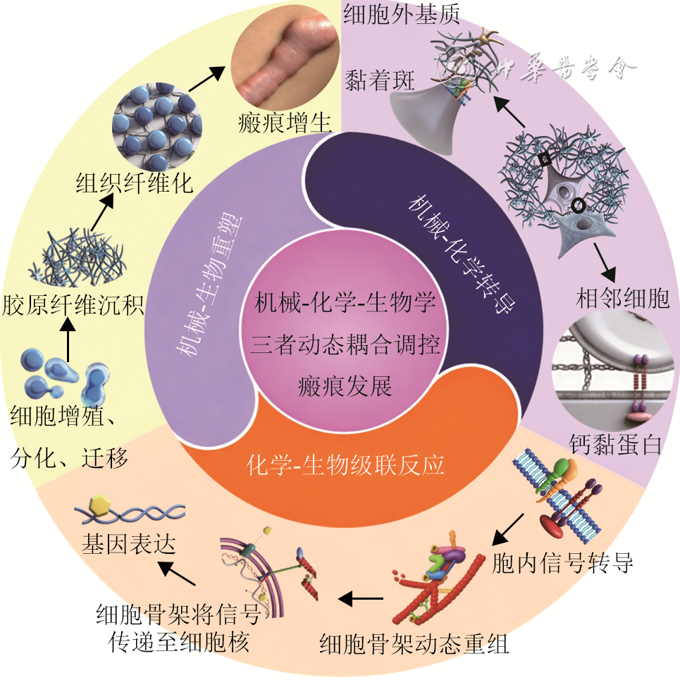
FengXQ, LiB, LinSZ, et al. Mechano-chemo-biological theory of cells and tissues: review and perspectives[J]. Acta Mechanica Sinica, 2025, 41(7):625315.DOI: 10.1007/s10409-025-25315-x.
|
| [40] |
GeM, ZhengW, YaoP, et al. Progress in tension-relieving suturing surgery: revolutionary surgical techniques and patient prognosis evaluation methods[J]. Front Surg, 2025,12:1587582. DOI: 10.3389/fsurg.2025.1587582.
|
| [41] |
ZitelliJA, MoyRL. Buried vertical mattress suture[J]. J Dermatol Surg Oncol, 1989,15(1):17-19. DOI: 10.1111/j.1524-4725.1989.tb03107.x.
|
| [42] |
XieY, ChenY, HongY, et al. Effect of trapezoidal excision combined with modified embedded vertical mattress suture technique on postoperative scar formation after cesarean section[J]. Am J Transl Res, 2024, 16(8):3812-3821.DOI: 10.62347/MGKQ5295.
|
| [43] |
ZhangX, DiaoJS, GuoSZ, et al. Wedge-shaped excision and modified vertical mattress suture fully buried in a multilayered and tensioned wound closure[J]. Aesthetic Plast Surg, 2009,33(3):457-460. DOI: 10.1007/s00266-009-9311-6.
|
| [44] |
SuX, ZhouX, TangY, et al. Modified buried vertical mattress suture combined with tension-reducing tape in forearm tattoo resection-a retrospective study[J]. J Cosmet Dermatol, 2025, 24(6):e70266.DOI: 10.1111/jocd.70266.
|
| [45] |
YangD, YaoL, ZhanY, et al. Application of remote buried dermal super-tension-reducing sutures for incisional scar prevention[J]. Clin Cosmet Investig Dermatol, 2025, 18:2749-2756.DOI: 10.2147/CCID.S549932.
|
| [46] |
SeeA, SmithHR. Partially buried horizontal mattress suture: modification of the Haneke-Marini suture[J]. Dermatol Surg, 2004,30(12Pt 1):1491-1492. DOI: 10.1111/j.1524-4725.2004.30508.x.
|
| [47] |
AlamM, GoldbergLH. Utility of fully buried horizontal mattress sutures[J]. J Am Acad Dermatol, 2004,50(1):73-76. DOI: 10.1016/s0190-9622(03)02097-8.
|
| [48] |
MengF, AndreaS, ChengS, et al. Modified subcutaneous buried horizontal mattress suture compared with vertical buried mattress suture[J]. Ann Plast Surg, 2017,79(2):197-202. DOI: 10.1097/SAP.0000000000001043.
|
| [49] |
MinP, ZhangS, SinakiDG, et al. Using Zhang's supertension-relieving suture technique with slowly-absorbable barbed sutures in the management of pathological scars: a multicenter retrospective study[J/OL]. Burns Trauma, 2023,11:tkad026[2025-10-13]. https://pubmed.ncbi.nlm.nih.gov/37334139/. DOI: 10.1093/burnst/tkad026.
|
| [50] |
ChenJ, MoY, ChenY, et al. Application and effect of tension-reducing suture in surgical treatment of hypertrophic scar[J]. BMC Surg, 2024,24(1):119. DOI: 10.1186/s12893-024-02390-7.
|
| [51] |
刘航, 胡铭, 饶明军, 等. 阶梯状递进式超减张缝合法闭合胸背部及四肢高张力创面[J].中国修复重建外科杂志,2024, 38(12):1505-1509. D0I:10.7507/1002-1892.202409048.
|
| [52] |
ChenW, JiangT, ZhongZ, et al. The effect of double W tension-reduced suture technique on the abdominal scars following the da Vinci robot-assisted gastrectomy for severely obese patients[J]. BMC Surg, 2023,23(1):115. DOI: 10.1186/s12893-023-01979-8.
|
| [53] |
WuF, TianY, WangF, et al. The suture effect of butterfly suture combined with the looped, broad, and deep buried suture in patients with pigmented naevus receiving surgery excision[J]. Arch Dermatol Res, 2025,317(1):433. DOI: 10.1007/s00403-025-03957-x.
|
| [54] |
ZhangY, LeiZ, LinB, et al. Split-level folding, step-type tension-relieving suture technique, and the evaluation on scar minimization[J]. J Cosmet Dermatol, 2024,23(6):2199-2208. DOI: 10.1111/jocd.16236.
|
| [55] |
HuangC, LiuOG. Using a zipper device to minimize scarring after excision of facial nevi in pediatric patients[J/OL]. J Craniofac Surg, 2024(2024-08-22)[2025-10-13].https://pubmed.ncbi.nlm.nih.gov/39171936/. DOI: 10.1097/SCS.0000000000010531. [published online ahead of print].
|
| [56] |
GaoY, WangY, LiW, et al. Clinical efficacy analysis of cosmetic suture technique combined with tension reducer in the treatment of facial skin trauma[J]. Medicine (Baltimore), 2024,103(52):e41040. DOI: 10.1097/MD.0000000000041040.
|
| [57] |
石璐璐,张汝锋,肖虎. 白细胞介素6/信号转导及转录激活因子3通路及β连环蛋白在机械应力致小鼠增生性瘢痕形成中的作用[J]. 中华烧伤杂志,2021,37(7):647-653.DOI: 10.3760/cma.j.cn501120-20200417-00231.
|
| [58] |
章一新, 柴筠. 瘢痕压力治疗的机制与临床应用[J]. 中华烧伤与创面修复杂志, 2025,41(4):316-324. DOI: 10.3760/cma.j.cn501225-20250215-00064.
|
| [59] |
中华医学会烧伤外科学分会. 儿童瘢痕预防与治疗临床实践指南(2025版)[J]. 中华烧伤与创面修复杂志,2025,41(11):1011-1028.DOI: 10.3760/cma.j.cn501225-20250630-00285.
|
| [60] |
De DeckerI, BeeckmanA, HoeksemaH, et al. Pressure therapy for scars: myth or reality? A systematic review[J]. Burns, 2023,49(4):741-756. DOI: 10.1016/j.burns.2023.03.007.
|
| [61] |
GieleHP, LiddiardK, CurrieK, et al. Direct measurement of cutaneous pressures generated by pressure garments[J]. Burns, 1997,23(2):137-141. DOI: 10.1016/s0305-4179(96)00088-5.
|
| [62] |
NedelecB, De OliveiraA, CalvaV, et al. Longitudinal evaluation of pressure applied by custom fabricated garments worn by adult burn survivors[J]. J Burn Care Res, 2020, 41(2):254-262.DOI: 10.1093/jbcr/irz154.
|
| [63] |
LaiCHY, Li-TsangCWP. Validation of the Pliance X System in measuring interface pressure generated by pressure garment[J]. Burns, 2009,35(6):845-851. DOI: 10.1016/j.burns.2008.09.013.
|
| [64] |
Kness-KnezinskisE, SheckleyM, HostlerAC, et al. Translational approaches manipulating mechanobiology to promote scarless healing in humans[J]. J Plast Reconstr Aesthet Surg, 2026, 112:25-33.DOI: 10.1016/j.bjps.2025.06.041.
|
| [65] |
ZhuH, LiuX, WangJ, et al. Traction-regulated persistence governs durotaxis across cell types[J]. Eur J Cell Biol, 2025, 104(4):151515.DOI: 10.1016/j.ejcb.2025.151515.
|
| [66] |
LewisCJ, DouglasH, MartinL, et al. Carbon dioxide laser treatment of burn-related scarring: results of the ELIPSE (Early Laser Intervention Promotes Scar Evolution) prospective randomized controlled trial[J]. J Plast Reconstr Aesthet Surg, 2023, 84:368-376.DOI: 10.1016/j.bjps.2023.06.012.
|
| [67] |
MaY, BarnesSP, ChenYY, et al. Influence of scar age, laser type and laser treatment intervals on adult burn scars: a systematic review and meta-analysis[J]. PLoS One, 2023,18(9):e0292097. DOI: 10.1371/journal.pone.0292097.
|
| [68] |
GhassemiM, MireshghollahP, JafarzadehA, et al. Evaluating the combination and comparison of ablative fractional lasers (CO2, Erbium-YAG) with pulsed dye laser (PDL) for treating hypertrophic scars: a systematic review[J]. Lasers Med Sci, 2025, 40(1):129.DOI: 10.1007/s10103-025-04382-2.
|
| [69] |
PengW, ZhangX, KongX, et al. The efficacy and safety of fractional CO2 laser therapy in the treatment of burn scars: a meta-analysis[J]. Burns, 2021, 47(7):1469-1477.DOI: 10.1016/j.burns.2021.08.010.
|
| [70] |
KimS. Clinical trial of a pinpoint irradiation technique with the CO2 laser for the treatment of atrophic acne scars[J]. J Cosmet Laser Ther, 2008,10(3):177-180. DOI: 10.1080/14764170801930080.
|
| [71] |
LiuXJ, LeiY, GoldMH, et al. Efficacy of pulsed dye laser combined with fractional CO2 laser in the treatment of pediatric burn scars[J]. Lasers Surg Med, 2023, 55(5):464-470.DOI: 10.1002/lsm.23648.
|
| [72] |
ClementiA, CannarozzoG, GuarinoL, et al. Sequential fractional CO2 and 1540/1570 nm lasers: a narrative review of preclinical and clinical evidence[J]. J Clin Med, 2025, 14(11):3867.DOI: 10.3390/jcm14113867.
|
| [73] |
LiangYY, ShenJC, LiW. Evolution of compressive mechanical properties of early hypertrophic scar during laser treatment[J]. J Biomech, 2021,129:110783. DOI: 10.1016/j.jbiomech.2021.110783.
|
| [74] |
NaouriM, AtlanM, PerrodeauE, et al. Skin tightening induced by fractional CO2 laser treatment: quantified assessment of variations in mechanical properties of the skin[J]. J Cosmet Dermatol, 2012,11(3):201-206. DOI: 10.1111/j.1473-2165.2012.00627.x.
|
 线性瘢痕水平埋没皮内缝合操作示例.mp4
|

|
Figures(5)
Copyright © Chinese Journal of Burns京ICP备07035254号-14
E-mail:shaoshangzazhi@163.com
Supported by:
Beijing Renhe Information Technology Co. Ltd


 DownLoad: