Current Issue

2026 Vol. 42, No. 2

Guideline and Consensus
Expert consensus on the prevention and management of orthopedic surgical site infection wounds (2026 edition)
2026, 42(2): 101-118. doi: 10.3760/cma.j.cn501225-20250515-00228
Abstract:
Orthopedic surgical site infection (SSI) is a major complication after orthopedic surgery, with incidence rates differing based on the specific type of procedure, typically ranging from 0.4% to 16.1%, and potentially exceeding 50% in high-risk scenarios. When orthopedic SSI advances to a stage necessitating intervention—characterized by wound dehiscence, tissue necrosis, or exposed implants—it not only substantially prolongs the recovery period but also increases the medical burden. Presently, there is an absence of standardized prevention and management protocols for such wounds both nationally and internationally, resulting in significant differences in clinical practice. In order to augment postoperative safety for orthopedic patients, diminish the incidence of orthopedic SSI wounds, and enhance the quality of diagnosis and treatment for these wounds, the Wound Repair Professional Committee of Chinese Medical Doctor Association has convened a multidisciplinary panel of experts to formulate this consensus. This consensus encompasses a range of topics, including prevention strategies, clinical manifestations, diagnostic evaluation, wound management and repair, functional reconstruction, and rehabilitation therapy for orthopedic SSI wounds. The objective is to furnish a comprehensive reference for the prevention and treatment of these wounds.
Expert Forum
Burn infection and immune disorder: new translational medicine horizons from pathogenesis to precision medicine
Yao Yongming, Ren Chao
2026, 42(2): 119-125. doi: 10.3760/cma.j.cn501225-20251104-00457
Abstract:
The "pathological vicious cycle" of skin barrier disruption and immune disorders following severe burns renders infection a primary complication and main cause of death of patients, with burn sepsis accounting for more than 50% of all burn-associated deaths. Currently, the prevention and treatment of burn infection are confronted with multiple challenges: the superimposed effects of uncontrolled inflammatory response and immune disorder triggered by infection, the rapid progression from local wound to systemic damage, and the massive colonization of multidrug-resistant bacteria, which significantly increases the difficulty of antibacterial therapy. This paper conducted an in-depth analysis of the mechanisms underlying immune dysfunction after burn infection from phenotypic manifestations to intrinsic regulation. It systematically elaborated on the crucial roles and mechanisms of immune evasion by which multidrug-resistant bacteria, imbalance of immune responses, intestinal flora, and novel subtypes of immune regulatory cells play in the development of immune disorder. This paper further reported the emerging technologies for infection assessment and immune monitoring among burn patients by integrating the latest advancements in intelligent wound detecting system and novel biomarkers. Focusing on innovative therapeutic strategies for targeted burn immune regulation and local microenvironment remodeling, it further analyzed the challenges in clinical translation and future development directions of innovative therapeutic strategies.
Discussion on key issues in the treatment of pediatric burns
Liu Yan, Yu Jia'ao, Zhong Shan
2026, 42(2): 126-132. doi: 10.3760/cma.j.cn501225-20250713-00302
Abstract:
Due to the immaturity of skin tissue and organ function, pediatric burn patients exhibit distinct characteristics in etiology, shock resuscitation, wound characteristics, infection susceptibility, and prognosis. There are still aspects that need further discussion in the current treatment protocols for pediatric burns. This paper focuses on the key issues in the treatment of pediatric burns, with an emphasis on the fluid resuscitation for burn shock, the management of burn wounds, and the early identification of burn sepsis. The aim is to provide insight into current practices and future development in the management of pediatric burns.
Mechanisms of surgical incision scar and technological innovations for scar tension reduction based the mechano-chemo-biological theory
Lyu Kaiyang, Li Yashu
2026, 42(2): 133-142. doi: 10.3760/cma.j.cn501225-20251013-00424
Abstract:
Surgical incisions often heal with linear scars, which may further develop into hypertrophic scars or keloids. Research indicates that biomechanical factors, particularly mechanical forces and matrix stiffness, regulate scar formation by influencing cellular behavior and extracellular matrix remodeling. Our team proposes that the dynamic coupling and synergistic interactions among mechanical, chemical, and biological factors jointly regulate scar development. We emphasize that sustained tension control following wound closure is crucial to prevent scar widening and hyperplasia. Currently used tension-reducing methods have limitations: the effect of intraoperative sutures is short-lived; external tension-reducing devices (e.g., tension-reducing tape or zipper) are prone to falling off and causing skin irritation, and patient compliance is low; fractional laser may still cause the scar to widen when used alone in high-tension areas. Therefore, our team proposes a scheme utilizing an intradermal suturing technique based on slow-absorbing sutures with in-situ backstitch, which aims to achieve long-term and effective tension management. Preliminary results from clinical applications showed that this scheme achieved continuous tension reduction in surgical incisions, and its combination with fractional laser therapy is expected to synergistically improve scar width, overcoming the shortcomings of conventional methods. Moving forward, further research will optimize this scheme and promote its standardization and widespread adoption, and gradually establish a scar prevention system centered on continuous tension reduction, ultimately promoting the ideal healing of surgical incisions.
Original Article · Infection and Immunity
Establishing a rat model of sepsis by seawater immersion combined with Vibrio vulnificus infection after scald
Deng Bihan, Cheng Xinyue, Zhu Xiaomei, Wang Jun, Yao Yongming
2026, 42(2): 143-152. doi: 10.3760/cma.j.cn501225-20251017-00432
Abstract:
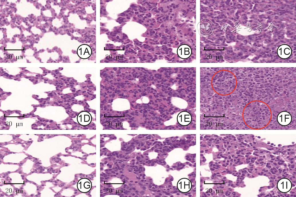
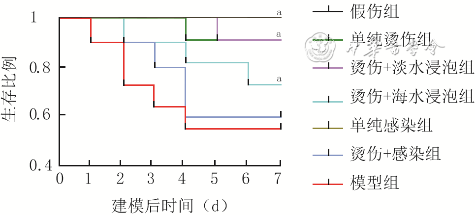
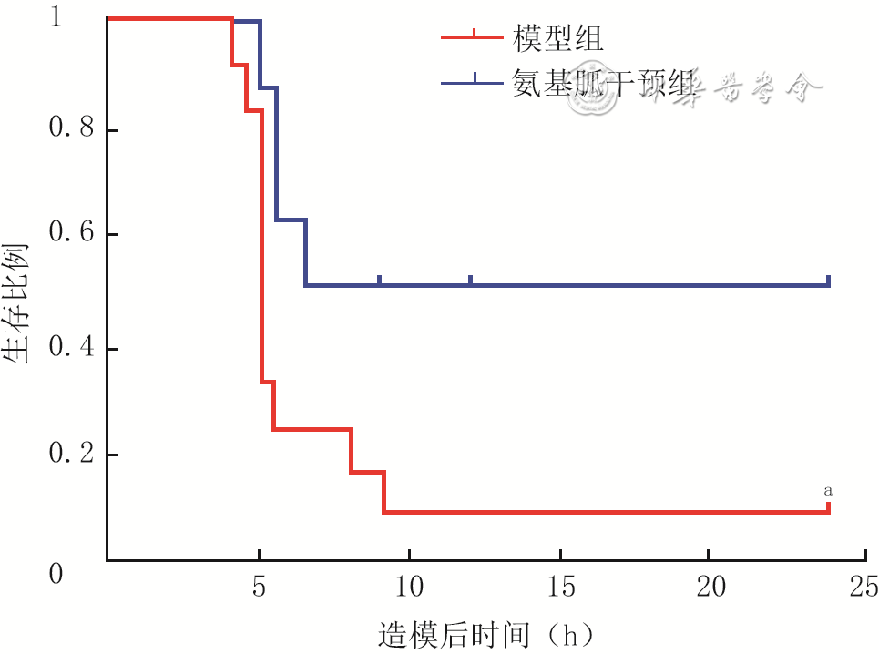
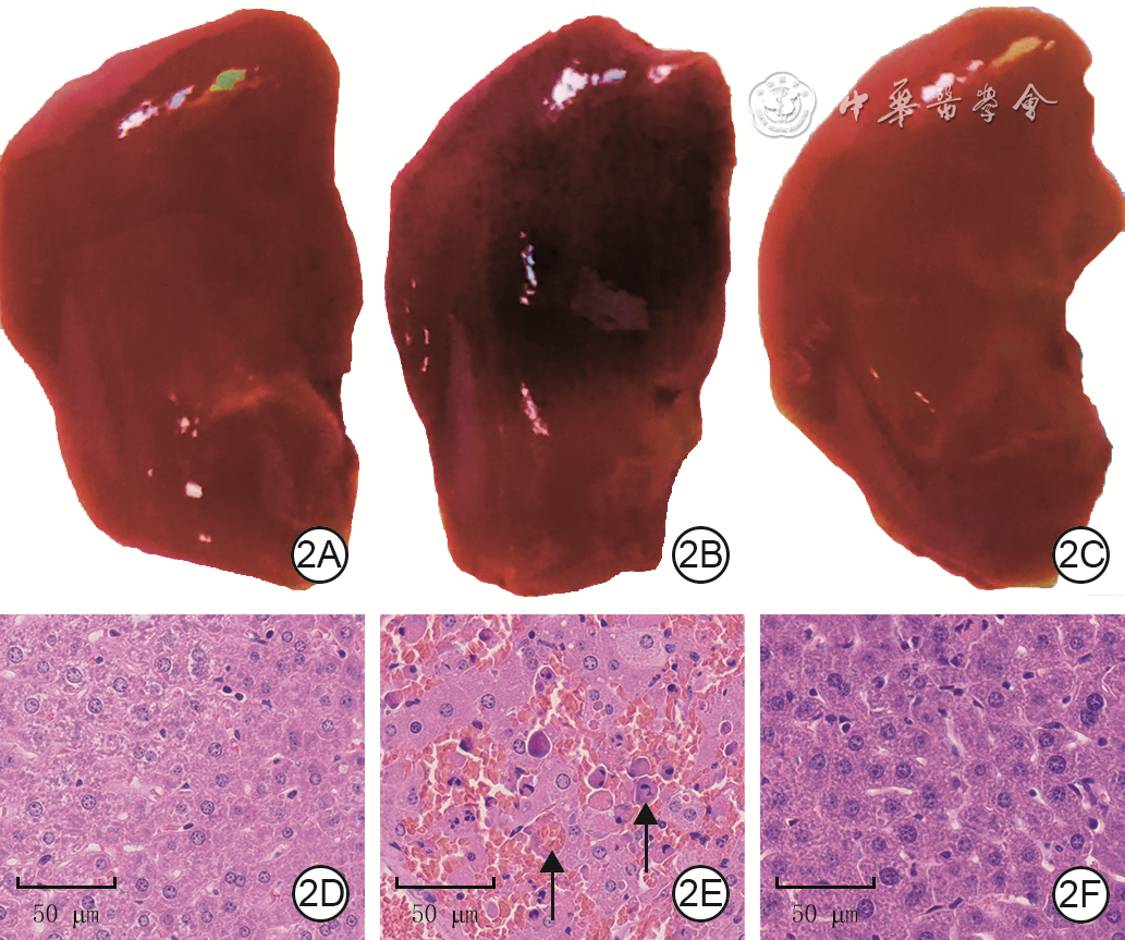
  Objective  To establish a rat model of sepsis by seawater immersion combined with Vibrio vulnificus infection after scald, providing an experimental basis for research on marine burn trauma-associated sepsis.  Methods  This study employed factorial design and was an experimental study. One hundred and fifteen male Sprague Dawley rats aged 8 weeks were allocated into three groups using a random number table method (the same grouping method applied below): model group (n=45), scald-only group (n=40), and sham injury group (n=30). Rats in the first two groups received dorsal scald injury, followed by either artificial seawater immersion for 30 min+injection of Vibrio vulnificus or injection of normal saline, respectively. In sham injury group, the dorsal region was immersed in warm water to induce sham injury, followed by injection of normal saline. On day 1, 3, and 5 after modeling, the pathological changes in the liver, kidney, lung, and heart in the three groups of rats were assessed using hematoxylin-eosin staining, and the pathological damage of the aforementioned organs was evaluated using a semi-quantitative scoring system. According to the instructions of the kit, a microplate reader was used to detect the serum levels of aspartate aminotransferase (AST), alanine aminotransferase (ALT), urea nitrogen, creatinine, creatine kinase isoenzyme (CK-MB), lactate dehydrogenase (LDH), and myeloperoxidase (MPO) in the three groups of rats. Fresh lung tissue in the three groups of rats was weighed and then dried to a constant weight to calculate the lung wet-to-dry weight ratio. In addition, the proportions of helper T cells, B cells, and cytotoxic T cells in peripheral blood of rats in scald-only group and model group were determined by flow cytometry. Serum levels of inflammatory factors in the three groups of rats, including interleukin-6 (IL-6), IL-10, IL-1β, and tumor necrosis factor-α (TNF-α), were measured using enzyme-linked immunosorbent assay method. An additional 70 male Sprague Dawley rats aged 8 weeks were assigned into seven groups (with 10 rats in each group): sham group, scald-only group, scald+freshwater immersion group, scald+seawater immersion group, scald+infection group, infection-only group, and model group. The rats in model group, scald-only group, and sham injury group were treated as before. The rats in scald+freshwater immersion group and scald+seawater immersion group were first received dorsal scald injury, followed by 30 min immersion in freshwater or artificial seawater, respectively. Rats in infection-only group received subcutaneous injection of Vibrio vulnificus. Rats in scald+infection group first received dorsal scald injury and then injected with Vibrio vulnificus 30 min after injury. Within 7 days after modeling, the survival status of the rats was observed daily and their survival rates were calculated.  Results  On day 1, 3, and 5 after modeling, the tissue structures of the liver, kidney, lung, and heart of rats in sham injury group were basically normal, and no obvious pathological damage was observed; the tissue of the aforementioned organs of rats in scald-only group had mild to moderate inflammatory reactions, with loose cytoplasm and obvious cellular edema, but the overall structure was basically normal; the tissue of the aforementioned organs of rats in model group showed obvious pathological changes, with the most severe changes on day 3 after modeling, mainly manifested as severe inflammatory reactions, tissue damage, and even necrosis. Compared with those in sham injury group, the pathological injury scores for the liver, kidney, lung, and heart of rats in model group were significantly increased on day 1, 3, and 5 after modeling (P<0.05). Compared with those in scald-only group, pathological injury scores for the liver, kidney, lung, and heart of rats in model group were significantly increased on day 1 after modeling (P<0.05), and pathological injury scores for the liver, kidney, and lung were significantly increased on day 3 and 5 after modeling (P<0.05). Compared with those in sham injury group, the serum levels of AST, ALT, urea nitrogen, creatinine, CK-MB, LDH, and MPO, as well as the lung wet-to-dry weight ratio of rats in model group were significantly increased on day 1 after modeling (P<0.05), and the serum levels of AST, ALT, urea nitrogen, creatinine, LDH, and MPO, as well as the lung wet-to-dry weight ratio were significantly increased on day 3 and 5 after modeling (P<0.05). Compared with those in scald-only group, the serum levels of AST, ALT, urea nitrogen, creatinine, CK-MB, LDH, and MPO, as well as the lung wet-to-dry weight ratio of rats in model group were significantly increased on day 1 after modeling (P<0.05), the serum levels of AST, ALT, creatinine, and LDH were significantly increased on day 3 after modeling (P<0.05), and the serum levels of AST, ALT, creatinine, LDH, and MPO were significantly increased on day 5 after modeling (P<0.05). On day 1, 3, and 5 after modeling, compared with those in sham injury group, the proportions of helper T cells, B cells, and cytotoxic T cells in peripheral blood of rats in model group were significantly decreased (P<0.05). On day 1, 3, and 5 after modeling, the serum levels of IL-6, IL-10, IL-1β, and TNF-α of rats in model group were significantly higher than those in both sham injury group and scald-only group (P<0.05). On day 7 after modeling, the survival rate of rats in model group was only 5/10, whereas it was 10/10 in both sham injury group and infection-only group. Within 7 days after modeling, the survival rate of rats in model group was significantly lower than that in sham group, scald-only group, scald+freshwater immersion group, scald+seawater immersion group, and infection-only group, respectively (with χ2 values of 19.31, 12.11, 12.33, 9.01, and 17.61, respectively, P values all <0.05), but was comparable to that in scald+infection group (P>0.05).  Conclusions  Seawater immersion combined with Vibrio vulnificus infection after scald successfully established a rat model of sepsis. This model exhibited marked pathological changes in major organs, significantly elevated inflammatory cytokine levels, decreased proportions of immune cells including helper T cells, B cells, and cytotoxic T cells, and a markedly reduced survival rate, indicating that it is a reliable experimental animal model.
Latent profile analysis of the relationship between immune subtypes in sepsis patients and response to glucocorticoid treatment and prognosis
Hong Dejiang, Zeng Wanting, Wang Wei, Hu Jinhao, Luo Lindi, Zhu Yingbo, Zhao Guangju
2026, 42(2): 153-162. doi: 10.3760/cma.j.cn501225-20251030-00451
Abstract:
  Objective  To explore the relationship between immune subtypes in sepsis patients and their response to glucocorticoid (GC) treatment and prognosis, so as to provide a reference for immune typing and treatment of burn and trauma patients with sepsis.  Methods  The study was a retrospective cohort study. From January 1, 2021 to June 20, 2024, 499 sepsis patients were admitted to the emergency intensive care unit (EICU) of the First Affiliated Hospital of Wenzhou Medical University, including 304 males and 195 females, aged 67.0 (55.0, 75.0) years. The patients were divided into survival group (n=395) and death group (n=104) according to the death within 30 days after admission (hereinafter referred to as 30-day death). The clinical characteristics of two groups of patients were compared, including age, body mass index, and other basic data, chronic lung, kidney, and liver diseases and other complications, sequential organ failure assessment (SOFA) score, acute physiology and chronic health evaluation Ⅱ (APACHE Ⅱ) score, mechanical ventilation, and hemodialysis within 24 hours after admission, and the intravenous administration of GC within 48 hours of hospitalization, i.e., early GC treatment, and length of hospital stay. Based on the 11 immune indicators of all patients within 48 hours after admission, latent profile analysis (LPA) was used to identify the immune subtypes of patients. The clinical characteristics of patients with different immune subtypes were compared. The impact of immune subtypes on the 30-day death risk of patients and the impact of early GC treatment on the 30-day death risk of patients with different immune subtypes were evaluated.  Results  There were statistically significant differences in age, body mass index, SOFA score and APACHE Ⅱ score within 24 hours after admission, length of hospital stay, complications of chronic lung, kidney, and liver diseases, mechanical ventilation and hemodialysis within 24 hours after admission, and early GC treatment between patients in survival group and death group (with U values of 15 316.00, 24 534.00, 16 981.50, 12 242.00, and 40 685.00, respectively, χ2 values of 7.66, 9.47, 5.17, 35.70, 20.76, and 6.57, respectively, P<0.05). LPA identified 4 immune subtypes in 499 patients, including 287 patients with immune stable type, 78 patients with immune activated type, 44 patients with immune suppressed type, and 90 patients with immune paralyzed type. There were statistically significant differences in SOFA score and APACHE Ⅱ score within 24 hours after admission, complication of chronic kidney disease, mechanical ventilation and hemodialysis within 24 hours after admission, and early GC treatment among the four immune subtypes of patients (with H values of 46.82 and 22.55, respectively, χ2 values of 12.56, 17.77, 13.81, and 14.84, respectively, P<0.05). Among patients with immune paralyzed type, the 30-day death ratio of patients with early GC treatment was significantly higher than that of patients without early GC treatment (χ2=5.95, P<0.05). After adjusting for age, gender, body mass index, complications, SOFA score, and APACHE Ⅱ score, the 30-day death risk of patients with immune stable type was significantly lower than that of patients with immune paralyzed type (HR=0.53, with 95% CI of 0.33-0.86, P<0.05), and early GC treatment for patients with immune paralyzed type had a significant impact on the increased 30-day death risk (HR=2.92, with 95% CI of 1.16-7.32, P<0.05).  Conclusions  There are 4 immune subtypes in sepsis patients. Patients with different subtypes exhibit unique clinical features, prognoses, and varying responses to early GC treatment. Early GC treatment has a significant impact on the increased risk of death in patients with immune paralyzed type.
Effects and mechanism of aminoguanidine on acute liver injury in mice
Xiao Hongyan, Su Shan, An Jiawei, Liu Guochen, Chen Yuping, Chen Yi, Zhu Junyu, Ouyang Yibin
2026, 42(2): 163-172. doi: 10.3760/cma.j.cn501225-20251016-00431
Abstract:
  Objective  To explore the effects and mechanism of aminoguanidine on acute liver injury in mice, providing a theoretical basis for the study of burn acute liver injury.  Methods  This study employed a group design and was an experimental study. Sixty male C57BL/6J mice aged 6 to 8 weeks were divided using a random number table method into blank control group, model group, aminoguanidine control group, and aminoguanidine intervention group, with 15 mice in each group. Mice in model group and aminoguanidine intervention group were intraperitoneally injected with lipopolysaccharide+D-galactosamine to induce acute liver injury to establish the model, and mice in aminoguanidine intervention group were intraperitoneally injected with aminoguanidine 12 hours before modeling; mice in aminoguanidine control group were only intraperitoneally injected with aminoguanidine; mice in blank control group were intraperitoneally injected with phosphate buffered saline. At 6 hours after modeling, hematoxylin-eosin staining was used to detect the pathological conditions of liver tissue in blank control group, model group, and aminoguanidine intervention group. According to the kit instructions, a microplate reader was used to determine the levels of aspartate aminotransferase (AST) and alanine aminotransferase (ALT) in the serum of mice in blank control group, model group, and aminoguanidine intervention group, as well as the content of malondialdehyde (MDA), glutathione (GSH), and iron ions in the liver tissue. TdT-mediated dUTP nick-end labeling staining was used to detect the cell apoptosis in liver tissue of mice in blank control group, model group, and aminoguanidine intervention group. Real-time fluorescence quantitative reverse transcription polymerase chain reaction was used to detect the mRNA levels of inflammatory genes such as nitric oxide synthase 2 (NOS2), interleukin-18 (IL-18), IL-1β, and NOD-like receptor pyrin domain-containing protein 3 (NLRP3) in liver tissue of mice in blank control group, model group, aminoguanidine control group, and aminoguanidine intervention group. Western blotting was used to detect the relative expression levels of ferroptosis marker proteins acyl-CoA synthetase long-chain family member 4 (ACSL4), GSH peroxidase 4 (GPX4), and inflammation-related pathway protein NLRP3 and ratio of phosphorylated P65 (p-P65) to P65 in liver tissue of mice in blank control group, model group, aminoguanidine control group, and aminoguanidine intervention group.  Results  At 6 hours after modeling, the liver lobule structure of mice in blank control group was intact, the arrangement of hepatocyte cords was regular, and there was no inflammatory cell infiltration or hepatocyte necrosis. The liver lobule structure of mice in model group was abnormal, the arrangement of hepatocyte cords was disordered, hepatocytes were necrotic, and there was a large amount of inflammatory cell infiltration and liver sinus dilation and congestion. The degree of pathological damage in aminoguanidine intervention group was between that of blank control group and model group. At 6 hours after modeling, compared with those in blank control group, the levels of AST and ALT in the serum of mice in model group were significantly increased (P<0.05); compared with those in model group, the levels of AST and ALT in the serum of mice in aminoguanidine intervention group were significantly decreased (P<0.05). At 6 hours after modeling, compared with that in blank control group, the content of MDA and iron ions in liver tissue of mice in model group was significantly increased (P<0.05), and the content of GSH was significantly decreased (P<0.05); compared with that in model group, the content of MDA in liver tissue of mice in aminoguanidine intervention group was significantly decreased (P<0.05), and the content of GSH was significantly increased (P<0.05). At 6 hours after modeling, the proportion of apoptotic cells in the liver tissues of mice in model group was (26.93±8.33)%, significantly higher than (0.43±0.29)% in blank control group (P<0.05) and (0.37±0.05)% in aminoguanidine intervention group (P<0.05). At 6 hours after modeling, there was no statistically significant difference in the mRNA levels of NOS2, IL-18, IL-1β, and NLRP3 in the liver tissue of blank control group and aminoguanidine control group (P>0.05); compared with those in blank control group, the mRNA levels of aforementioned genes in liver tissue of mice in model group were significantly increased (P<0.05); compared with those in model group, the mRNA levels of aforementioned genes in liver tissue of mice in aminoguanidine intervention group were significantly decreased (P<0.05). At 6 hours after modeling, there was no statistically significant difference in the relative expression levels of ACSL4, NLRP3, and GPX4 or ratio of p-P65 to P65 in liver tissue of mice in blank control group and aminoguanidine control group (P>0.05); compared with those in blank control group, the relative expression levels of ACSL4 and NLRP3 and ratio of p-P65 to P65 in the liver tissue of mice in model group were significantly increased (P<0.05), while the relative expression level of GPX4 was significantly decreased (P<0.05); compared with those in model group, the relative expression levels of ACSL4 and NLRP3 and ratio of p-P65 to P65 in aminoguanidine intervention group were significantly decreased (P<0.05), and the relative expression level of GPX4 protein was significantly increased (P<0.05).  Conclusions  Aminoguanidine can improve liver function and alleviate acute liver injury induced by lipopolysaccharide+D-galactosamine in mice by down-regulating inflammatory response and inhibiting ferroptosis.
Original Article
Establishment and efficacy evaluation of a rabbit lower limb partial-thickness scald model
Guan Hao, Zhang Hao, Zhang Wanfu, Chen Yang, Yang Yunshu, Han Fei, Tong Lin, Zhou Qin
2026, 42(2): 173-181. doi: 10.3760/cma.j.cn501225-20241028-00418
Abstract:
  Objective  To establish a rabbit lower limb partial-thickness scald model and evaluate its efficacy.  Methods  This study was a repeated measures designed and group designed experimental study. Forty healthy male New Zealand rabbits aged 6 to 8 months were selected and divided into scald group (30 rabbits) and normal control group (10 rabbits) according to the random number table method. After depilation and anesthesia, rabbits in the scald group were subjected to a lower limb partial-thickness scald model by immersing their right lower limb in 70 ℃ hot water for 12 s using a bucket-type constant temperature water bath pot and a self-made rabbit scald fixation bracket, followed by fluid resuscitation within 6 h post-injury. The rabbits in normal control group were subjected to hair removal and anesthesia as scald group at the same time point except for scald and fluid resuscitation. Ten rabbits were randomly selected from scald group, and the wound tissue morphology was observed after hematoxylin-eosin staining before injury and at 6 and 24 h after injury, respectively. Another 10 rabbits were randomly selected from scald group for observation of skin blood perfusion volume in right lower limb using laser Doppler flowmetry before injury and at 0 (immediately), 1, 3, 6, 12, 24, and 48 h after injury, respectively. The remaining 10 rabbits in scald group were measured for subcutaneous and deep muscle temperatures in right lower limb using non-contact infrared temperature sensors and contact temperature sensors before injury and at 10 s, 20 s, 40 s, 2 min, 10 min, 20 min, 40 min, 1 h, 2 h, 3 h, 4 h, 5 h, and 6 h after injury, respectively. The 10 rabbits for blood perfusion volume observation in scald group were measured for right lower limb circumference before injury and at 1, 2, 3, 4, 5, 6, 24, 48, and 72 h after injury, respectively, and wound healing time of dorsal foot, plantar foot, and crural regions were recorded. Ten rabbits from normal control group and 10 rabbits after temperature measurement from scald group were taken to detect the serum levels of interleukin-1β (IL-1β), IL-6, tumor necrosis factor-α (TNF-α), superoxide dismutase (SOD), and malondialdehyde by enzyme-linked immunosorbent assay at 24 h after injury.  Results  Before injury, epidermis and skin appendages of the right lower limb of rabbits in scald group were intact. At 6 h after injury, epidermal continuity was interrupted, partial skin appendages disappeared, interstitial spaces increased, and edema increased. At 24 h after injury, skin appendages atrophied and disappeared, tissue edema increased significantly, and inflammatory cell infiltration increased in superficial dermis. The skin blood perfusion volume of right lower limb of rabbits in scald group before injury and at 0, 1, 3, 6, 12, 24, and 48 h after injury was (1.00±0.26), (1.03±0.29), (1.04±0.29), (1.19±0.37), (1.30±0.50), (1.36±0.99), (1.39±0.22), and (0.72±0.21) perfusion units, respectively (main effect of time factor, F=12.55, P<0.05). The subcutaneous temperature in right lower limb of rabbits in scald group before injury and at 10 s, 20 s, 2 min, 10 min, 20 min, 40 min, 1 h, 2 h, 3 h, 4 h, 5 h, 6 h after injury was significantly lower than that at 40 s after injury (P<0.05). The deep muscle temperature in right lower limb of rabbits in scald group before injury and at 10 s, 20 s, 40 s, 2 min, 20 min, 40 min, 1 h, 2 h, 3 h, 4 h, 5 h, and 6 h after injury was significantly lower than that at 10 min after injury (P<0.05). The right lower limb circumference in rabbits in scald group at 24 h after injury was significantly larger than that before injury and at 1, 2, 3, 4, 5, 6, 48, and 72 h after injury (with P values all <0.05). The wound healing time of plantar foot of rabbits in scald group was significantly shorter than that of dorsal foot and crural region (with P values both <0.05). Compared with those in normal control group, serum levels of IL-1β, IL-6, TNF-α, and malondialdehyde in rabbits in scald group were significantly increased (with t values of 21.92, 7.48, 12.58, and 117.34, respectively, P<0.05), while serum level of SOD was significantly decreased at 24 h after injury (t=6.63, P<0.05).  Conclusions  The rabbit lower limb partial-thickness scald model established in this study can safely and stably simulate the pathophysiological process and clinical characteristics of human limb scald, thus is a better model for studying partial-thickness scald of human limbs.
Efficacy of free multiple lateral crural perforator flaps from one lower limb under CDU precise localization for repairing multiple wounds at different sites of the finger
Cheng Heyun, Ju Jihui, Zhao Qiang, Hou Ruixing, Cheng Junnan, Liu Shuang, Wang Benyuan, Guo Quanwei, Zhou Wei
2026, 42(2): 182-188. doi: 10.3760/cma.j.cn501225-20241210-00484
Abstract:
  Objective  To explore the efficacy of free multiple lateral crural perforator flaps from one lower limb under color Doppler ultrasound (CDU) precise localization for repairing multiple wounds at different sites of the finger.  Methods  This study was a retrospective study of case series. From April 2018 to October 2022, 20 patients with 3 or 4 wounds at different sites of the finger who met the inclusion criteria were admitted to Suzhou Ruihua Orthopedic Hospital. There were 15 males and 5 females, aged 26 to 59 years. Preoperative CDU-guided precise perforator localization was performed. After intraoperative debridement, the single wound area ranged from 1.8 cm×1.2 cm to 8.0 cm×4.0 cm. Free multiple lateral crural perforator flaps from one lower limb were harvested to repair the wounds, with the area of single flap resected ranging from 2.0 cm×1.3 cm to 9.0 cm×4.2 cm. The donor site wounds were closed by direct suturing. Intraoperative measurement of the distance between perforator entry point and CDU-marked perforator site, perforator artery diameter, pedicle length, tunica intima condition, and perforator origin were recorded. After surgery, the occurrence of vascular crisis and flap survival were recorded. The recovery of recipient and donor areas were recorded during follow-up. At the final follow-up, the sensory function of the flaps was assessed by the sensory function evaluation standard of the British Medical Research Council, and the flap repair outcome was evaluated using a flap comprehensive assessment scale.  Results  The intraoperatively measured distances between perforator entry point and CDU-marked perforator site ranged from 0 to 5 mm, the perforator artery diameters ranged from 0.3 to 0.7 mm, the pedicle lengths ranged from 3 to 8 cm, and the tunica intima was smooth, flat, and elastic. Fifty-one perforators originated from the superficial peroneal artery, 8 perforators originated from the anterior tibial artery, and 6 perforators originated from the peroneal artery. After surgery, two flaps developed arterial crisis, but both survived after surgical exploration and arterial reanastomosis. The other flaps survived. During follow-up of 4 to 14 months, the flaps exhibited good texture, no significant swelling, and no noticeable color difference from the recipient site; neither donor nor recipient sites showed pain or significant scar hyperplasia. At the final follow-up, the sensory function of the flaps was graded as S2 in 31 flaps and S3 in 34 flaps, the flap repair outcome was evaluated as excellent in 21 flaps and good in 44 flaps.  Conclusions  The lateral cutaneous perforators of the lower leg have several advantages, including a large number of vessels, large vessel diameter, long pedicle, high-quality endothelium, and multiple source arteries. The application of free multiple lateral crural perforator flaps from one lower limb under CDU precise localization has low surgical risk, excellent flap repair outcome, and minimal number and damage of donor site in repairing multiple wounds at different sites of the finger.
Review
Selection strategies and progress of donor and recipient sites for vascularized lymph node transfer
Chen Jiayang, Hu Weijun, Zhang Weihua, Han Linxuan, Lan Rongyu
2026, 42(2): 189-195. doi: 10.3760/cma.j.cn501225-20250220-00076
Abstract:
Lymphedema is a chronic and progressive disease resulted from the accumulation of protein-rich interstitial fluid in subcutaneous tissue and skin due to dysfunction of the lymphatic system. Lymphedema not only severely causes damage to the limb function of patients but also affects their mental health. Although there are many treatment methods available at present, it is difficult to completely improve long-term lymphedema. In recent years, vascularized lymph node transfer based on microsurgical techniques has become a research hotspot for the treatment of lymphedema and has been proven to be effective in reducing the volume of affected limbs and improving the quality of life of patients. However, there is still a lack of unified standards regarding the selection of donor site tissue flaps and the determination of recipient sites. This article starts with the selection strategies of donor and recipient sites for vascularized lymph node transfer, and reviews the latest research progress, aiming to provide evidence-based medical evidence for clinical practice and promote the standardized development of lymphedema treatment.
Research progress on energy metabolism disorders in diabetic foot ulcers
Hu Yujie, Wang Xiaoyang, Jiang Duyin
2026, 42(2): 196-202. doi: 10.3760/cma.j.cn501225-20241015-00387
Abstract:
Diabetic foot ulcer (DFU) represents a common and difficult-to-treat complication in diabetic patients. During the initiation and progression of DFU, the impaired energy metabolism in patients leads to a spectrum of pathological processes, including vasculopathy and neuropathy. Through pseudo-hypoxia, oxidative stress, and chronic inflammation, the energy metabolism disorders can result in metabolic reprogramming and functional dysregulation of immune and reparative cells, ultimately leading to ulcer formation and delayed wound healing. This review summarized the intrinsic relationship between common energy substance metabolism disorders in DFU patients and the onset and progression of DFU, the impact of energy metabolism disorders on wound cells in DFU, as well as the foundational and clinical research progress on targeted energy metabolism therapy for DFU.