Abstract:
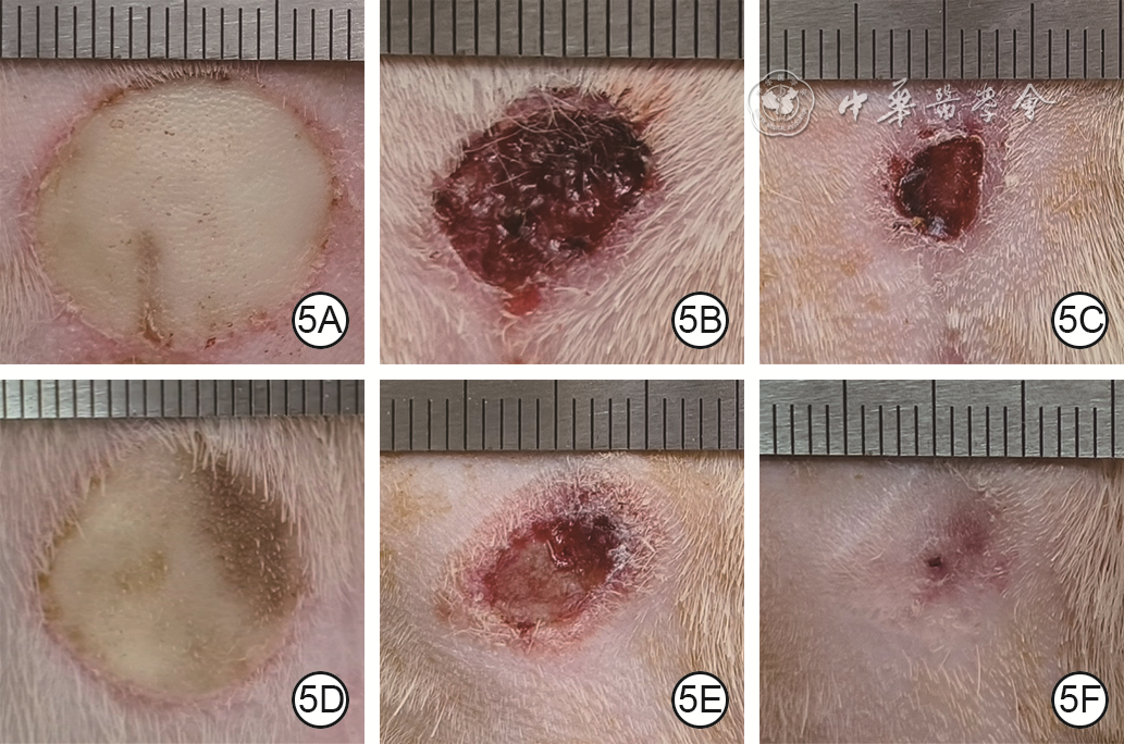
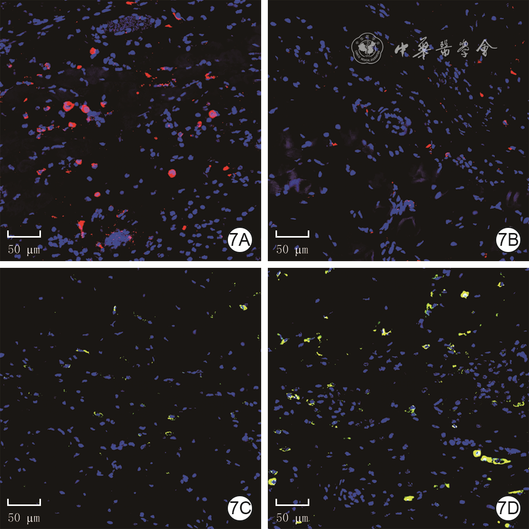
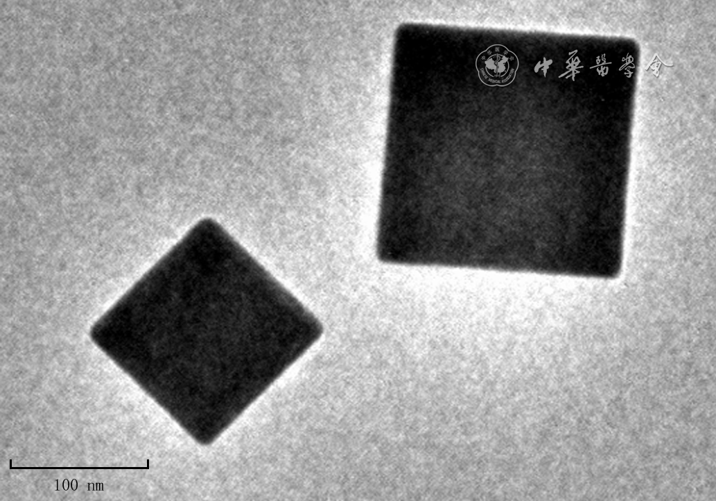
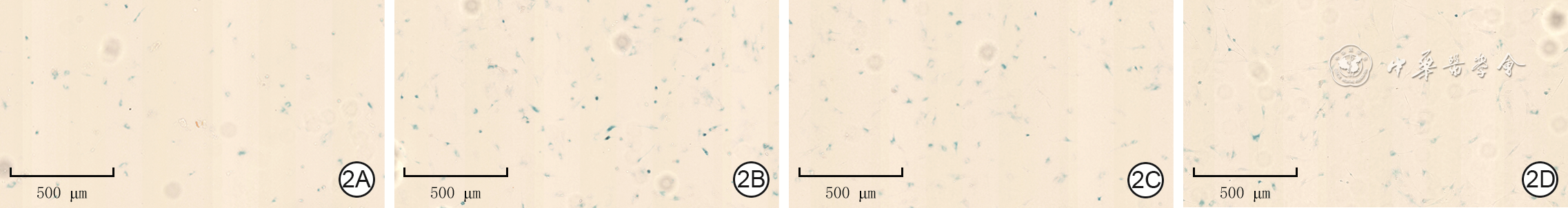
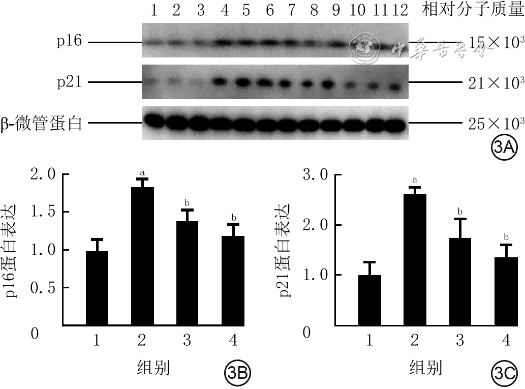
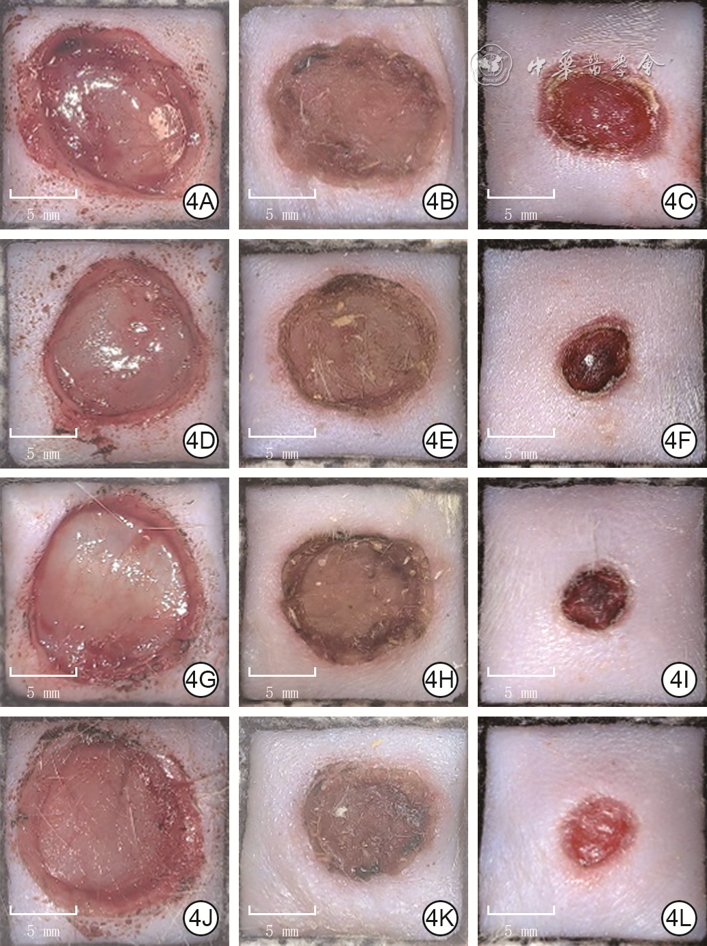
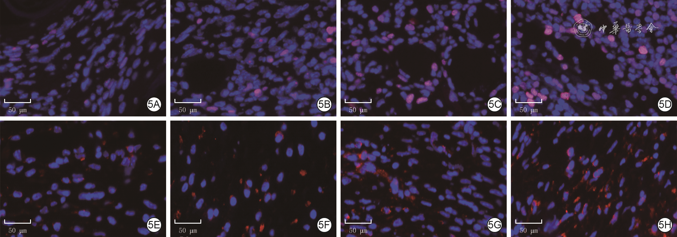
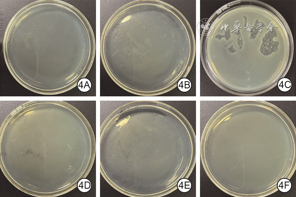
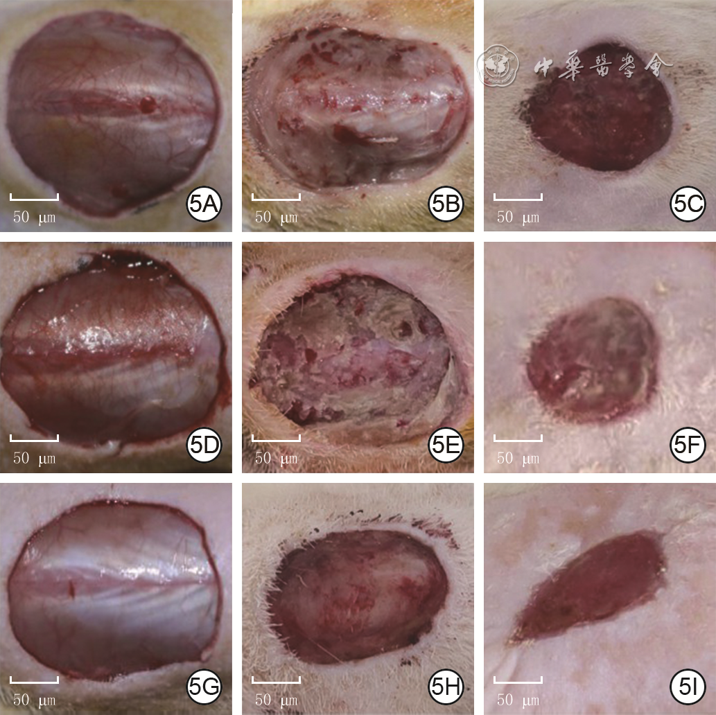
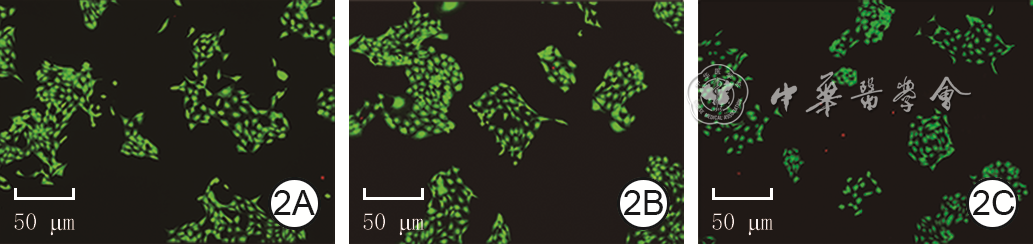
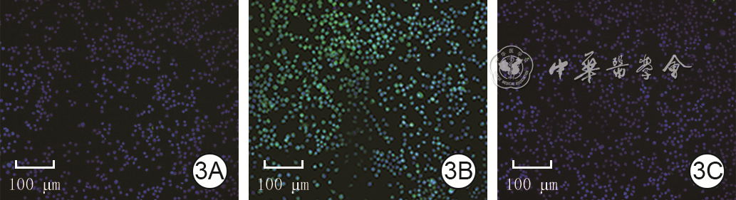
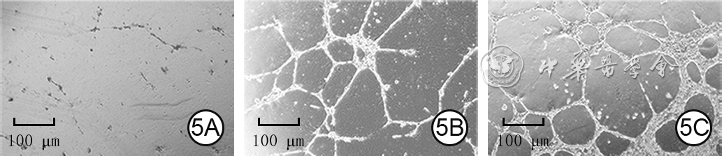
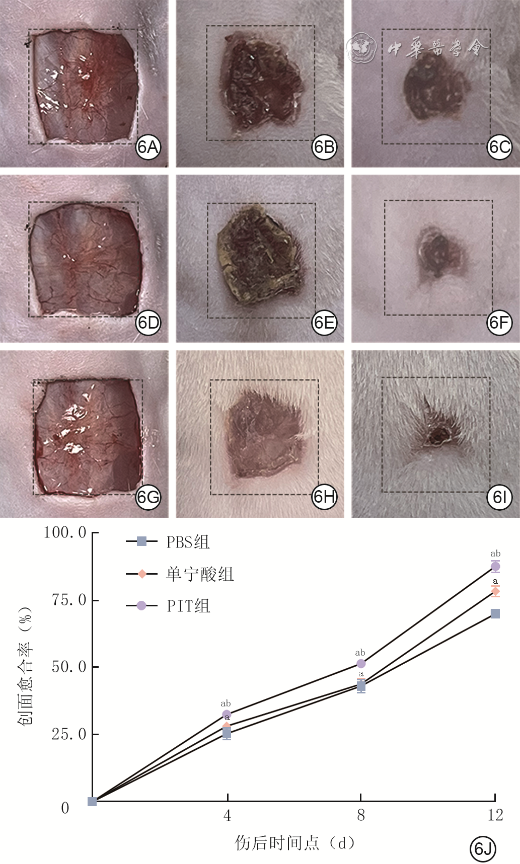
Objective To investigate the effects and mechanisms of polyvinyl alcohol/ionic liquid-tannic acid composite hydrogel (PIT) on wound healing of full-thickness skin defects in diabetic mice. Methods This study adopted a grouped design and repeated measurement design for experimental research. An ionic hydrogel matrix crosslinked by polyvinyl alcohol-4-(1H)-vinylimidazole-1-methylene benzoic acid and oxidized hyaluronic acid was prepared, and tannic acid was loaded via Cu2+ chelation to construct PIT. A 1,1-diphenyl-2-picrylhydrazyl (DPPH) solution was prepared, which was reacted with tannic acid and PIT respectively for 24 hours. An ultraviolet spectrophotometer was used to detect the DPPH radical scavenging rate. According to the random number table method (the same grouping method below), mouse macrophage RAW264.7 cells were divided into a phosphate buffered saline (PBS) group cultured with PBS, as well as a hydrogen peroxide group and a PIT group, in which cells were first treated with hydrogen peroxide for 12 hours and then respectively cultured under routine condition and with PIT. After 24 hours of culture, the 2',7'-dichlorodihydrofluorescein diacetate fluorescent probe method was adopted to detect the intracellular reactive oxygen species (ROS) level. Escherichia coli ATCC 25922, Staphylococcus aureus ATCC 25923, methicillin-resistant Staphylococcus aureus (MRSA) BNCC 337371, and human umbilical vein endothelial cells (HUVEC) were collected, and all of them were divided into PBS group, tannic acid (TA) group, and PIT group, which were cultured with PBS, TA solution, and PIT, respectively. After 12 hours of bacterial culture, the plate colony counting method was used to count bacterial colonies; after 24 hours of cell culture, the tube formation assay was performed to measure the total tube length, the number of branching nodes, and the number of branches. The sample size of all the above experiments was 3. Eighteen 8-week-old male Kunming mice were selected and divided into PBS group, TA group, and PIT group (with 6 mice in each group) to build a full-thickness skin defect wound model of diabetes. At post injury day (PID) 0 (immediately), the wounds of mice in PBS group, TA group, and PIT group were treated with PBS, TA solution, and PIT by dropping, respectively, and then the dressing was changed every day. The wound healing status was observed at PID 0, 4, 8, and 12, and the wound healing rates at PID 4, 8, and 12 were calculated. At PID 12, wound tissue was harvested. Hematoxylin-eosin staining was performed to observe the status of wound re-epithelialization and measure the thickness of newborn epithelium. Masson staining was conducted to observe the deposition of collagen fibers in wounds and calculate the proportion of collagen fiber-positive area. Results After 24 hours of reaction, the DPPH radical scavenging rate of PIT was significantly higher than that of TA (t=16.35, P<0.05). After 24 hours of culture, the ROS level of RAW264.7 cells in hydrogen peroxide group was significantly higher than that in PBS group (P<0.05), and the ROS level of RAW264.7 cells in PIT group was significantly lower than that in hydrogen peroxide group (P<0.05). After 12 hours of culture, the bacterial colony counts of Escherichia coli, Staphylococcus aureus, and MRSA in PIT group were significantly less than those in PBS group and TA group (P<0.05). After 24 hours of culture, compared with those in PBS group and TA group, the total tube length of HUVECs in PIT group was significantly prolonged, and the number of branching nodes and the number of branches increased significantly (P<0.05). From PID 0 to 12, the wounds of mice in all three groups healed gradually. At PID 4, 8, and 12, the wound healing rates of mice in PIT group were (31.6±2.0)%, (51.8±2.5)%, and (97.9±1.5)%, respectively, which were significantly higher than (18.6±0.6)%, (39.5±2.0)%, and (74.6±2.0)% in PBS group and (21.5±1.1)%, (40.7±0.8)%, and (85.3±2.1)% in TA group (P<0.05). At PID 12, the re-epithelialization of wounds in mice of PBS group was incomplete, and collagen fibers were sparsely distributed with disordered arrangement; the degree of wound re-epithelialization of mice in TA group was higher than that in PBS group, and collagen fibers were distributed in bundles with loose arrangement; the degree of wound re-epithelialization of mice in PIT group was higher than that in TA group, and collagen fibers were densely and orderly arranged in layers. At PID 12, compared with those in PBS group and TA group, the thickness of newborn epithelium in wounds of mice in PIT group increased significantly, and the proportion of collagen fiber-positive area enlarged significantly (P<0.05). Conclusions PIT relies on multiple metal ion chelation-mediated mechanisms, including antibacterial activity, antioxidant activity, and angiogenesis promotion. It significantly accelerates the wound healing of full-thickness skin defects in diabetic mice, and improves the quality of tissue repair.
Zong Yuange,Huang Wanqi,Zhang Ze,et al.Effects and mechanisms of PIT on wound healing of full-thickness skin defect in diabetic mice[J].Chin J Burns Wounds,2026,42(7):1-10.DOI: 10.3760/cma.j.cn501225-20260127-00049.


 Abstract
 PDF
PDF