2025 Vol. 41, No. 9

Guideline and Consensus
Expert consensus update on the clinical diagnosis and treatment of inhalation injury in adults (2025 edition)
2025, 41(9): 811-824. doi: 10.3760/cma.j.cn501225-20250502-00202
Abstract:
Inhalation injury (INI) is one of the common complications of burns and a major factor contributing to increased mortality and reduced quality of life of patients after burn injury. In 2018, the first expert consensus on INI was released in China to provide guidance for the clinical diagnosis and treatment of INI, however, there were still many unclear issues. With the growing demand for medical services and the emergence of high-quality clinical research in related disciplines, this consensus incorporated new clinical evidence to supplement and update certain decisions and issues in the management of INI, aiming to further standardize the management of INI and provide support for its clinical decision-making.
Expert consensus on the assessment and treatment of acute traumatic wounds in the elderly (2025 edition)
2025, 41(9): 825-839. doi: 10.3760/cma.j.cn501225-20250417-00179
Abstract:
Due to the special physiological changes in the elderly, the assessment and treatment of acute traumatic wounds in the elderly often require comprehensive consideration. Currently, there remains a dearth of unified protocols for the assessment and treatment of acute traumatic wounds in the elderly, both domestically and internationally. To address this gap, the Burns and Trauma Branch of Chinese Geriatrics Society convened a panel of national experts in related fields to form a consensus writing group. Building on relevant domestic and international research, and taking into full consideration the pathophysiological characteristics of the elderly, multiple rounds of discussions were conducted on clinical problems in the assessment and treatment of acute traumatic wounds in the elderly, and the scope of the consensus and five primary clinical questions were finally determined. Following literature retrieval and evidence evaluation, 19 recommendations were formulated after comprehensive discussions at two consensus meetings. This consensus serves as a valuable reference for professionals engaged in fields such as trauma, burns, and wound repair during their routine clinical practice, aiming to enhance the treatment level for acute traumatic wounds in the elderly in China.
Expert Forum
Intelligent intensive care unit construction and severe burn management: integration and innovation
Zhou Feihu, Mao Zhi
2025, 41(9): 840-846. doi: 10.3760/cma.j.cn501225-20250715-00307
Abstract:
The development of intelligent intensive care unit (ICU) has promoted the optimal treatment of severely burned patients. Intelligent ICUs, based on the Internet of Things technology, enable automatic data acquisition and sharing through device connection, integrate multimodal burn data and carry out prediction and evaluation of diseases, and promote remote monitoring and treatment. Faced with challenges such as the complexity and diversity of severe burn patients' conditions, insufficient model interpretability, and inadequate data security, the future construction of intelligent burn ICUs will still require continuous exploration and innovation in areas including software and hardware infrastructure, multimodal information integration, intelligent predictive modeling, virtual reality-assisted diagnosis and treatment, telemedicine linkage, clinical guideline updates, and multi-disciplinary integration.
Original Article · Severe Burns
Roles and mechanism of mitophagy in myocardial injury in mice following delayed resuscitation after severe burns
Zhang Zejin, Li Dawei, Liu Liwei, Jia Hao, Wei Wenhui, Li Meizhuo, Li Duo, Shen Chuan'an
2025, 41(9): 847-856. doi: 10.3760/cma.j.cn501225-20250514-00226
Abstract:
  Objective  To explore the roles and mechanism of mitophagy in myocardial injury in mice following delayed resuscitation after severe burns.  Methods  This study was an experimental study. Forty-five 6-8-week-old male C57BL/6J mice were divided into sham injury group, 0 h post-injury rehydration group, 3 h post-injury rehydration group, 6 h post-injury rehydration group, and no post-injury rehydration group, with 9 mice in each group, according to the random number table (the same grouping method below). The mice in sham injury group were subjected to a sham injury, while the mice in the remaining 4 groups were subjected to full-thickness scald (hereinafter referred to as burn) of 30% total body surface area. The mice in 0 h post-injury rehydration group, 3 h post-injury rehydration group, and 6 h post-injury rehydration group received rehydration starting from 0 (immediately), 3, and 6 h post-injury, respectively. The mice in sham injury group and no post-injury rehydration group did not receive rehydration after injury. At 12 h post-injury, the serum levels of lactate dehydrogenase (LDH) and creatine kinase isoenzyme (CK-MB) were detected using an automated biochemical analyzer. Myocardial histopathological scoring was performed using Kishimoto scores after hematoxylin-eosin staining. The protein expressions of mitophagy-related proteins, including translocase of outer mitochondrial membrane 20 (TOM20), microtubule-associated protein 1 light chain 3B (LC3B), P62, and Beclin-1 in myocardial tissue were detected by Western blotting, and the ratio of LC3B-Ⅱ/LC3B-Ⅰ was calculated. Twenty-seven 6-8-week-old male C57BL/6J mice were divided into 6 h post-injury rehydration alone group, 6 h post-injury rehydration+3-methyladenine (3-MA) group, and 6 h post-injury rehydration+rapamycin group, with 9 mice in each group. One week before burn modeling, mice in 6 h post-injury rehydration+3-MA group and 6 h post-injury rehydration+rapamycin group were injected intraperitoneally with the mitophagy inhibitor 3-MA or mitophagy activator rapamycin. All the mice in 3 groups received rehydration starting from 6 h post-injury following severe burns. At 12 h post-injury, the serum levels of LDH and CK-MB were detected, myocardial histopathological scoring was performed, and the protein expressions of mitochondrial autophagy-related proteins were detected as before. The mitochondrial ultrastructure changes in myocardial tissue were observed under a transmission electron microscope after uranium-lead double staining, and Flameng scoring was performed.  Results  At 12 h post-injury, compared with those in 0 h post-injury rehydration group the serum levels of LDH and CK-MB in mice in 6 h post-injury rehydration group and no post-injury rehydration group were significantly increased (P<0.05). At 12 h post-injury, compared with that in 0 h post-injury rehydration group, the myocardial histopathological score in mice in sham injury group was significantly decreased (P<0.05), and the myocardial histopathological scores in mice in 3 h post-injury rehydration group, 6 h post-injury rehydration group, and no post-injury rehydration group were significantly increased (P<0.05). At 12 h post-injury, compared with those in 0 h post-injury rehydration group, the protein expression of Beclin-1 was significantly decreased (P<0.05), while the protein expressions of TOM20 and P62 were significantly increased (P<0.05) in myocardial tissue of mice in sham injury group; the protein expression of Beclin-1 was significantly increased (P<0.05), while the protein expressions of TOM20 and P62 were significantly decreased (P<0.05) in myocardial tissue of mice in 3 h post-injury rehydration group; the protein expressions of Beclin-1 and ratios of LC3B-Ⅱ/LC3B-Ⅰwere significantly increased (P<0.05), while the protein expressions of TOM20 and P62 were significantly decreased (P<0.05) in myocardial tissue of mice in 6 h post-injury rehydration group and no post-injury rehydration group. At 12 h post-injury, compared with those in 6 h post-injury rehydration alone group, the serum levels of LDH and CK-MB in mice in 6 h post-injury rehydration+3-MA group were significantly decreased (P<0.05), while the serum levels of LDH and CK-MB in mice in 6 h post-injury rehydration+rapamycin group were significantly increased (P<0.05). At 12 h post-injury, compared with that in 6 h post-injury rehydration alone group, the myocardial histopathological score in mice in 6 h post-injury rehydration+3-MA group was significantly decreased (P<0.05), while the myocardial histopathological score in mice in 6 h post-injury rehydration+rapamycin group was significantly increased (P<0.05). At 12 h post-injury, compared with those in 6 h post-injury rehydration alone group, the protein expression of Beclin-1 and ratio of LC3B-Ⅱ/LC3B-Ⅰ were significantly decreased (P<0.05), while the protein expressions of TOM20 and P62 were significantly increased (P<0.05) in myocardial tissue of mice in 6 h post-injury rehydration+3-MA group; the protein expression of Beclin-1 and ratio of LC3B-Ⅱ/LC3B-Ⅰwere significantly increased (P<0.05), while the protein expressions of TOM20 and P62 were significantly decreased (P<0.05) in myocardial tissue of mice in 6 h post-injury rehydration+rapamycin group. At 12 h post-injury, compared with 2.67±0.11 in 6 h post-injury rehydration alone group, the mitochondrial Flameng score (2.07±0.11) in myocardial tissue of mice in 6 h post-injury rehydration+3-MA group was significantly decreased (P<0.05), while the mitochondrial Flameng score (3.60±0.20) in myocardial tissue of mice in 6 h post-injury rehydration+rapamycin group was significantly increased (P<0.05).  Conclusions  Delayed rehydration leads to myocardial injury and excessive activation of mitophagy in mice with severe burns. The inhibition of mitophagy can mitigate myocardial injury caused by delayed rehydration after severe burns.
Multicenter retrospective analysis of early coagulation characteristics and risk factors for prognosis of adult patients with severe burns
Shen Tuo, Ma Qimin, Chu Zhigang, Zhang Yi, Tang Wenbin, Cui Pei, Li Xiaoliang, Chang Liu, Chen Zhaohong, Chang Fei, Liu Yongji, Wu Choulang, Guo Guanghua, Zhu Feng
2025, 41(9): 857-866. doi: 10.3760/cma.j.cn501225-20250530-00250
Abstract:
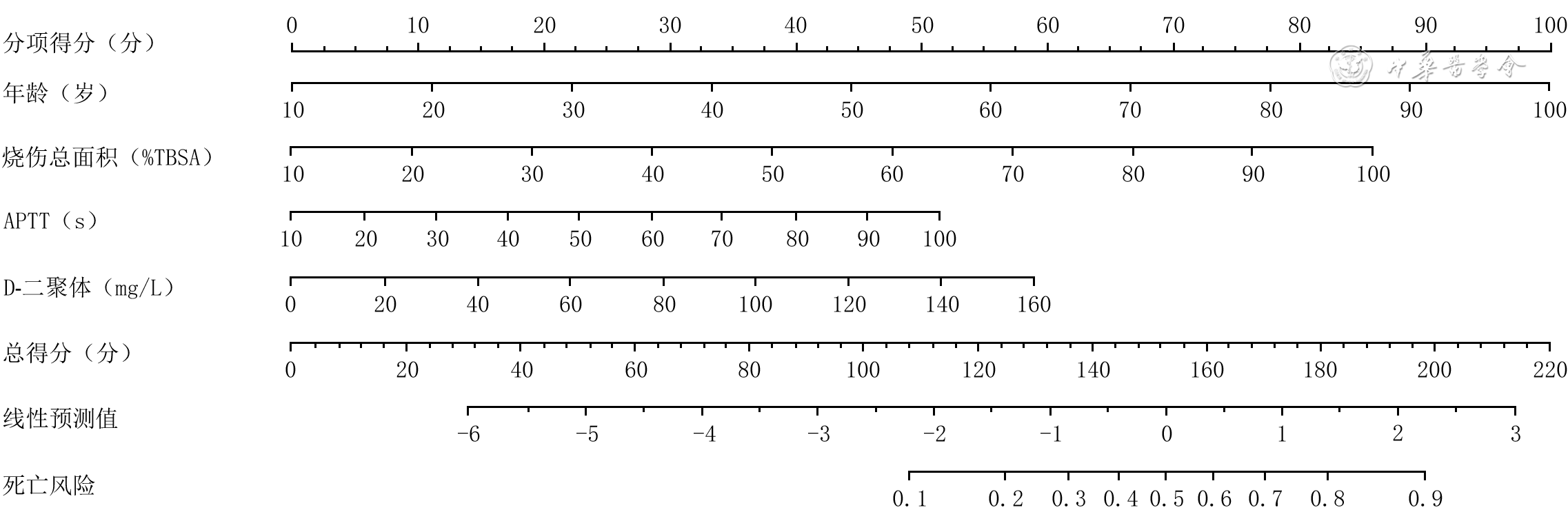
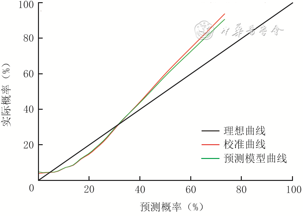
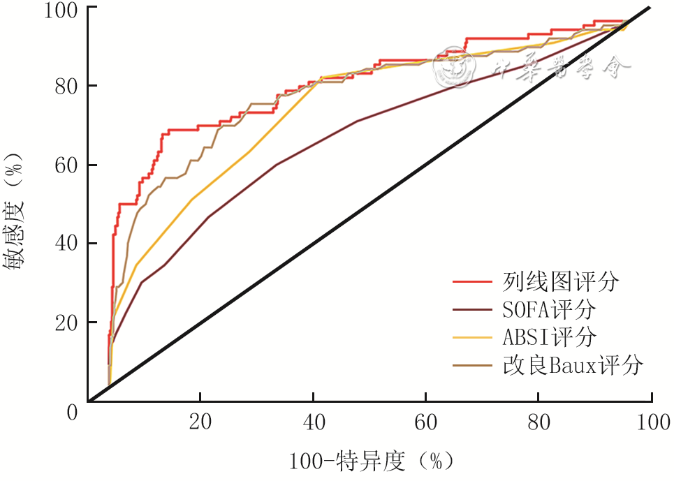
  Objective  To investigate the early coagulation characteristics and risk factors for prognosis of adult patients with severe burns.  Methods  This study was a retrospective study of case series. A total of 583 adult patients with severe burns who met the inclusion criteria were admitted to the 12 hospitals in China from January 2015 to December 2020, including 75 cases from the First Affiliated Hospital of Naval Medical University, 64 cases from Tongren Hospital of Wuhan University & Wuhan Third Hospital, 48 cases from the Affiliated Hospital of Nantong University, 146 cases from the Guangzhou Red Cross Hospital of Jinan University, 55 cases from the 924th Hospital of PLA, 46 cases from Zhengzhou First People's Hospital, 35 cases from the Fourth People's Hospital of Dalian, 20 cases from Fujian Medical University Union Hospital, 18 cases from Zhangjiagang First People's Hospital, 7 cases from Heilongjiang Provincial Hospital, 18 cases from Taizhou Hospital of Zhejiang Province, and 51 cases from the First Affiliated Hospital of Nanchang University. The general clinical characteristics (including gender, age, total burn area, degree of inhalation injury, and cause of burn) and coagulation function indicators within 24 hours after injury (including prothrombin time (PT), activated partial thromboplastin time (APTT), thrombin time (TT), international normalized ratio (INR), and D-dimer, fibrinogen (FIB), platelets (PLT), and serum total calcium levels) of patients were statistically analyzed. Based on the data of the above indicators, the sepsis-related organ failure assessment (SOFA) score, modified Baux score, and abbreviated burn severity index (ABSI) score of patients were calculated. According to the survival outcome within 28 days after injury, the patients were divided into survival group (499 cases) and death group (84 cases). General clinical characteristics and coagulation function indicators were compared between the 2 groups of patients. According to the severity of burns, patients were divided into three groups: severe burn group (185 cases ) with a total burn area <50% total body surface area (TBSA), extremely severe burn group (251 cases) with a total burn area ≥50% and <80%TBSA, and critical burn group (147 cases) with a total burn area ≥80%TBSA. Coagulation function indicators were compared among the 3 groups of patients. Based on the specific time of admission at the early stage of burns, patients with admission time <2 h, ≥2 h and <4 h, ≥4 h and <8 h, ≥8 h and <16 h, and ≥16 h and <24 h after burns were divided respectively into early burn group 1 (207 cases), early burn group 2 (158 cases), early burn group 3 (129 cases), early burn group 4 (54 cases), and early burn group 5 (35 cases), and the coagulation function indicators were compared among the 5 groups of patients. The independent risk factors affecting mortality 28 days after injury in 583 adult patients with severe burns were screened. A predictive nomogram was constructed based on the independent risk factors. Receiver operating characteristic (ROC) curves were constructed and the area under the curves (AUCs) was calculated for prediction models based on SOFA score, ABSI score, and modified Baux score. The AUC based on the nomogram score was compared with those of the aforementioned prediction models. The efficacy of the aforementioned prediction models was assessed.  Results  Among the 583 patients, there were 409 males and 174 females, aged 18-97 years, with total burn area of 60.00% (44.50%, 80.00%) TBSA and 138 patients with moderate-to-severe inhalation injury. Compared with those in survival group, patients in death group had larger age, total burn area, and INR, longer PT and APTT, higher D-dimer and PLT levels, and proportion of moderate-to-severe inhalation injury (with Z values of 6.47, 7.48, 3.48, 2.89, 2.79, 5.33, and 2.59, respectively, χ2=11.30, all P values<0.05). Only D-dimer level in the 2 group of patients was above the upper limit of the normal range. There were statistically significant differences among severe burn group, extremely severe burn group, and critical burn group of patients in terms of PT, APTT, INR, and D-dimer, PLT, serum total calcium levels (with H values of 17.85, 19.78, 26.89, 52.64, 14.21, and 12.90, respectively, P<0.05). There were statistically significant differences among early burn group 1, early burn group 2, early burn group 3, early burn group 4, and early burn group 5 of patients in terms of PT, APTT, INR, and D-dimer, FIB, PLT, serum total calcium levels (with H values of 29.66, 60.13, 25.51, 28.24, 14.38, 11.41, and 42.96, respectively, P<0.05). Multivariate logistic regression analysis showed that age, total burn area, APTT, and D-dimer level were independent risk factors for mortality 28 days after injury in adult patients with severe burns (with odds ratios of 1.056, 1.048, 1.029, and 1.018, respectively, 95% cofidence intervals (CIs) of 1.036-1.076, 1.033-1.063, 1.005-1.053, and 1.002-1.035, respectively, P<0.05). The ROC curves showed that the AUCs of the predictive models based on the SOFA score, ABSI score, and modified Baux score were 0.66, 0.76, and 0.80, respectively, with 95% CIs of 0.61-0.75, 0.71-0.82, and 0.74-0.86, respectively, which were all lower than 0.81 (95% CI of 0.76-0.87) of the nomogram score-based predictive model. The DeLong test showed that the predictive ability of the nomogram score-based model for predicting mortality risk in adult patients with severe burns 28 days after injury was significantly better than those of the models based on the SOFA score and ABSI score (both P values<0.05), but similar to that of the prediction model based on the modified Baux score (P>0.05).  Conclusions  In the early stage of adult patients with severe burns, only D-dimer level was above the upper limit of the normal range. Age, total burn area, APTT, and D-dimer level are all independent risk factors for mortality in adult patients with severe burns 28 days after injury.
Original Article
Effects of the pretreatment of human umbilical cord mesenchymal stem cells with tumor necrosis factor-α and interleukin-1β on the healing of ischemic wounds in rats
Liu Zhiyuan, Pan Wenjie, Wang Zhen, Shu Yue, Chen Siyu, Qi Fang, Xu Guangchao, Wei Zairong, Deng Chengliang
2025, 41(9): 867-876. doi: 10.3760/cma.j.cn501225-20241008-00377
Abstract:
  Objective  To investigate the effects of the pretreatment of human umbilical cord mesenchymal stem cells (hUMSCs) with tumor necrosis factor-α (TNF-α) and interleukin-1β (IL-1β) on the healing of ischemic wounds in rats.  Methods  This study was an experimental research. The umbilical cord tissue was collected from three healthy full-term neonates delivered by cesarean section from delivery women aged 22-35 years in the Affiliated Hospital of Zunyi Medical University between December 2022 and April 2023. The hUMSCs were isolated, and after the scratch test, the cells were divided into pretreatment group pretreated with TNF-α and IL-1β of both final mass concentrations of 20 ng/mL and control group cultured conventionally according to the random number table method (the same grouping method below). The cell migration rates were calculated at 8, 16, and 32 h after scratch, with sample size of 3. Five 9-week-old male Sprague-Dawley (SD) rats were used to construct a bipedicled flap with an area of 11.0 cm×2.5 cm on the back, two ischemic wounds with diameter of 0.6 cm were created on the flaps, and two non-ischemic wounds with diameter of 0.6 cm were created on normal dorsal skin. The wound healing rates were calculated at 6, 12, and 18 d after modeling to validate the reliability of the ischemic wound model. Another 15 male SD rats aged 9 weeks were subjected to the same ischemic wound model as above and divided into pretreated hUMSC group, conventional hUMSC group, and phosphate-buffered saline (PBS) group, with 5 rats in each group, which were respectively injected with pretreated hUMSCs as before, conventionally cultured hUMSCs, and PBS at the wound margin. The wound healing rates were calculated at 3, 6, 9, and 12 d after injection. At 12 d after injection, the wound tissue was collected for hematoxylin-eosin staining to assess epithelialization of wounds, for Masson staining to observe collagen fiber formation in wound tissue, and for immunofluorescence staining to detect the number of blood vessels, arginase-1 (Arg1)-positive cells, and inducible nitric oxide synthase (iNOS)-positive cells in wound tissue.  Results  At 16 and 32 h after scratch, the cell migration rates in pretreatment group were significantly higher than those in control group (with Z values of -2.61 and -2.61, respectively, P<0.05). At 6, 12, and 18 d after modeling, the healing rates of non-ischemic wounds of rats were significantly higher than those of ischemic wounds (with Z values of 2.61, 2.79, and 2.79, respectively, P<0.05). At 6, 9, and 12 d after injection, the wound healing rates in pretreated hUMSC group of rats were 6.83% (3.13%, 12.22%), 66.83% (60.09%, 95.68%), and 96.98% (91.67%, 99.80%), respectively, which were significantly higher than 1.54% (0.30%, 3.47%), 1.54% (0.60%, 5.90%), and 3.08% (0.90%, 7.64%) in PBS group (with Z values of 7.60, 9.40, and 10.00, respectively, P<0.05). At 9 d after injection, the wound healing rate in pretreated hUMSC group of rats was significantly higher than 5.86% (4.72%, 8.12%) in conventional hUMSC group (Z=5.60, P<0.05). At 12 d after injection, the wound epithelialization basically completed in pretreated hUMSC group of rats, and the collagen fiber formation in wound tissue was markedly better than that in conventional hUMSC group and PBS group. At 12 d after injection, the number of blood vessels and Arg1-positive cells in wound tissue in pretreated hUMSC group of rats was significantly more than that in conventional hUMSC group and PBS group (P<0.05), and the number of blood vessels and Arg1-positive cells in wound tissue in conventional hUMSC group of rats was significantly more than that in PBS group (P<0.05); the number of iNOS-positive cells in pretreated hUMSC group of rats was significantly less than that in conventional hUMSC group and PBS group (with both P values <0.05), and the number of iNOS-positive cells in conventional hUMSC group of rats was significantly less than that in PBS group (P<0.05).  Conclusions  The pretreatment of hUMSCs with TNF-α and IL-1β can enhance the abilities of migration, immunomodulation, and promoting angiogenesis of the cells, thereby promoting the healing of ischemic wounds.
Clinical application effect of vascular imaging technology in repairing limb wounds with free transplantation of superficial circumflex iliac artery perforator flaps
Zhang Lu, Li Dong, Fan Anwei
2025, 41(9): 877-886. doi: 10.3760/cma.j.cn501225-20250228-00100
Abstract:
  Objective  To investigate the clinical application effect of vascular imaging technology in repairing limb wounds with free transplantation of superficial circumflex iliac artery (SCIA) perforator flaps.  Methods  This study was a retrospective cohort study. Forty patients (26 males and 14 females, aged 19-60 years) with skin and soft tissue defects of the limbs who met the inclusion criteria were admitted to Xingtai General Hospital of North China Medical Health Group from March 2019 to August 2023. According to the exploration method for blood vessels in the flap area to be cut, the patients were divided into color Doppler flow imaging (CDFI) group with CDFI exploration and three-dimensional computed tomography angiography (3D-CTA) group with 3D-CTA exploration, with 20 cases in each group. Preoperatively, based on the CDFI results, the vessel diameter at the origin and the length of SCIA and superficial circumflex iliac vein (SCIV) were measured in CDFI group of patients, and the resulting values were recorded as the estimated values. Preoperatively, the vessel diameter at the origin and the length of SCIA and SCIV were measured in 3D-CTA group of patients based on the 3D-CTA results, and the resulting values were recorded as the estimated values; the anatomic relationship between the SCIA and the deep circumflex iliac artery as well as the superficial inferior epigastric artery in the inguinal region was identified, and the SCIA perforators were classified into 5 types (type 1, 2, 3, 4, and 5) based on this. The wound area of the limbs ranged from 3.0 cm×3.0 cm to 16.0 cm×8.0 cm after debridement. SCIA perforator flaps were designed and harvested to repair the wounds. During surgery, the preoperatively located perforator vessels (detected by 3D-CTA or CDFI) were identified, the anatomic relationship between the SCIA and the deep circumflex iliac artery as well as the superficial inferior epigastric artery was observed, the actual vessel diameter at the origin and the length of the SCIA and SCIV were measured, and the resulting values were recorded as the actual measured values. The area of the harvested flaps ranged from 4.0 cm×4.0 cm to 17.0 cm×9.0 cm. The wounds at donor sites were closed directly. In 3D-CTA group, the SCIA perforators classification of patients identified by preoperative 3D-CTA technology was statistically analyzed and compared with the intraoperative findings. The estimated values of vessel diameter at the origin and the length of SCIA and SCIV detected by either CDFI or 3D-CTA in the two groups of patients were recorded and compared with the actual values measured intraoperatively. Postoperatively, the flap survival was observed in the two groups of patients. During follow-up, flap appearance and sensory recovery were evaluated in the two groups of patients, and the wounds healing at donor site was observed.  Results  Preoperative 3D-CTA exploration in the 3D-CTA group of patients showed the SCIA perforators were type 1 in 6 cases, type 2 in 5 cases, type 3 in 4 cases, type 4 in 3 cases, and type 5 in 2 cases, which was consistent with the results in intraoperative observation. Compared with those in CDFI group, the estimated values of vessel diameter at the origin of SCIA and SCIV in 3D-CTA group of patients were significantly smaller (with t values of 2.83 and 2.23, respectively, P<0.05). Compared with the estimated values, the actual measured values of vessel diameter at the origin of SCIA and SCIV in CDFI group of patients were significantly smaller (with t values of 3.12 and 2.16, respectively, P<0.05), and the mean differences between the groups (95% confidence intervals) were 0.25 (0.12-0.38) and 0.26 (0.14-0.38) mm, respectively. Compared with those in CDFI group, the estimated values of vessel length of SCIA and SCIV in 3D-CTA group of patients were significantly shorter (with t values of 2.43 and 2.18, respectively, P<0.05). Compared with the estimated values, the actual measured values of vessel length of SCIA and SCIV in CDFI group of patients were significantly shorter (with t values of 2.06 and 2.10, respectively, P<0.05), and the mean differences within groups (95% confidence intervals) were 1.5 (1.0-2.0) and 1.5 (1.0-2.0) cm, respectively. In CDFI group, 2 patients developed partial flap necrosis postoperatively, which healed well after dressing change, and the remaining patients exhibited satisfactory flap survival. All flaps in 3D-CTA group of patients survived completely. During postoperative follow-up of 12 to 18 months, both groups of patients showed favorable flap appearance and sensory functional recovery. The wounds in donor sites healed well, with only linear scars remaining.  Conclusions  Preoperative application of 3D-CTA technology for vascular exploration can clarify the type of SCIA perforators and the vessel diameter at the origin and the length of SCIA, thereby guiding the design of the SCIA perforator flap and enabling its precise harvest.
Effects of microneedle injection of rat epidermal stem cells on wound healing of full-thickness skin defects in rats
Wang Xue, Cheng Wei, Liu Xiaojun, Wang Shunsheng, Wu Zhuofan, Su Jiandong
2025, 41(9): 887-894. doi: 10.3760/cma.j.cn501225-20241031-00427
Abstract:
  Objective  To investigate the effects of microneedle injection of rat epidermal stem cells (ESCs) on wound healing of full-thickness skin defects in rats.  Methods  This study was an experimental study. The hollow silicon-based microneedle injection device was developed successfully by utilizing laser-etching technology. ESCs were isolated and cultured from the skin of two 2-day-old female Sprague-Dawley rats and identified successfully by using flow cytometry and immunofluorescence method. A total of 18 eight-week-old female Sprague-Dawley rats were divided into control group, spray group, and microneedle group according to the random number table method, with 6 rats in each group. All rats were inflicted with a full-thickness skin defect wound on the dorsum. On day 0 (immediately after injury), the wounds in control group and spray group were sprayed with 0.5 mL phosphate-buffered solution and 0.5 mL rat ESCs suspension, respectively, and the wounds in microneedle group of rats were injected with 0.5 mL rat ESCs suspension through the hollow silicon-based microneedle injection device. On days 3, 7, and 14 after injury, the wound healing was observed and the wound healing rate was calculated. On day 14 after injury, the wound tissue of rats were harvested for hematoxylin and eosin staining to observe the epithelialization of wounds, for Masson staining to detect the collagen fiber content in wound tissue, and for immunohistochemical staining to detect the neovascular number in wound tissue.  Results  Within 14 days after injury, the wounds in three groups of rats progressively healed. On day 3 after injury, there was no statistically significant difference in the overall comparison of the wound healing rate among the three groups of rats (P>0.05). On days 7 and 14 after injury, the wound healing rates in microneedle group of rats were (77.0±4.6)% and (95.0±2.1)%, respectively, which were significantly higher than (66.5±8.6)% and (88.3±3.1)% in spray group and (44.5±5.7)% and (78.8±6.3)% in control group (P<0.05); the wound healing rates in spray group of rats were significantly higher than those in control group (P<0.05). On day 14 after injury, the degree of wound epithelialization in the microneedle group of rats was higher than that in spray group and control group, and hair follicles developed in the newly regenerated skin tissue. On day 14 after injury, the collagen fiber content in the wound tissue in microneedle group and spray group of rats was significantly higher than that in control group (P<0.05), and the collagen fiber content in the wound tissue in microneedle group of rats was significantly higher than that in spray group (P<0.05); the neovascular number in the wound tissue in microneedle group and spray group of rats was significantly more than that in control group (P<0.05), and the neovascular number in the wound tissue in microneedle group of rats was significantly more than that in spray group (P<0.05).  Conclusions  ESCs can promote neovascularization and collagen fiber formation in the wound tissue, thereby accelerating the wound healing and improve the quality of wound healing in rats with full-thickness skin defects. Injecting rat ESCs suspension through hollow silicon-based microneedle injection device shows better wound healing effects than using conventional approaches, making it an effective cell delivery method.
Clinical effects of superior epigastric artery perforator fascial flaps in repairing huge chest wall wounds after breast tumor surgery
Yuan Ying, Zhou Min, Qi Baiwen, Yu Aixi, Jian Chao, Wu Yifan
2025, 41(9): 895-901. doi: 10.3760/cma.j.cn501225-20240930-00367
Abstract:
  Objective  To investigate the clinical effects of superior epigastric artery perforator fascial flaps in repairing huge chest wall wounds after breast tumor surgery.  Methods  This study was a retrospective observational research. From July 2018 to June 2024, 8 patients with breast tumor were admitted to Zhongnan Hospital of Wuhan University. The areas of skin and soft tissue defects of chest wall caused by extended resection of tumor reached 20 cm×18 cm to 24 cm×20 cm, and all of them were repaired by superior epigastric artery perforator fascial flaps. The flaps were excised along the surface of the rectus abrectus muscle on the same side of the chest wall defects, with the harvested flap area of 20 cm×8 cm to 18 cm×10 cm. The inferior abdominal vessels and their large perforators were retained at the distal end of the flaps. The inferior abdominal wall vessels at the distal end of the flaps were anastomosed with the internal thoracic vascular branches in the recipient areas in 4 patients, while no vessels were anastomosed in the other 4 patients. The wounds in the donor areas were sutured directly in all patients. The survival of the flaps, and the healing of the wounds and the occurrence of complications in the donor and recipient areas were observed after the operation. Postoperative follow-up was conducted to observe the thoracic tumor recurrence, the flap appearance, and the occurrence of complications in the donor area.  Results  The flaps in all patients survived well, without occurrence of vascular crisis. The scattered ecchymosis appeared at the distal end of the flaps in two patients after surgery, which did not progress to flap necrosis after symptomatic and supportive treatment, and the ecchymosis subsided within 4 weeks after surgery. All the wounds in the donor and recipient areas healed without complications. The follow-up for 3 to 6 months showed no local recurrence of the tumor, satisfactory appearance of the flap, mild linear scar hyperplasia left in the donor area, and no abdominal hernia or abdominal wall weakness.  Conclusions  The superior abdominal artery perforator fascial flap is simple in resection operation, has a high survival rate, causes less damage to the donor area, and can effectively repair the huge skin and soft tissue defect wounds of the chest wall after operation of breast tumor.
Contention
2025, 41(9): 902-904. doi: 10.3760/cma.j.cn501225-20250223-00081
Abstract:
Review
Research advances on the application of fibroblasts and their derivatives in the treatment of diabetic foot ulcers
Wang Xinlu, Gao Shaoying, Wei Zairong
2025, 41(9): 905-910. doi: 10.3760/cma.j.cn501225-20241204-00475
Abstract:
Diabetic foot ulcer (DFU) is one of the serious complications of diabetes mellitus, which is characterized by susceptibility to infection, difficult to cure, and easy to relapse. At present, the treatment methods for DFUs include standard nursing care, surgical treatment, and medication, but the clinical efficacy is suboptimal. Fibroblasts (Fbs), as one of the key cells for wound healing, can not only secrete fibronectin and collagen to construct scaffolds for cell migration, proliferation, and colonization of keratinocytes and epithelial cells, but also secrete inflammatory factors, growth factors, and cytokines to reverse the chronic inflammation of DFUs, promote cell proliferation, and improve the wound microenvironment. It is one of the cell types currently used in DFU wound treatment. This article reviews the application of Fbs and their derivatives in DFU treatment from the perspective of Fbs, in order to provide a reference for DFU treatment.