2025 Vol. 41, No. 10

Expert Forum
Researches and clinical translation of smart dressings
Lyu Guozhong, Zhao Peng
2025, 41(10): 911-917. doi: 10.3760/cma.j.cn501225-20250807-00348
Abstract:
The research and application of smart dressings are a focal point in fields such as burn and wound repair. Breakthroughs in miniaturized electronic components, fiber optics and wireless communication technologies, and flexible circuit, and the research and development of novel biomaterials have laid foundation for the development and application of smart dressings. Real-time, rapid-response, multifunctional smart dressings are emerging, showing great potential in aspects such as early detection of wound infections, assessment of inflammatory responses, monitoring changes in wound characteristics, and estimating wound healing direction and duration. They also provide opportunities for precise and controlled local drug delivery to wounds and microcurrent stimulation therapy. This article introduces the classification, research progress, and clinical trials of smart dressings, and analyzes the functions, advantages, and limitations of sensor-based smart dressings by categorizing them into active and passive types. Additionally, this article introduces three types of broadly defined, non-sensor-based smart dressings that can identify changes in specific wound parameters and respond accordingly such as changing color, releasing drugs, or exhibiting microcurrent. Based on this, the article also provides an outlook on the future of smart dressings.
Wound repair strategies of natural polysaccharide hydrogels based on microenvironmental regulation
Xu Fujian, Li Yang
2025, 41(10): 918-927. doi: 10.3760/cma.j.cn501225-20250722-00316
Abstract:
The efficiency of wound repair and the function of regenerated tissue are governed by the precise regulation of the wound microenvironment. At the physiological level, wounds undergo intricate cascades of signaling events, including persistent amplification of inflammation, remodeling of cytokine networks, disruption of redox homeostasis, and succession of microbial communities. The spatiotemporal dynamics of these signals directly determine the course and quality of wound repair. At the physical level, impaired exudate management, compromised microbial barriers, stress concentration at wound edges, and local thermal imbalance further exacerbate tissue injury and delay wound repair. Conventional wound therapies often fail to address the individualized features and dynamic pathological changes of wounds, underscoring the urgent need for a new generation of intelligent dressings capable of real-time sensing pathological signals and adaptive modulation of the wound microenvironment. Natural polysaccharides, characterised by their structural diversity and modifiability, provide a versatile design space for constructing multifunctional hydrogels. The integration of these platforms with exogenous stimulus-responsive platforms further expands the functional potential of wound dressings. This article systematically reviews the latest advances in precise regulation of wound microenvironment by natural polysaccharide hydrogels, focusing on the intelligent response mechanisms to physiological microenvironment and the functional design strategies for physical microenvironment. It aims to provide theoretical guidance for the rational design and clinical application of natural polysaccharide hydrogels.
Research on the pathogenesis and treatment of diabetic foot ulcer wounds
Wang Xin, Wang Yinyu, Qi Jianwu
2025, 41(10): 928-936. doi: 10.3760/cma.j.cn501225-20250124-00039
Abstract:
With the improvement of living standards and dietary conditions of civilians, the incidence of diabetes mellitus is also increasing gradually. Diabetic foot ulcers (DFU), a serious complication of diabetes mellitus, are associated with high rates of amputation and mortality, placing huge psychological and economic burdens on patients. The complex pathogenesis and difficult-to-heal wounds pose significant challenges to the prognosis of DFU patients. This article will elaborate on the pathogenesis of DFU wounds by integrating the latest research progress at home and abroad and summarize the current treatment schemes for different types of DFU wounds, aiming to provide guidance for clinical treatment of DFU wounds.
Original Article·Advanced Materials and Wound Repair
Effects of mitochondrial transplantation on full-thickness skin defects in diabetic rats
Li Yuqian, Zhang Tingting, Zou Guilian, Zhou Haowei, Zhou Sitong, Zhao Hanwen, Guo Baolin, Li Jing
2025, 41(10): 937-948. doi: 10.3760/cma.j.cn501225-20250721-00315
Abstract:
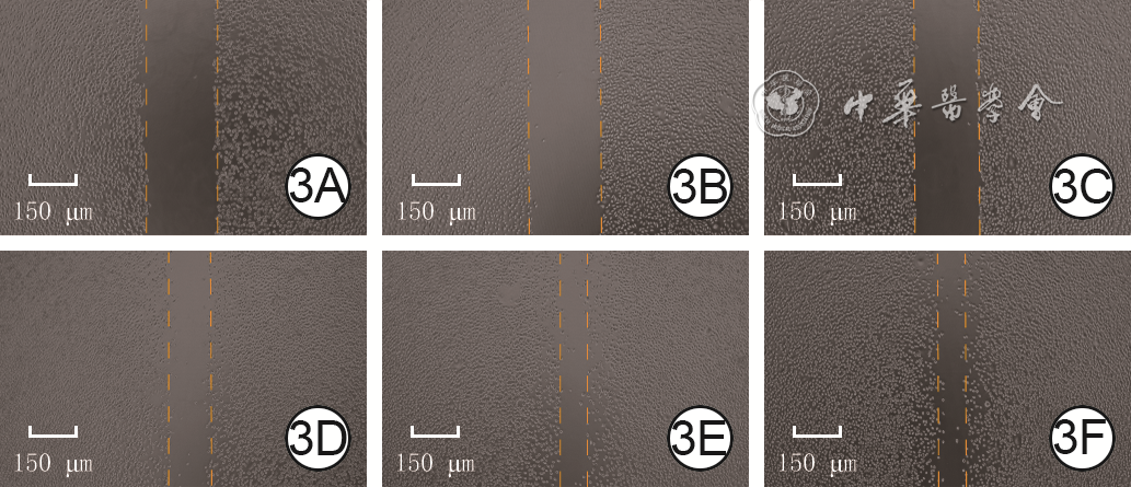
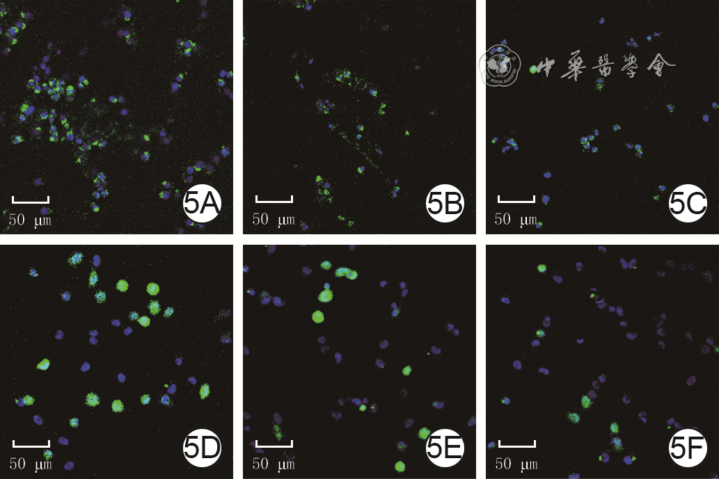
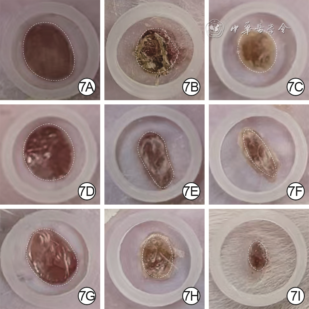
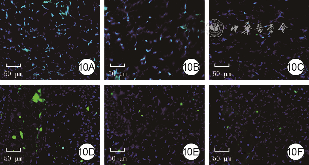
  Objective  To explore the effects of mitochondrial transplantation on full-thickness skin defects in diabetic rats.  Methods  This study was an experimental study. Functionally intact mitochondria were extracted from the liver tissue of 6-8-week-old male Sprague Dawley rats (the same age and sex below). Mouse L929 cells and human umbilical vein endothelial cells (HUVECs) were cultured in medium containing high-glucose (50 mmol/L) for 24 hours to induce high-glucose injury. According to the random number table method (the same grouping method below), they were then divided into control group (Ctrl group, treated with conventional medium), growth factor group (GF group, treated with medium containing 20 U/mL recombinant human epidermal growth factor), and mitochondrial group (Mito group, treated with medium containing 12.5 μg/mL exogenous mitochondria). The scratch assay was performed to evaluate the migration rate of mouse L929 cells at 6 hours after scratching (n=3). The length and number of branch nodes of tube formed by HUVECs were measured after 2 and 6 hours of culture (n=3). After 24 hours of culture, the reactive oxygen species (ROS) levels and mitochondrial membrane potential in aforemetioned two types of cells were detected according to the kit instructions (n=6). Eighteen Sprague Dawley rats were selected and a type 1 diabetic rat model was successfully established. Then, the full-thickness skin defects with a diameter of 1 cm were created on their backs. The rats were divided into Ctrl group, GF group, and Mito group (with each group of 6 rats). At post-injury days (PID) 0 (immediately), 3, and 6, the wounds were subcutaneously injected with normal saline (Ctrl group), topically sprayed with an equal amount of rhEGF solution at a dose of 40 U/cm² (GF group), or subcutaneously injected with equal amount of mitochondrial suspension at a dose of 5 μg/g (Mito group), respectively. The percentage of remaining wound area of rats was calculated at PID 3, 6, and 12. At PID 12, the epithelialization and collagen deposition in the wound of rats were detected by hematoxylin and eosin staining and Masson's staining, respectively. Immunofluorescence staining was used to detect the expression of CD31 (a marker for neovascularization) and neurofilament 200 (a marker for nerves) in the wound of rats. The ROS levels, number of apoptotic cells, ATP content in the wound of rats were detected according to the kit instructions. The levels of tumor necrosis factor-α (TNF-α), interleukin-1β (IL-1β), and IL-6 in the wound of rats were detected by enzyme-linked immunosorbent assay.  Results  At 6 hours after scratching, compared with that in Ctrl group, the migration rate of mouse L929 cells in GF group was significantly increased (P<0.05). compared with that in GF group, the migration rate of mouse L929 cells in Mito group was significantly increased (P<0.05); After 2 and 6 hours of culture, compared with those in Ctrl group, the numbers of branch nodes and the lengths of tube formed by HUVECs in both GF group and Mito group were significantly increased (P<0.05). Compared with those in GF group, the numbers of branch nodes and the lengths of tube formation of HUVECs after 2 and 6 hours of culture in Mito group were significantly increased (P<0.05). After 24 hours of culture, compared with those in Ctrl group, the ROS levels in both mouse L929 cells and HUVECs in GF group and Mito group were significantly decreased (P<0.05), while the mitochondrial membrane potentials were significantly increased (P<0.05); compared with those in GF group, the ROS levels in both mouse L929 cells and HUVECs in Mito group were significantly decreased (P<0.05), while the mitochondrial membrane potentials were significantly increased (P<0.05). At PID 3, 6, and 12, the percentages of remaining wound area of rats in Mito group ((46±3)%, (37±3)%, (18±3)%) were significantly lower than those in Ctrl group ((71±4)%, (63±4)%, (47±5)%) and GF group((62±3)%, (54±3)%, (33±4)%), P<0.05. At PID 12, in the wounds of rats in Mito group, the status of epithelialization and collagen deposition, as well as the conditions of angiogenesis and nerve repair, were superior to those in growth factor and control groups. Compared with those in Ctrl group, the ROS levels and number of apoptotic cells in the wounds of rats in GF group and Mito group were significantly decreased (P<0.05), the ATP content significantly increased (P<0.05), and the levels of TNF-α, IL-1β, and IL-6 were all significantly reduced (P<0.05). Compared with those in GF group, the ROS levels and number of apoptotic cells in the wounds of rats in Mito group were significantly decreased (P<0.05), the ATP content significantly increased (P<0.05), and the levels of TNF-α, IL-1β, and IL-6 were all significantly reduced (P<0.05).  Conclusions  Mitochondrial transplantation enhances mitochondrial ATP production and reduces the level of oxidative stress in cells under high-glucose injury, which improves the migration capacity of mouse L929 cells and the angiogenesis capacity of HUVECs. Simultaneously, it facilitates epithelialization and collagen deposition in full-thickness skin defect wounds of diabetic rats, reduces the levels of inflammatory cytokines, and inhibits cell apoptosis, thereby accelerating wound healing.
Performance of hyaluronic acid biogel and its effect on healing of infected burn wounds in mice
Shi Mingsheng, Liang Xiaoyang, Zhang Rui, Wang Yongyu, Fang Luhang, Li Yang, Xu Fujian
2025, 41(10): 949-957. doi: 10.3760/cma.j.cn501225-20250422-00186
Abstract:
  Objective  To explore the performance of hyaluronic acid biogel and its effect on healing of infected burn wounds in mice.  Methods  This study was an experimental study. The hyaluronic acid biogel prepared in phosphate buffered saline (PBS) was soaked in PBS. Then the liquid absorption rate of hyaluronic acid biogel at 1, 2, 3, 4, and 5 min of soaking was calculated. After mixing hyaluronic acid biogel in powder form with PBS to form a gel, the mechanical modulus of the hyaluronic acid biogel was tested using a rotational rheometer. The hyaluronic acid biogel solution at final mass concentrations of 5, 10, and 20 g/L along with PBS were added to Staphylococcus aureus and Escherichia coli bacteria culture solution, respectively, then after incubation for 12 h, dilution plating was performed on Lysogeny Broth agar medium. The colony growth on the medium was observed by a colony counter after culture for 24 h. Twenty-four male BALB/c mice aged 8-10 weeks underwent dorsal burn wound creation followed by necrotic tissue excision. A infected burn wound model was established by locally instilling Staphylococcus aureus bacteria culture solution. With the successful modeling being confirmed at 24 h after incubation, the mice were divided into 4 groups (n=6) according to the random number table method. The wounds in normal saline group received normal saline, while the wounds in Jingwanhong ointment group, hydrocolloid dressing group, and hyaluronic acid biogel group received Jingwanhong ointment, hydrocolloid dressing, and hyaluronic acid biogel, respectively, for 14 d. At treatment day 7, 10, and 14, the remaining wound area was measured and the percentage of residual wound area was calculated. At treatment day 7, the number of Staphylococcus aureus colonieson wounds were counted by a colony counter. All the experiments used a sample size of 3.  Results  The hyaluronic acid biogel achieved the highest liquid absorption rate of 387.9% within 2 min of soaking in PBS, and stabilized at over 300.0% within 5 min of soaking. Frequency scanning of the mechanical modulus revealed that the hyaluronic acid biogel's elastic modulus remained stable above 250 Pa and increased with rising angular frequency. Amplitude scanning of the mechanical modulus indicated that at an angular frequency of 10 rad/s, the linear viscoelastic range of the hyaluronic acid biogel was close to 1 000%. After culture for 24 h, the medium added with PBS was almost completely covered with Staphylococcus aureus and Escherichia coli. The medium added with hyaluronic acid biogel solution at final mass concentrations of 5, 10, and 20 g/L showed no significant growth of Staphylococcus aureus or Escherichia coli. At treatment day 7, 10, and 14, the percentage of residual wound area of mice in hyaluronic acid biogel group was significantly lower than that in normal saline, Jingwanhong ointment, and hydrocolloid dressing groups (with P values all <0.05). At treatment day 14, the percentage of residual wound area of mice in hydrocolloid dressing group was significantly lower than that in normal saline and Jingwanhong ointment groups (with P values both <0.05). At treatment day 7, the number of Staphylococcus aureus colonies on wounds of mice in hyaluronic acid biogel group ((4.3±0.6) colonies) was significantly lower than that in normal saline group ((2 400.7±225.4) colonies), Jingwanhong ointment group ((899.0±57.0) colonies), and hydrocolloid dressing group ((11.7±5.7) colonies), with P values all <0.05.  Conclusions  Hyaluronic acid biogel exhibits an excellent capacity to absorb liquid and form hydrogel, and promotes healing of infected burn wounds in mice by eliminating Staphylococcus aureus.
Influence of porosity and Young's modulus of gelatin methacrylate anhydride hydrogels on the biological behavior of mouse bone marrow mesenchymal stem cells
Kong Yue, Tian Feng, Liu Qinghua, Zhang Chao, Song Wei, Kong Yi, Li Zhao, Enhejirigala, Huang Sha
2025, 41(10): 958-967. doi: 10.3760/cma.j.cn501225-20250630-00286
Abstract:
  Objective  To investigate the influence of porosity and Young's modulus of gelatin methacrylate anhydride (GelMA) hydrogels on the biological behavior of mouse bone marrow mesenchymal stem cells (BMSCs), and to provide a theoretical basis for the development of tissue engineering scaffolds for skin wound repair.  Methods  This study was an experimental study. GelMA hydrogels that were prepared by photo-crosslinking 50 g/L GelMA solution for 30 and 60 seconds and 150 g/L GelMA solution for 30 and 60 seconds, were designated respectively as low-concentration short-time group, low-concentration long-time group, high-concentration short-time group, and high-concentration long-time group. The porosity of the 4 groups of hydrogels after freeze-drying for 48 hours was measured and calculated using Image J 1.54 software. The Young's modulus of the 4 groups of hydrogels was detected using a Young's modulus tester. The correlation between the porosity and Young's modulus was evaluated and a predictive model between them was established. The remaining mass percentages of the 4 groups of hydrogels after soaking in phosphate buffered saline for 7 days were calculated. BMSCs were isolated from the femurs of 10 C57BL/6 mice aged 1 day of unknown sex. Cell-loaded GelMA hydrogels were prepared by mixing BMSCs with 50 g/L GelMA solution and photo-crosslinking for 30 and 60 seconds, and by mixing BMSCs with 150 g/L GelMA solution and photo-crosslinking for 30 and 60 seconds, and were designated respectively as low-concentration short-time cell-loaded group, low-concentration long-time cell-loaded group, high-concentration short-time cell-loaded group, and high-concentration long-time cell-loaded group. The cell survival in the 4 cell-loaded groups of hydrogels after 7 days of culture was detected using a live/dead cell kit, and the cell survival rate was calculated. After 10 days of osteogenic induction culture of the hydrogels in the 4 cell-loaded groups, the mRNA expressions of osteogenic-related factors Runt-related transcription factor 2 (Runx2) and alkaline phosphatase (ALP), and stemness-related factors SRY-box transcription factor 2 (Sox2) and Nanog in the cells were detected by real-time fluorescence quantitative reverse transcription polymerase chain reaction; the protein expressions of Runx2 and Nanog in the cells were detected by immunofluorescence method. Except for the protein expression (qualitative observation), the sample size of each group for the other indicators was three.  Results  The porosity of hydrogels in low-concentration short-time group, low-concentration long-time group, high-concentration short-time group, and high-concentration long-time group after freeze-drying for 48 hours decreased successively, being (92.7±0.9)%, (85.0±1.8)%, (68.6±1.2)%, and (56.3±5.8)%, respectively; the Young's modulus increased successively, being (5.933±0.020), (7.803±0.089), (20.772±0.106), and (22.498±0.060) kPa, respectively. Except for the Young's modulus of hydrogels between the high-concentration long-time group and high-concentration short-time group (P>0.05), the pairwise comparisons of hydrogel porosity or Young's modulus among the remaining groups showed statistically significant differences (P<0.05). The porosity and Young's modulus of the hydrogels were significantly negatively correlated (R2=0.91, P<0.05), and a linear equation predictive model between them was established accordingly. After soaking for 7 days, the remaining mass percentages of hydrogels in low-concentration long-time group, high-concentration short-time group, and high-concentration long-time group were significantly higher than that in low-concentration short-time group (P<0.05), and the remaining mass percentage of hydrogels in high-concentration long-time group was significantly higher than that in low-concentration long-time group (P<0.05). After 7 days of culture, a large number of live cells was observed in the 4 cell-loaded groups of hydrogels, and there was no statistically significant difference in the overall comparison of cell survival rate among the groups (P>0.05). After 10 days of osteogenic induction culture, the mRNA expressions of Runx2 and ALP in the cells of hydrogels in high-concentration long-time cell-loaded group were significantly higher than those in the other three cell-loaded groups (P<0.05), and the mRNA expressions of Sox2 and Nanog in the cells of hydrogels in low-concentration short-time cell-loaded group were significantly higher than those in the other three cell-loaded groups (P<0.05); the protein expression of Runx2 in the cells of hydrogels in high-concentration long-time cell-loaded group was the highest, and the protein expression of Nanog in the cells of hydrogels in low-concentration short-time cell-loaded group was the highest.  Conclusions  The porosity and Young's modulus of GelMA hydrogel synergistically regulate the biological behavior of mouse BMSCs, where high porosity and low Young's modulus are conducive to maintaining stemness, while low porosity and high Young's modulus drive osteogenic differentiation.
Original Article·Diabetic Wounds
Clinical effect of tibial transverse transport technique combined with continuous low negative pressure wound therapy in the treatment of Wagner grade Ⅲ to Ⅴ chronic diabetic foot ulcers
Liao Yuandan, Chen Yu, Ma Xikun, Li Yaxing, Gan Tingjiang, Zhang Hui, Xie Huiqi
2025, 41(10): 968-976. doi: 10.3760/cma.j.cn501225-20241106-00436
Abstract:
  Objective  To explore the clinical effect of tibial transverse transport (TTT) technique combined with continuous low negative pressure wound therapy in the treatment of Wagner grade Ⅲ to Ⅴ chronic diabetic foot ulcers.  Methods  This study was a retrospective cohort study. From January 2016 to December 2022, 38 patients with Wagner grade Ⅲ to Ⅴ chronic diabetic foot ulcers who met the inclusion criteria were admitted to the Department of Orthopedics of West China Hospital of Sichuan University. According to the treatment method for the ulcer wounds, the patients were divided into a control group (20 cases, 10 males and 10 females, aged (64.0±2.3) years) treated with TTT technique alone and a combined treatment group (18 cases, 10 males and 8 females, aged (60.8±2.9) years) treated with TTT technique combined with continuous low negative pressure wound therapy. After debridement, the ulcer wound areas in control group and combined treatment group were (29±13) cm² and (30±18) cm², respectively. Postoperatively, ulcer wound healing and the occurrence of complications were observed. The incidence of complications, ulcer wound healing time, external fixator removal time, time to ambulation, and time to full weight-bearing of the affected limb in the two groups of patients were statistically analyzed and compared. Before surgery and at the last follow-up, the ankle-brachial index or differences in ankle-brachial index, skin temperature, and visual analogue scale (VAS) score of the affected foot were statistically analyzed and compared between the two groups of patients. The ulcer wound healing time was the primary observation indicator, and the other indicators were secondary observation indicators.  Results  Postoperatively, in combined treatment group, one patient developed a new ulcer at another site after the original ulcer wound healed; this ulcer wound healed after the patient was re-admitted for treatment with TTT technique combined with continuous low negative pressure wound therapy. One patient had a recurrence of the ulcer at the same site due to poor blood glucose control and underwent amputation. The ulcer wounds of the other patients healed well. In control group, four patients had recurrence of the ulcer wound due to poor blood glucose control, among whom one patient underwent amputation and three patients healed after treatment with TTT technique combined with continuous low negative pressure wound therapy. The incidence of complications in combined treatment group and control group of patients were 2/18 and 4/20, respectively, showing no statistically significant difference between the two groups (P>0.05). Postoperatively, compared with those in control group, the ulcer wound healing time, external fixator removal time, time to ambulation, and time to full weight-bearing of the affected limb in combined treatment group of patients were significantly shortened (with Z values of 2.52 and 4.90, respectively, and t values of 3.99 and 4.10, respectively, P<0.05). The difference in ulcer wound healing time between the two groups of patients was 6 (1, 11) days. Before surgery, there were no statistically significant differences in the VAS score, ankle-brachial index, and skin temperature of the affected foot between the two groups of patients (P>0.05). At the last follow-up, compared with those in control group, the VAS score of the affected foot in combined treatment group of patients was significantly decreased (Z=2.24, P<0.05), the differences in ankle-brachial index was significantly increased (t=3.53, P<0.05) and the skin temperature was significantly increased (Z=4.49, P<0.05).  Conclusions  The TTT technique combined with continuous low negative pressure wound therapy can significantly shorten the ulcer wound healing time, external fixator removal time, time to ambulation, and time to full weight-bearing of the affected limb in the treatment of Wagner grade Ⅲ to Ⅴ chronic diabetic foot ulcers, and effectively improve the ankle-brachial index, skin temperature, and pain of the affected foot, showing good clinical application prospects and safety.
Original Article
Clinical efficacy of free anterolateral thigh tissue flap transplantation with a single-vein dual-drainage system at recipient site in repairing scalp high-voltage electrical burn wounds
Nie Guangchen, Zheng Wang, Hao Xuguang, Li Zongyu, Shao Tiebin, Mou Bin, Liu Yong, Meng Xiangyue, Feng Jinghui, Wu Binan
2025, 41(10): 977-985. doi: 10.3760/cma.j.cn501225-20250220-00074
Abstract:
  Objective  To investigate the clinical efficacy of free anterolateral thigh (ALT) tissue flap transplantation with a single-vein dual-drainage system at recipient site in repairing scalp high-voltage electrical burn wounds.  Methods  This study was a retrospective observational study. From January 2015 to August 2024, five patients (4 males and 1 female, aged 33-64 years) with scalp defects caused by high-voltage electrical burns who met the inclusion criteria were treated at the Department of Burns, Plastic and Wound Repair of the Fifth Hospital of Harbin. After debridement, the scalp soft tissue defects of patients ranged from 16 cm×15 cm to 21 cm×18 cm. A tissue flap consisting of skin, subcutaneous tissue, deep fascia, and part of the vastus lateralis muscle was harvested and referred to as ALT tissue flap. Among the patients, the perforators included in the tissue flap were musculocutaneous perforators in three cases and septocutaneous perforators in two cases. The source vessel was consistently the descending branch of the lateral circumflex femoral artery, and each tissue flap contained two accompanying veins. The size of the harvested ALT skin flap ranged from 17 cm×12 cm to 22 cm×13 cm, with the vastus lateralis muscle area accounting for approximately 1/4 to 1/3 of the total wound area. The descending branch of the lateral circumflex femoral artery carried by the tissue flap was anastomosed end-to-end with the recipient artery. The recipient vein was transected horizontally. The vein with better conditions in tissue flap was anastomosed end-to-end anterogradely to the proximal end of the transected recipient vein, while the other vein was anastomosed end-to-end retrogradely to its distal end, thereby establishing a single-vein dual-drainage system. In all cases, the recipient artery used for anastomosis was the superficial temporal artery; the recipient veins included the posterior auricular vein in one case, the frontal branch of the superficial temporal vein in two cases, and the parietal branch of the superficial temporal vein in the other two cases. The skin paddle of the tissue flap was used to cover the main defect, while the vastus lateralis muscle was extended to cover the remaining wound, followed by wound edge suture. The stage Ⅰ wound repair was completed by directly pulling and suturing the donor site wound. At three weeks postoperatively, the stage Ⅱ procedure was performed. A split-thickness skin graft was harvested from the contralateral anterolateral thigh and used to cover the exposed muscle tissue at recipient site. The tissue flap harvesting time in the stage Ⅰ was recorded. After the stage Ⅰ surgery, the patient's tissue flap was observed for the occurrence of vascular crises and survival, the wound healing at donor sites was observed. After the stage Ⅱ surgery, the wound healing at recipient sites was observed. During follow-up, the tissue flap appearance of patients was evaluated, and sensory recovery was assessed. The complications such as infection at donor sites were recorded, and it was evaluated whether the function of the donor limb had been affected.  Results  The tissue flap harvesting time for the five patients in the stage Ⅰ surgery ranged from 32-41 (37±3) min. After the stage Ⅰ surgery, no arterial or venous vascular crisis occurred in any tissue flap, and all tissue flaps survived completely. The wounds at donor sites healed well. After the stage Ⅱ surgery, the wounds at recipient sites achieved complete healing. During the follow-up period of 7 to 24 months after surgery, all tissue flaps of patients exhibited no significant bulkiness, possessed soft texture, and survived completely with partial recovery of sensation. No complications such as infection were observed at donor sites, and the function of donor limbs remained unaffected.  Conclusions  For scalp soft tissue defects caused by high-voltage electrical burns with severely compromised recipient veins, the use of a free ALT tissue flap combined with a single-vein dual-drainage system at recipient site can effectively enhance venous outflow efficiency, reduce the risk of vascular crisis, shorten operative time, and decrease donor site morbidity.
Efficacy of precisely designed first dorsal metacarpal artery flap under the guidance of high-frequency color Doppler ultrasound in repairing small-sized skin and soft tissue defects in the thumb
Fu Jiansong, Liu Ying, Wang Hui, Gao Shunhong, Ma Tiepeng, Dong Huishuang, Yu Jun, Yao Jun, Zhou Tong
2025, 41(10): 986-993. doi: 10.3760/cma.j.cn501225-20241130-00468
Abstract:
  Objective  To explore the efficacy of precisely designed first dorsal metacarpal artery flap under the guidance of high-frequency color Doppler ultrasound in repairing small-sized skin and soft tissue defects in the thumb.  Methods  This study was a retrospective observational study. From January 2021 to August 2023, 29 patients with small-sized skin and soft tissue defects in the thumb who met the inclusion criteria were admitted to the Department of Hand Surgery of the Second Hospital of Tangshan. There were 16 males and 13 females, with ages ranging from 22 to 65 years. The wound area after debridement ranged from 2.5 cm×1.5 cm to 3.0 cm×2.0 cm. Before flap transplantation, the body surface projections of the anastomotic branches between the first metacarpal radial dorsal artery and the radial proper digital artery of the affected finger measured by high-frequency color Doppler ultrasound examination were recorded. During surgery, the first dorsal metacarpal artery flap was harvested to repair the defects in the thumb, with the harvested flap area of 3.0 cm×2.0 cm to 3.5 cm×2.5 cm. The superficial branch of radial nerve carried by the flap was anastomosed with the proper digital nerve in the recipient area. The wound in the flap donor site was directly sutured. It was observed whether the body surface projections of the anastomotic branches between the first metacarpal radial dorsal artery and the radial proper digital artery of the affected finger measured by preoperative high-frequency color Doppler ultrasound examination were consistent with the intraoperative findings. After surgery, the wound healing and suture removal time in the donor and recipient sites, as well as the flap survival were observed. At the last follow-up (6-12 months postoperatively), the function of the affected finger was evaluated according to the trial criteria for evaluation of partial function of upper extremity by the Hand Surgery Society of Chinese Medical Association, and the excellent and good rate was calculated. The satisfaction scoring of flap efficacy was performed. Additionally, the two-point discrimination distance of flap was measured, and Semmes-Weinstein monofilament test was performed to evaluate flap sensory function.  Results  The body surface projections of the anastomotic branches between the first metacarpal radial dorsal artery and the radial proper digital artery of the affected finger of 29 patients measured by preoperative high-frequency color Doppler ultrasound examination were all consistent with the intraoperative findings. After surgery, all wounds in the donor and recipient sites healed well, the suture removal time was 14-16 days postoperatively. The flaps survived completely in 26 patients, and the partial flap necrosis occurred in 3 patients. At the last follow-up, the functional score of the affected finger was 88±8, with the excellent and good rate of 96.6% (28/29). The satisfaction scoring of flap efficacy was 7.4±1.0, the two-point discrimination distance of flap was (6.9±0.7) mm, and the Semmes-Weinstein monofilament test result was (3.46±0.19) g.  Conclusions  The application of precisely designed first dorsal metacarpal artery flap under the guidance of high-frequency color Doppler ultrasound in repairing small-sized skin and soft tissue defects in the thumb has the advantages of high flap survival rate, good functional recovery of the affected finger, and high satisfaction with flap efficacy.
Discipline Construction
Exploration of the "Hainan Model" in the construction of specialized wound repair discipline
Cheng Shaowen
2025, 41(10): 994-997. doi: 10.3760/cma.j.cn501225-20250219-00071
Abstract:
Chronic refractory wounds have emerged as a significant public health challenge that needs to be addressed urgently in China, necessitating strengthened construction of wound repair discipline. Since the National Health Commission of China officially encouraged medical institutions to establish specialized wound repair discipline in 2019, diverse regional models of specialized discipline have emerged nationwide, effectively advancing the construction and development of wound repair discipline system in China. Leveraging Hainan's unique regional characteristics, the First Affiliated Hospital of Hainan Medical University has innovatively integrated the wound repair discipline into the construction of the emergency medical rescue system, successfully creating the distinctive "Hainan Model" for specialized wound repair discipline. This model not only addresses the wound treatment needs of local populations, but also provides robust support for the implementation of national strategic initiatives, achieving dual value in both disciplinary development and social benefits and highlighting its core strengths and advantages. This paper systematically elaborates on the developmental trajectory of Hainan's wound repair system and the exploration of the "Hainan Model" in construction of specialized wound repair discipline, aiming to offer valuable reference for building a China-specific wound repair discipline system and promote its high-quality development.
Review
Research advances on the application of artificial intelligence technology in the diagnosis and treatment of sepsis patients
Zhang Yuwen, Bai Yinglu, Zhang Xuemin, Wen Yue, Yang Zhuoya, Lu Mingye, Xu Xiaolong, Liu Qingquan
2025, 41(10): 998-1003. doi: 10.3760/cma.j.cn501225-20250708-00292
Abstract:
In recent years, with the rapid advancement of artificial intelligence technology, this technology has been extensively applied in the field of emergency and critical care. Artificial intelligence demonstrates significant potential in clinical decision support, offering novel tools for the auxiliary diagnosis and treatment of sepsis. The sepsis prediction model developed based on artificial intelligence technology can efficiently capture and integrate information from diverse modalities, enhance the accuracy of early sepsis prediction, and establish a prognostic evaluation system, thereby guiding clinical precision treatment and ultimately improving the survival rates of sepsis patients. This paper reviews the research advances on the application of artificial intelligence technology in the early prediction and subtype identification of sepsis, prognosis assessment, and prediction of related organ dysfunction. It aims to provide a reference and inspiration for the further application of this technology in the field of emergency and critical care.
Current status, representative devices, and future development trends of wound measurement technologies
Zhong Shuyan, Shu Maoguo, Du Huicong
2025, 41(10): 1004-1010. doi: 10.3760/cma.j.cn501225-20241231-00516
Abstract:
Wound measurement plays a critical role in wound repair and chronic disease management, its accuracy directly influences the development of personalized treatment plans and the evaluation of wound healing progress. Although traditional one-dimensional measurement methods (such as the ruler method and the probe method) are simple to use, they are unable to meet modern clinical demands due to insufficient accuracy and consistency. In recent years, two-dimensional imaging methods, three-dimensional imaging methods, and the corresponding intelligent measurement devices have become mainstream of wound measurement. By employing digital image processing, three-dimensional modeling, and artificial intelligence technologies, the measurement accuracy has been significantly improved, providing multidimensional data support for the assessment of complex wounds. This article systematically reviews the current development status of wound measurement technologies, representative devices, and their clinical applications. It also explores future directions for optimization, including the integration of artificial intelligence, multi-modal data fusion, and privacy protection. The aim is to provide practical guidance and technical references for clinicians and researchers.