Abstract:
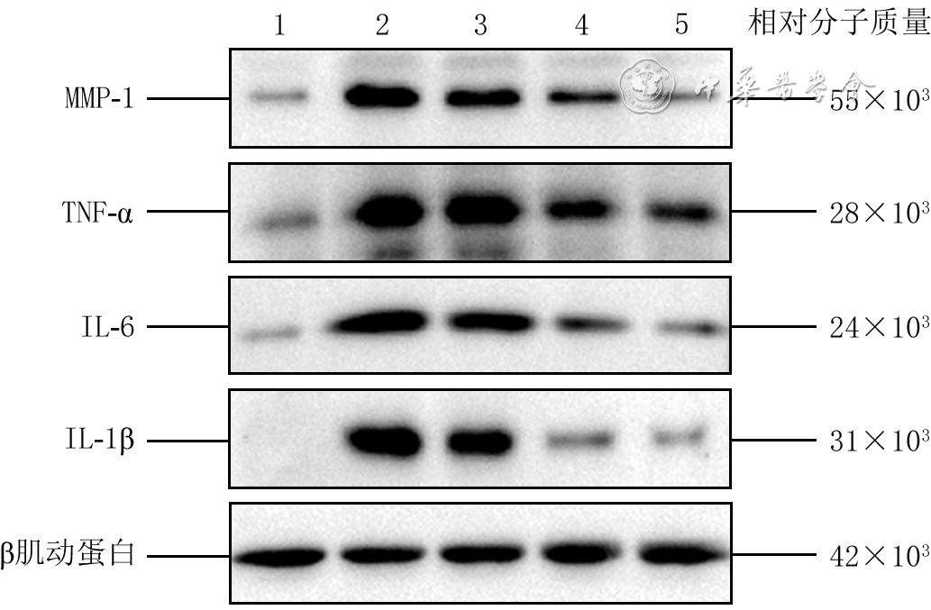
Objective To investigate the effects of glutathione on acute cutaneous photodamage in mice and its mechanism. Methods This study was a group-designed experimental study. Fifteen 8-week-old male C57BL/6 mice were shaved on the dorsal skin and divided into blank control group, model group, low-dose intervention group, medium-dose intervention group, and high-dose intervention group using a random number table, with 3 mice in each group. Mice in model group received daily combined irradiation of ultraviolet B and ultraviolet A on the dorsal skin to cause acute photodamage, followed by intraperitoneal injection of phosphate-buffered saline (PBS). Mice in blank control group were subjected to sham injury without ultraviolet irradiation and received daily intraperitoneal injection of PBS only. Mice in low-dose intervention group, medium-dose intervention group, and high-dose intervention group received daily ultraviolet irradiation as the model group, followed by intraperitoneal injection of 50, 100, and 200 mg/kg of glutathione, respectively. Two hours after the last injection on day 7 post injury (hereinafter referred to as day 7 post injury), the color and morphology of the dorsal skin of mice in each group were observed grossly. Then, dorsal skin tissue was excised for the following assays. Hematoxylin-eosin staining was used to examine the structure of the stratum corneum, epidermis, and dermis, the morphology of appendages (hair follicles, sweat glands, and sebaceous glands), the presence of hemorrhage, inflammatory cell infiltration, and the thickness of epidermis were also measured. Masson staining was used to detect collagen fiber deposition in the skin tissue. Western blotting was used to detect the protein expressions of inflammation-related proteins (interleukin-1β (IL-1β), IL-6, tumor necrosis factor-α (TNF-α), and matrix metalloproteinase 1 (MMP-1)) in the skin tissue. Results On day 7 post injury, compared with that in blank control group, the skin of mice in model group showed extensive scaling, redness, swelling, and crusting. Compared with that in model group, the severity of photodamage of the skin of mice in low-dose intervention group, medium-dose intervention group, and high-dose intervention group was alleviated successively. On day 7 post injury, compared with that in blank control group, the skin tissue structure of mice in model group was disorganized, characterized by thickened and detached stratum corneum, increased number and disordered arrangement of epidermal cell layers, dermal edema, abnormal morphology of appendages (hair follicles, sweat glands, sebaceous glands), scattered hemorrhagic foci, and extensive inflammatory cell infiltration. Compared with that in model group, the degree of tissue disorganization in the skin of mice in low-dose intervention group, medium-dose intervention group, and high-dose intervention group was alleviated successively. On day 7 post injury, the epidermal thickness of the skin of mice in model group was (116.4±6.4) μm, which was significantly greater than (20.9±1.6) μm in blank control group (P<0.05). Compared with that in model group, the epidermal thickness of the skin of mice in medium-dose intervention group and high-dose intervention group ((77.7±5.6) μm and (56.9±0.8) μm, respectively) was significantly decreased (with P valuesboth <0.05). On day 7 post injury, compared with that in blank control group, the skin tissue of mice in model group showed a significant increase in collagen fiber content and disorganized fiber arrangement, indicating a certain degree of collagen fiber proliferation. Compared with that in model group, the arrangement of collagen fibers in the skin tissue of mice in low-dose intervention group, medium-dose intervention group, and high-dose intervention group became progressively more regular, and the overall tissue structure gradually recovered to normal. On day 7 post injury, compared with those in blank control group, the protein expression levels of IL-1β, IL-6, TNF-α, and MMP-1 in the skin tissue of mice in model group were significantly increased (with P values all <0.05). Compared with those in model group, the protein expression levels of IL-1β and IL-6 in the skin tissue of mice in low-dose intervention group were significantly decreased (with P values both <0.05), while the protein expression levels of IL-1β, IL-6, TNF-α, and MMP-1 in the skin tissue of mice in medium-dose intervention group and high-dose intervention group were significantly decreased (with P values all <0.05). Conclusions Glutathione can significantly alleviate acute cutaneous photodamage induced by combined irradiation of ultraviolet B and ultraviolet A in mice, with a clear dose-dependent protective effect, and its mechanism may be related to inhibiting the expression of inflammatory factors, reducing MMP-1-mediated collagen degradation, and improving dermal collagen structure.
Wang Q,Zhou L,Deng Y,et al.Effects of glutathione on acute cutaneous photodamage in mice and its mechanism[J].Chin J Burns Wounds,2026,42(5):477-485.DOI: 10.3760/cma.j.cn501225-20250628-00284.



 Abstract
 PDF
PDF