2024 Vol. 40, No. 5

Expert Forum
Exploring wound healing from the perspective of circadian rhythm regulation
Wang Dali, Qi Fang
2024, 40(5): 401-406. doi: 10.3760/cma.j.cn501225-20240122-00027
Abstract:
Wound healing is a complex and finely regulated biological process involving a variety of cell types, such as immune cells, fibroblasts, keratinocytes, endothelial cells, and stem cells. In recent years, scientific research has unveiled the significant role of circadian rhythms in the regulation of wound healing process. Although the molecular mechanisms by which circadian rhythms precisely regulate wound healing are not yet fully understood, accumulating evidence has revealed that some key signals and cellular functions play crucial roles in the circadian rhythm regulation of wound healing. A deeper understanding of these mechanisms is important not only to uncover the basic biological processes of wound healing, but also to develop new therapeutic strategies, such as regulating circadian rhythms to guide medication and optimize trauma treatment. By reviewing the current research results on circadian rhythm regulation of wound healing, this paper aims to provide a comprehensive perspective on this emerging field and provide valuable insights for future research directions.
Coordinating tissue repair: molecular pathways controlling the function of harmful and repairing neutrophils
He Weifeng, Yan Lingfeng
2024, 40(5): 407-414. doi: 10.3760/cma.j.cn501225-20240306-00089
Abstract:
Neutrophils are the most abundant circulating white blood cells and play an indispensable role as first responders of damaged tissue and infected sites in the early inflammatory response of healing. Neutrophils provide immediate host defense by engulfing and destroying pathogens, releasing cytotoxic enzymes and metabolites, and spreading inflammatory networks. However, if left uncontrolled, these defense mechanisms can cause significant collateral damage. Focusing on the triggers of harmful neutrophil inflammation and immunomodulatory deficits, as well as grasping the specific drivers of harmful inflammation, is of great significance for recalibrating inflammation to promote endogenous tissue repair. This article, starting from the causes of neutrophil inflammation imbalance, elaborated the main mechanism of neutrophil-mediated tissue injury and related pathological manifestations, and highlighted the therapeutic targets with promising applications.
Original Article · Wound Healing Mechanism
Role and mechanism of Vγ4 T cell depletion in epidermal tissue repair after ultraviolet damage to mouse skin
Li Yashu, He Weifeng, Lyu Kaiyang
2024, 40(5): 415-424. doi: 10.3760/cma.j.cn501225-20240121-00026
Abstract:
  Objective  To explore the role and mechanism of Vγ4 T cell depletion in epidermal tissue repair after ultraviolet damage to mouse skin.  Methods  The study was an experimental study. Fifty-four female C57BL/6J wild-type mice aged 6 to 8 weeks were divided into Vγ4 T cell depletion group and control group (27 mice in each group) according to the random number table, and the Armenian hamster anti-mouse Vγ4 T cell receptor (TCR) monoclonal antibody of 200 µg and an equal amount of homologous control IgG antibody were intraperitoneally injected, respectively. At one week after injection (the same time point to harvest mice below), dermal cells and lymph node cells were respectively extracted from the back skin tissue, armpit and inguinal lymph nodes of 3 mice in each group (mice in following study were all taken from these 2 groups), and the proportions of Vγ4 T cells in dermal cells and lymph node cells were detected by flow cytometry. Five mice from each group were harvested for observation of skin on the back and skin tissue structure was observed and the epidermal tissue thickness was measured after hematoxylin-eosin (HE) staining. Five mice from each group were harvested for detection of proportion of dendritic epidermal T cells (DETCs) in epidermal cells by flow cytometry after extracted. Three mice were taken from each group and recruited in Vγ4 T cell depletion+5 times ultraviolet irradiation (UVR) group and control+5 times UVR group, respectively, then UVR was administered once per day for 5 times, and the condition of skin on the back was observed immediately after daily irradiation. Five mice were taken from each group and divided into Vγ4 T cell depletion+1 UVR group and control+1 UVR group, respectively. Immediately after one UVR treatment, the epidermal tissue thickness was measured after HE staining. Three mice from each group were selected and recruited in Vγ4 T cell depletion alone group and control alone group, then 3 mice from each group rwere recruited in Vγ4 T cell depletion+1 time UVR group and control+1 time UVR group, respectively, and were treated as before. The mRNA expressions of insulin-like growth factor-Ⅰ (IGF-Ⅰ), keratinocyte growth factor (KGF), Vγ5 TCR, interleukin-15 (IL-15), IL-1β, IL-23, natural killer group 2 member D (NKG2D), histocompatibility antigen 60 (H60), mouse UL16-binding protein-like transcript 1 (Mult1), and retinoic acid early inducible protein 1 (Rae1) in the epidermal tissue were detected by real-time fluorescent quantitative reverse transcription polymerase chain reaction.  Results  At one week after injection, the proportions of Vγ4 T cells in dermal cells and lymph node cells of mice in Vγ4 T cell depletion group were significantly lower than those in control group (with t values of 27.99 and 13.12, respectively, P<0.05); there were no statistically significant differences in the skin general condition and tissue structure of mice between Vγ4 T cell depletion group and control group; the epidermal tissue thickness of mice between Vγ4 T cell depletion group and control group was similar (P>0.05); the proportion of DETCs in epidermal cells of mice in Vγ4 T cell depletion group was (3.9±0.8)%, which was significantly higher than (1.6±0.5)% in control group (t=4.84, P<0.05). Compared with that in control+5 times UVR group, the skin scale increased after one UVR treatment, scaly scab appeared after 2 times of irradiation, and scaly scab increased significantly after 3 to 5 times of irradiation in Vγ4 T cell depletion+5 times UVR group. Immediately after UVR treatment, the epidermal tissue thickness of mice in Vγ4 T cell depletion+1 time UVR group was significantly increased compared with that in control+1 time UVR group (t=11.50, P<0.05). Compared with those in control alone group, the mRNA expression of Vγ5 TCR in the epidermal tissue of mice in Vγ4 T cell depletion alone group was up-regulated (t=41.16, P<0.05), while the mRNA expression of IL-23 was down-regulated (t=6.52, P<0.05); compared with those in control alone group, the mRNA expressions of Vγ5 TCR and KGF in the epidermal tissue of mice in control+1 time UVR group were significantly up-regulated (with t values of 15.22 and 13.22, respectively, P<0.05), while the mRNA expressions of IGF-Ⅰ and IL-23 were significantly down-regulated (with t values of 3.71 and 4.95, respectively, P<0.05); compared with those in Vγ4 T cell depletion alone group, the mRNA expressions of IGF-Ⅰ and KGF in the epidermal tissue of mice in Vγ4 T cell depletion+1 time UVR group were significantly up-regulated (with t values of 11.40 and 18.88, respectively, P<0.05), while the mRNA expression of IL-1β was significantly down-regulated (t=4.42, P<0.05); compared with those in control+1 time UVR group, the mRNA expressions of Vγ5 TCR, IGF-Ⅰ, and KGF in the epidermal tissue of mice in Vγ4 T cell depletion+1 time UVR group were significantly up-regulated (with t values of 4.52, 15.24, and 9.43, respectively, P<0.05); the mRNA expression of IL-15 in the epidermal tissue of mice in these 4 groups was generally similar (P>0.05). Compared with those in control alone group, the mRNA expressions of NKG2D and Rae1 in the epidermal tissue of mice in Vγ4 T cell depletion alone group were significantly up-regulated (with t values of 3.67 and 47.40, respectively, P<0.05), the mRNA expressions of NKG2D, Mult1, and Rae1 in the epidermal tissue of mice in control+1 time UVR group were significantly up-regulated (with t values of 5.30, 6.50, and 9.16, respectively, P<0.05); compared with those in Vγ4 T cell depletion alone group, the mRNA expressions of NKG2D, H60, Mult1, and Rae1 in the epidermal tissue of mice in Vγ4 T cell depletion+1 time UVR group were significantly down-regulated (with t values of 4.57, 4.13, 4.67, and 27.36, respectively, P<0.05); compared with those in control group+1 time UVR group, the mRNA expressions of NKG2D, H60, Mult1, and Rae1 in the epidermal tissue of mice in Vγ4 T cell depletion+1 time UVR group were significantly down-regulated (with t values of 5.77, 8.18, 12.90, and 8.08, respectively, P<0.05).  Conclusions  The clearance of Vγ4 T cells is conducive to the proliferation and down-regulation of cytotoxicity of DETCs, and may promote the repair of mouse epidermal damage after UVR.
Effects of recombinant human metallothionein-Ⅲ combined with wound dressing on wound healing of full-thickness skin defects in mice
Shen Xin, Sun Zuoyi, Zhang Rui, Xue Yuying
2024, 40(5): 425-432. doi: 10.3760/cma.j.cn501225-20231031-00164
Abstract:
  Objective  To investigate the effects of recombinant human metallothionein-Ⅲ (rh-MT-Ⅲ) combined with wound dressing on wound healing of full-thickness skin defects in mice.  Methods  This study was an experimental study. Twenty-four half male and half female 6 weeks old Institute of Cancer Research mice were taken, and two symmetrical round full-thickness skin defect wounds were prepared on the back of each mouse. The mice were stratified and randomly divided into normal saline group, dressing group, rh-MT-Ⅲgroup (applying the corresponding solution on the wounds), and combined treatment group (applying a mixture of rh-MT-Ⅲ and wound dressing on the wounds) according to sex and body weight, with 6 mice in each group. From 1 to 7 d after injury, all mice were observed daily for changes in activity, diet, and fur growth, their body weights and wound areas were recorded, and the relative wound area percentages were calculated. On 7 d after injury, the wound tissue of mice was taken for hematoxylin-eosin staining to observe the newborn granulation tissue, for Masson staining to observe collagen fiber formation, and for immunofluorescence staining to detect cell proliferation (denoted as Ki67 relative fluorescence intensity) and cell apoptosis (denoted as TdT-mediated dUTP nick end labeling (TUNEL) relative fluorescence intensity). The sample size in the above experiments was 6.  Results  There were no abnormalities in activity, diet, or fur growth in the 4 groups of mice within 7 d after injury. There were no statistically significant differences in the overall comparison of the body weights of mice in the 4 groups from 1 to 7 d after injury (P>0.05). The relative wound area percentages of mice in combined treatment group were significantly lower than those in normal saline group and rh-MT-Ⅲ group on 2, 3, 4, 5, 6, and 7 d after injury (P<0.05), and the relative wound area percentages of mice in combined treatment group were significantly lower than those in dressing group on 3, 4, 5, 6, and 7 d after injury (P<0.05). The relative wound area percentages of mice in dressing group on 6 and 7 d after injury and in rh-MT-Ⅲ group on 7 d after injury were significantly lower than those in normal saline group (P<0.05). On 7 d after injury, a large number of capillaries and fibroblasts could be seen in wound tissue of mice in combined treatment group, and the growth of new epithelial tissue at the upper edge of the wound was better than that of the other three groups; the collagen fibers in the wound tissue of mice in combined treatment group had higher degree of density and arrangement in a more orderly manner than those of the other three groups. On 7 d after injury, the Ki67 relative fluorescence intensity in the wound tissue of mice in dressing group, rh-MT-Ⅲ group, and combined treatment group was (289±35)%, (197±17)%, and (389±56)%, which was significantly higher than (100±15)% in normal saline group, respectively (P<0.05), and the Ki67 relative fluorescence intensity in the wound tissue of mice in combined treatment group was significantly higher than that in dressing group and rh-MT-Ⅲ group, respectively (with both P values <0.05). On 7 d after injury, the TUNEL relative fluorescence intensity in the wound tissue of mice in dressing group, rh-MT-Ⅲ group, and combined treatment group was (55.5±5.7)%, (66.7±8.0)%, and (20.0±2.2)%, which was significantly lower than (100.0±12.9)% in normal saline group, respectively (P<0.05), and the TUNEL relative fluorescence intensity in the wound tissue of mice in combined treatment group was significantly lower than that in dressing group and rh-MT-Ⅲ group, respectively (with both P values <0.05).  Conclusions  rh-MT-Ⅲ combined with wound dressing can promote the growth of granulation tissue around the wound as well as collagen deposition, increase the cell proliferation vitality, reduce cell apoptosis, and promote the re-epithelialization of skin at the edge of the wounds, thus accelerating the healing of full-thickness skin defect wounds in mice.
Visualized analysis of research hotspots and evolutionary trends in the field of wound repair mechanism research
Zhao Jiuhong, Lyu Yehui, Lei Yihan
2024, 40(5): 433-442. doi: 10.3760/cma.j.cn501225-20240118-00022
Abstract:
  Objective  To explore the research hotspots and evolutionary trends in the field of wound repair mechanism research.  Methods  This study was a bibliometric analysis study. English literature related to wound repair mechanism published in the core collection of Web of Science database from the establishment of the database to December 26th, 2023 that met the inclusion criteria were retrieved. The annual number of publications and their citations were counted, and the change trend was analyzed. Based on the aforementioned annual publication volume, the relevant literature in the core collection of Web of Science database in this field from the first 5 years when the publication growth in this field was rapidly turning, to December 31st, 2023 was searched again, and the total number of publications was recorded and annual growth rate of published literature was calculated; and based on the trend line of annual publication volume, the publication volume in this field in 2024 was predicted. The CiteSpace 6.2.R4 software was used for visualized analysis of the literature from the second retrieval, including the source journals, the cited literature, and the keywords, to discuss the current research status and the evolution of hotspots of wound repair mechanism.  Results  The first search retrieved a total of 3 992 literature related to the research on wound repair mechanisms, among which the annual number of publications and their citations increased rapidly from 2015 to 2023. The time limit for the second retrieval was set to be from January 1st, 2011 to December 31st, 2023, during which a total of 3 206 literature was published, with an average annual growth rate of 13.30%. According to the publication trend line at this stage, it was predicted that the number of publications in this field will reach 500 in 2024. The literature from the second retrieval was published in 717 journals. The research directions of the top 10 journals with the most published literature (accounting for 18.75% (601/3 206) of the total number of publications) mainly focused on trauma, molecules, pharmacology of Chinese medicine, and stem cells, with the United Kingdom and the United States, etc. as the main publishing countries. There were 7 journals with impact factors >5 and 6 journals belonging to the Q1 or Q2 areas of the Chinese Academy of Sciences. There were 906 nodes and 9 large clusters for the keywords of cited literature of literature from the second retrieval (Q=0.64, S=0.82). The main clusters of cited literature of literature from the second retrieval were #2 matrix metalloproteinases and #3 transforming growth factor β1 from 2006 to 2015, #1 macrophage polarization, #4 mesenchymal stem cells, #6 antibacterial, and #7 plant extraction from 2016 to 2023. During 2021-2023, the main clusters of cited literature of literature from the second retrieval being #1 macrophage polarization, #4 mesenchymal stem cells, #6 antibacterial, and #7 plant extraction had the most closely related co-occurrence. The analysis of the top 5 cited literature of literature from the second retrieval with high citation value, high centrality value, and high Sigma value showed that the main research directions were the influence of macrophages and inflammation regulation on wound repair, the influence of fibroblasts on wound repair, and the influence of growth factors and cytokines on wound repair. The keywords of literature from the second retrieval formed 636 nodes and 7 clusters, that being #0 antibacterial, #1 mesenchymal stem cells, #2 cell migration, #3 wound repair, #4 exosomes, #5 negative pressure wound treatment, and #6 diabetic foot ulcer (Q=0.59, S=0.80). For the literature from the second retrieval, the main clusters from 2016 to 2023 were #0 antibacterial, #1 mesenchymal stem cells, and #4 exosomes, and the main clusters before 2015 were #2 cell migration and #3 wound repair. A total of 110 burst keywords (hereinafter referred to as burst words) were formed for the keywords of literature from the second retrieval, and the top 10 burst words in terms of intensity were mouse, gene expression, skin injury, epithelial cells, signaling pathways, biomaterials, exosomes, molecular docking, hydrogels, and macrophage polarization, with different start and end time periods. Among them, the high-intensity burst words from 2021 to 2023 were hydrogel (belonging to cluster #0 antibacterial), exosome (belonging to cluster #1 mesenchymal stem cells), molecular docking (belonging to cluster #0 antibacterial), and macrophage polarization (belonging to cluster #0 antibacterial).  Conclusions  In the future, the development of wound repair mechanism research will still be at a steady phase. The research hotspots in this field have shifted from growth factors and wound repair physiology to antibacterial and stem cells. Future research directions in this field may include using molecular docking technology and network pharmacology to screen drugs that promote wound repair and study their underlying mechanisms, the regulation of macrophage polarization by exosomes, and the mechanism by which hydrogels promote wound healing through antibacterial effects.
Original Article
Clinical efficacy of membrane induction technique combined with local myocutaneous flap in repairing sinus cavity pressure injury in the greater trochanteric region
Tong Lin, Zhang Wanfu, Han Fei, Guan Hao
2024, 40(5): 443-450. doi: 10.3760/cma.j.cn501225-20231029-00144
Abstract:
  Objective  To investigate the clinical efficacy of membrane induction technique combined with local myocutaneous flap in repairing sinus cavity pressure injury in the greater trochanteric region.  Methods  The study was a retrospective case series study. From January 2020 to January 2023, 12 patients with sinus cavity pressure injury in the greater trochanteric region combined with varying degrees of infection who met the inclusion criteria were admitted to the Department of Burns and Cutaneous Surgery of the First Affiliated Hospital of Air Force Medical University, including 8 males and 4 females, aged 42-76 years. There were 9 patients with unilateral greater trochanteric pressure injury and 3 patients with bilateral greater trochanteric pressure injury. Three patients were complicated with sepsis. The external wound opening area of pressure injury before debridement was 1.5 cm×1.0 cm-3.0 cm×3.0 cm, and the internal cavity area measured during intraoperative debridement was 10.0 cm×8.5 cm-20.0 cm×10.0 cm. After the general condition of the whole body was improved, the covering/filling with antibiotic bone cement after debridement was performed in stage Ⅰ, the wound was repaired with local myocutaneous flap with the area of 10.0 cm×9.0 cm-22.5 cm×11.5 cm in stage Ⅱ, and the wound in the donor area was sutured directly. The levels of inflammatory indexes including white blood cell count, C-reactive protein, procalcitonin, and erythrocyte sedimentation rate, as well as the positive proportions of bacterial culture in wound exudation samples of all patients before and at 7 days after stage Ⅰ surgery were compared. The mental status, body temperature, heart rate, and respiratory rate of patients complicated with sepsis before and at 3 days after stage Ⅰ surgery were recorded. The survival of local myocutaneous flap and wound healing were observed in all patients after stage Ⅱ surgery. The recurrence of pressure injury and the appearance and texture of the myocutaneous flap were followed up in all patients.  Results  Compared with those before stage Ⅰ surgery, the white blood cell count, C-reactive protein level, procalcitonin level, and erythrocyte sedimentation rate of 12 patients at 7 days after stage Ⅰ surgery were significantly decreased (with t values of 6.67, 7.71, 2.72, and 3.52, respectively, P<0.05). The proportion of positive bacterial culture in wound exudation samples at 7 days after stage Ⅰ surgery was 2/12, which was significantly lower than 11/12 before stage Ⅰ surgery (P<0.05). The mental state of 3 patients complicated with sepsis improved significantly at 3 days after stage Ⅰ surgery, which was improved as compared with that before stage Ⅰ surgery, their body temperature returned to normal, heart rate was <90 times/min, and respiratory rate was <20 times/min. A total of 15 wounds were repaired by local myocutaneous flaps, 14 local myocutaneous flaps survived well after stage Ⅱ surgery and the wounds were healed, while a partial necrosis occurred at the distal end of one local myocutaneous flap, which was healed at 14 days after bedside debridement and suturing. Follow-up for 3 to 24 months after stage Ⅱ surgery showed that the pressure injury was not recurrent in any patient, the flap was not bloated, the color of the myocutaneous flap was similar to the surrounding skin tissue, and the myocutaneous flap was soft in texture.  Conclusions  Membrane induction technique combined with local myocutaneous flap in the treatment of sinus cavity pressure injury in the greater trochanteric region can decrease the systematic levels of inflammatory indexes of patients and reduce the bacterial load of the wound by covering or filling with antibiotic bone cement, and form the induction membrane to provide a good basis for later wound repair. The local myocutaneous flap shows good clinical effects including a high survival rate, few complications, good appearance, and low recurrence rate of postoperative pressure injury.
Effects of advanced platelet-rich fibrin/chitosan thermosensitive hydrogel on full-thickness skin defect wound healing in diabetic rats
Xun Haoyi, Su Xiaowei, Hu Fangchao, Liu Xiangyu, Wu Yushou, Liu Tian, Sun Ran, Duan Hongjie, Chi Yunfei, Chai Jiake
2024, 40(5): 451-460. doi: 10.3760/cma.j.cn501225-20231020-00127
Abstract:
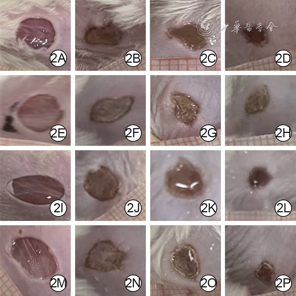
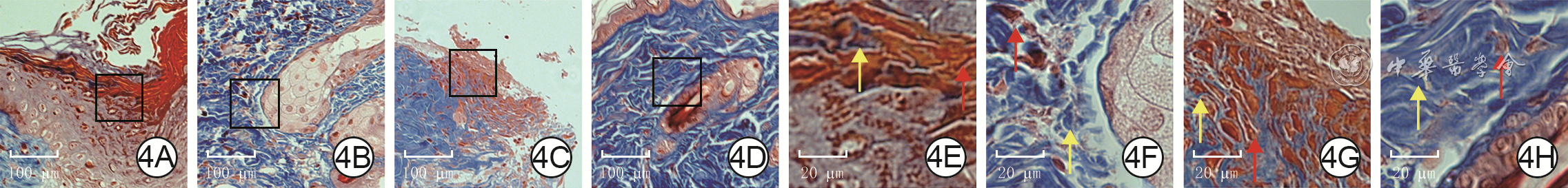
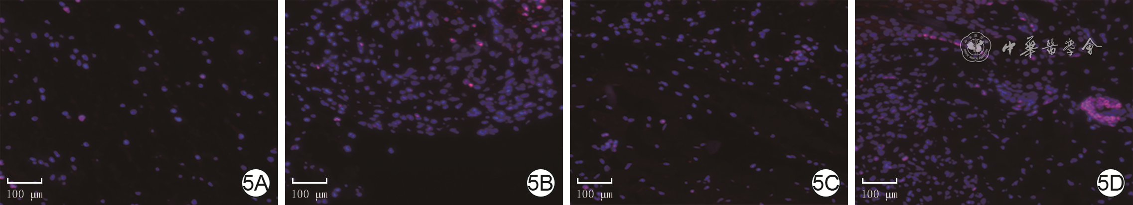
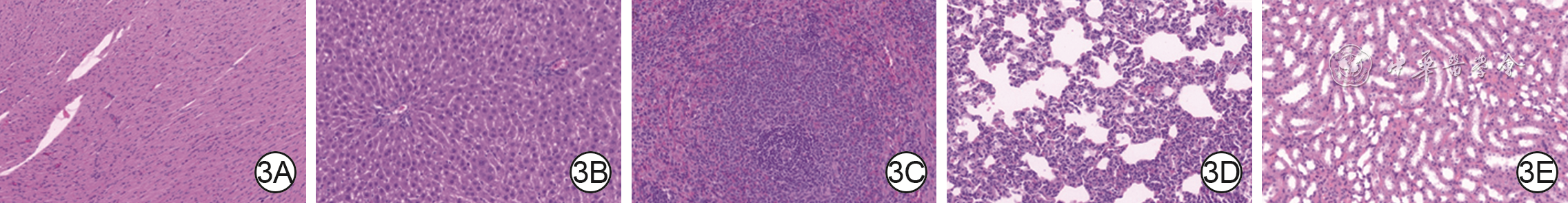
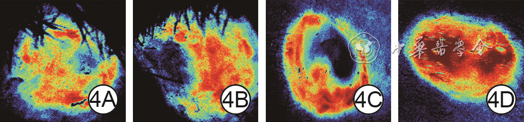
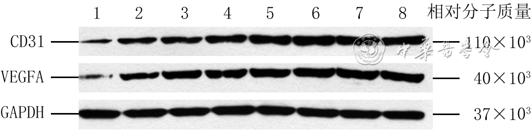
  Objective  To prepare advanced platelet-rich fibrin (A-PRF)/chitosan thermosensitive hydrogel (hereinafter referred to as composite hydrogel) and explore the effects of composite hydrogel on full-thickness skin defect wound healing in diabetic rats.  Methods  This study was an experimental study. The composite hydrogel with porous mesh structure and thermosensitive characteristics was successfully prepared, containing A-PRF with mass concentrations of 10, 15, 20, 50, and 100 g/L. Diabetic model was successfully established in male Sprague-Dawley rats aged 6-8 weeks by intraperitoneal injection of streptozotocin, and 4 full-thickness skin defect wounds were established on the back of each rat (finally the model was successfully established in 36 rats). Three wounds of each rat were divided into blank group (no drug intervention), positive control group (dropping recombinant human granulocyte-macrophage stimulating factor gel), and chitosan hydrogel group (dropping chitosan hydrogel solution). Thirty rats were collected, and the remaining one wound of each rat (totally 30 wounds) was divided into 10, 15, 20, 50, and 100 g/L composite hydrogel groups, with 6 wounds in each group, which were dropped with composite hydrogel solution containing 10, 15, 20, 50, and 100 g/L A-PRF, respectively. Taking the remaining six rats, the remaining one wound from each rat was dropped with composite hydrogel solution containing 100 g/L A-PRF. On 14 d after injury, 6 rats with one wound dropped with composite hydrogel containing 100 g/L A-PRF were selected for hematoxylin-eosin (HE) staining to observe the inflammation, hemorrhage, or necrosis of the heart, liver, spleen, lung, and kidney. On 10 d after injury, 6 rats with one wound dropped with composite hydrogel containing 15 g/L A-PRF were selected to observe the blood perfusion of wounds in the four groups (with sample size of 6). On 7 and 14 d after injury, the wound healing rates in the eight groups were calculated. On 14 d after injury, the wound tissue in the eight groups was taken for HE and Masson staining to observe the formation of new epithelium and collagen formation, respectively; the positive expressions of CD31 and vascular endothelial growth factor A (VEGFA) were detected by immunohistochemistry, and the percentages of positive areas were calculated; the protein expressions of CD31 and VEGFA were detected by Western blotting; the mRNA expressions of CD31 and VEGFA were detected by real-time fluorescent quantitative reverse transcription polymerase chain reaction method (with all sample sizes of 4).  Results  On 14 d after injury, no obvious inflammation, hemorrhage, or necrosis was observed in the heart, liver, spleen, lung, and kidney in the 6 rats. On 10 d after injury, the blood perfusion volume of wound in 15 g/L composite hydrogel group was significantly more than that in blank group, positive control group, and chitosan hydrogel group, respectively (with P values all <0.05). On 7 and 14 d after injury, the wound healing rates of blank group were (26.0±8.9)% and (75.0±1.8)%, which were significantly lower than those of positive control group, chitosan hydrogel group, and 10, 15, 20, 50, and 100 g/L composite hydrogel groups, respectively ((45.8±3.2)%, (49.8±3.7)%, (51.2±2.9)%, (68.5±2.4)%, (68.8±1.5)%, (72.7±2.1)%, (75.0±3.7)% and (79.1±1.9)%, (77.2±1.7)%, (82.3±1.3)%, (89.6±1.9)%, (89.8±1.3)%, (87.3±1.1)%, (87.9±1.3)%), P<0.05; the wound healing rates of positive control group, chitosan hydrogel group, and 10 g/L composite hydrogel group were significantly lower than those of 15, 20, 50, and 100 g/L composite hydrogel groups (P<0.05). On 14 d after injury, the wound epithelialization degrees of 15, 20, 50, and 100 g/L composite hydrogel groups were higher than those of the other 4 groups, the new microvascular situation was better, and the collagen was more abundant and arranged more neatly. On 14 d after injury, the percentages of CD31 and VEGFA positive areas in wounds in positive control group and the percentage of VEGFA positive area in wounds in chitosan hydrogel group were significantly higher than those in blank group (P<0.05), the percentage of VEGFA positive area in wounds in 10 g/L composite hydrogel group was significantly higher than that in blank group, chitosan hydrogel group, and positive control group (with P values all <0.05), and the percentages of CD31 and VEGFA positive areas in wounds in 15, 20, 50, and 100 g/L composite hydrogel groups were significantly higher than those in blank group, positive control group, chitosan hydrogel group, and 10 g/L composite hydrogel group (P<0.05). On 14 d after injury, the protein and mRNA expressions of CD31 and VEGFA in wound tissue in chitosan hydrogel group, positive control group, and 10 g/L composite hydrogel group were significantly higher than those in blank group (P<0.05); the protein expression of VEGFA in wound tissue in 10 g/L composite hydrogel group was significantly higher than that in positive control group (P<0.05), and the mRNA expressions of CD31 and VEGFA in wound tissue in 10 g/L composite hydrogel group were significantly higher than those in positive control group and chitosan hydrogel group (P<0.05); the protein and mRNA expressions of CD31 and VEGFA in wound tissue in 15, 20, 50, and 100 g/L composite hydrogel groups were significantly higher than those in blank group, positive control group, chitosan hydrogel group, and 10 g/L composite hydrogel group (P<0.05); the mRNA expressions of CD31 and VEGFA in wound tissue in chitosan hydrogel group were significantly lower than those in positive control group (P<0.05).  Conclusions  The composite hydrogel has high biological safety, can improve wound blood perfusion, effectively promote the formation of blood vessels and collagen in wound tissue, thus promoting the wound healing of full-thickness skin defects in diabetic rats. 15 g/L is the optimal mass concentration of A-PRF in composite hydrogel.
Effects of antibacterial absorbable suture closure in the repair of small range of bone defect wounds due to deep sternal wound infection after median thoracotomy
Li Hanhua, Xiong Bing, Liu Zu'an, Huang Zhifeng, Sun Chuanwei, Luo Hongmin, Ma Lianghua, Bian Huining, Zheng Shaoyi, Lai Wen
2024, 40(5): 461-467. doi: 10.3760/cma.j.cn501225-20231103-00178
Abstract:
  Objective  To investigate the effects of antibacterial absorbable suture closure in the repair of small range of bone defect wounds due to deep sternal wound infection after median thoracotomy.  Methods  This study was a retrospective non-randomized clinical controlled study. A total of 32 patients (20 males and 12 females, aged (58±11) years) who met the inclusion criteria and underwent closure with antibacterial absorbable sutures (hereinafter referred to as direct closure surgery) admitted to Guangdong Provincial People's Hospital of Southern Medical University (hereinafter referred to as our hospital) from October 2017 to December 2021 were included in direct closure group. A total of 39 patients (27 males and 12 females, aged (59±11) years) who met the inclusion criteria and received bilateral pectoralis major muscle flap packing repair admitted to our hospital from January 2015 to January 2020, were included in muscle flap packing group. In the two groups, sternal infected wounds were thoroughly debrided during stage Ⅰ surgery, followed by wound repair during stage Ⅱ surgery. The width of sternal cross-section defects after debridement was less than 1 cm for patients in the two groups. For patients in direct closure group, stage Ⅱ wound repair involved intermittent sutures to the anterior sternal plate or full-thickness sternum with a total of 6 or 7 double sternal sutures. Relevant data including the duration of the stage Ⅱ wound repair surgery and the volume of blood loss during surgery, length of hospital stay, and bacterial wound infection of patients in the two groups were recorded. The postoperative complications and wound healing of patients in the two groups were recorded. During follow-up, the wound infection or recurrence of patients in the two groups and the sternal healing of patients in direct closure group were observed.  Results  Compared with those in muscle flap packing group, the duration of stage Ⅱ wound repair surgery and length of hospital stay of patients in direct closure group were significantly shorter (with t values of 13.61 and 6.25, respectively, P<0.05), and there was no statistically significant difference in intraoperative blood loss volume of the stage Ⅱ wound repair surgery between the two groups (P>0.05). The main bacterial infection in the two groups was Staphylococcus. In direct closure group, one patient had exudation in the wound two weeks post-operation, however the wound healed well after two weeks of conservative dressing changes; the wounds of the other patients healed well. In muscle flap packing group, 5 patients had postoperative complications, of which one patient died, and the wounds of 4 patients healed after dressing change or reoperation; the wounds of the other patients healed well. There was no statistically significant difference in complication incidence of patients between the two groups (P>0.05). During the follow-up of 22-45 months, there was no re-infection or recurrence in the wound of patients in direct closure group and surviving patients in muscle flap packing group, the sternum of patients in the direct closure group achieved anatomical union.  Conclusions  Direct closure surgery can not only effectively repair sternal cross-sectional defects with width below 1 cm due to deep sternal wound infections after median thoracotomy, but can also significantly shorten the operation time and duration of hospitalization.
Analysis of effects and influencing factors of continuous renal replacement therapy in severe burn patients complicated with acute kidney injury
Heng Xue, Li Changmin, Liu Wei, Li Ning, Yuan Zhiqiang, Peng Yizhi, Li Haisheng, Luo Gaoxing
2024, 40(5): 468-475. doi: 10.3760/cma.j.cn501225-20240207-00052
Abstract:
  Objective  To preliminarily evaluate the effects and analyze the influencing factors of continuous renal replacement therapy (CRRT) in severe burn patients complicated with acute kidney injury (AKI).  Methods  This study was a retrospective case series study. From January 2010 to December 2020, 79 severe burn patients complicated with AKI who received CRRT and met the inclusion criteria were admitted to the First Affiliated Hospital of Army Medical University (the Third Military Medical University). The general data (the same below) of all patients were collected, including gender, age, body mass index, burn area, burn index, cause of injury, whether combined with inhalation injury, acute physiology and chronic health status evaluation Ⅱ (APACHE Ⅱ) score and sepsis-related organ failure assessment (SOFA) score on admission, admission time after burn, and time of AKI after admission. The total efficacy of CRRT, including overall effective rate, complete effective rate, partial effective rate, ineffective rate, and deterioration rate, creatinine, urea, cystatin C, and fluid overload rate before and after treatment, in-hospital mortality, predictive mortality based on Baux scoring model, the most common cause of death, and length of hospital stay were recorded. According to the effect of CRRT, the patients were divided into effective group (42 patients) and ineffective group (37 patients). The general information of patients, the time to initiate CRRT after the occurrence of AKI, the duration of CRRT, etiology of AKI, AKI stage before CRRT initiation, CRRT mode, anticoagulant type, and in-hospital mortality were compared between the two groups of patients. The independent influencing factors for CRRT in severe burn patients complicated with AKI were screened. According to the etiology of AKI, the patients were divided into prerenal group (22 patients) and renal group (57 patients). The general information of patients, the time to initiate CRRT after the occurrence of AKI, the duration of CRRT, and total efficacy of CRRT (except for the most common cause of death) were compared between the two groups of patients.  Results  Among the 79 patients, 73 cases were male and 6 cases were female, with age of (46±14) years, body mass index of (24.0±2.9) kg/m2, total burn area of (69±26)% total body surface area (TBSA), full-thickness burn area of (44±25)%TBSA, and burn index of 57 (36, 76). There were 36 cases of flame burns, 19 cases of electrical burns, 16 cases of hydrothermal burns, 6 cases of explosive burns, and 2 cases of chemical burns. Thirty-nine patients were complicated with inhalation injury. The APACHE Ⅱ score was 16 (12, 18) and the SOFA score was 11 (5, 13) on admission. The patients were admitted to the hospital on 0 (0, 2) d after burn, and AKI occurred on 0 (0, 6) d after admission. The overall effective rate of CRRT was 53.16% (42/79), the complete effective rate was 30.38% (24/79), the partial effective rate was 22.78% (18/79), the ineffective rate was 31.65% (25/79), and the deterioration rate was 15.19% (12/79). The creatinine and urea of patients after treatment were significantly lower than those before treatment (with Z values of -3.26 and -2.54, respectively, P<0.05); there were no statistically significant differences in the cystatin C and fluid overload rate of patients before and after treatment (P>0.05). The in-hospital mortality of patients was 17.72% (14/79), and the predictive mortality based on Baux scoring model was 75.10% (18.94%, 91.84%). The most common cause of death was multiple organ failure, and the length of hospital stay was 39.43 (11.52, 110.58) d. There were statistically significant differences in the full-thickness burn area, the duration of CRRT, and etiology of AKI of patients between effective group and ineffective group (with Z values of -1.99 and -2.90, respectively, χ2=5.58, P<0.05). There were no statistically significant differences in the other indicators (P>0.05). The etiology of AKI and full-thickness burn area were the independent influencing factors for CRRT in severe burn patients complicated with AKI (with odds ratios of 4.21 and 1.03, respectively, 95% confidence intervals of 1.20-14.80 and 1.00-1.05, respectively, P<0.05). There were statistically significant differences in the cause of injury, overall effective rate of CRRT, total burn area, burn index, admission time after burn, time of AKI after admission, the time to initiate CRRT after the occurrence of AKI, and predictive mortality based on Baux score model of patients between prerenal group and renal group (with χ2 values of 12.59 and 5.58, respectively, Z values of 2.46, 2.43, -2.43, -4.03, -3.01, and -2.31, respectively, P<0.05). Before treatment, urea and cystatin C of patients in renal group were significantly higher than those in prerenal group (with Z values of -2.98 and -2.77, respectively, P<0.05), and the liquid overload rate was significantly lower than that in prerenal group (Z=-2.99, P<0.05); after treatment, the cystatin C of patients in renal group was significantly higher than that in prerenal group (Z=-2.08, P<0.05); there were no statistically significant differences in the other indicators (P>0.05).  Conclusions  CRRT can significantly improve renal function, avoid fluid overload, and alleviate renal injury in severe burn patients complicated with AKI. Prerenal AKI is the main independent influencing factor leading to ineffective CRRT.
Clinical effects of free dorsal interosseous artery perforator flaps in repairing multi-finger skin and soft tissue defects
Zheng Yun, Cheng Liangkun, Cui Liuchao, Tan Yuzhong, Tian Lin
2024, 40(5): 476-481. doi: 10.3760/cma.j.cn501225-20231130-00221
Abstract:
  Objective  To investigate the clinical effects of free dorsal interosseous artery perforator flaps in repairing multi-finger skin and soft tissue defects.  Methods  The study was a retrospective observational study. From April 2020 to June 2022, 7 patients with multi-finger skin and soft tissue defects were admitted to the Department of Hand Microsurgery of the Chongqing Great Wall Orthopaedic Hospital, including 4 males and 3 females, aged 27 to 54 years. A total of 18 fingers were injured in 7 patients, of which 4 fingers were involved in 1 case, 3 fingers were involved in 2 cases, and 2 fingers were involved in 4 cases. The area of skin and soft tissue defects after stage Ⅰ debridement ranged from 3.0 cm×2.0 cm to 7.5 cm×3.0 cm. All the patients underwent stage Ⅰ debridement, stage Ⅱ interosseous dorsal artery perforator flap transplantation to repair the wound, and stage Ⅲ flap pedicle division and finger-split. The incision area of the flap was 4.0 cm×2.5 cm to 10.5 cm×3.5 cm. The interosseous dorsal artery was anastomosed with the proper digital artery by end to end, and the concomitant veins of 2 interosseous dorsal arteries were anastomosed with 2 superficial subcutaneous veins of the fingers by end to end. The donor area was treated by subcutaneous suture after full reduction of tension. The survival of flap after stage Ⅲ was observed. Follow-up was conducted once every 3 months after the stage Ⅲ operation to observe the appearance, texture, sensation of the operative areas in fingers, the range of motion of the finger joint, and the wound healing of the donor area. At the last follow-up, the function of fingers was assessed according to the trial standard of upper limb partial function assessment of the Hand Surgery Society of Chinese Medical Association.  Results  All the flaps in 7 patients survived after stage Ⅲ operation. During follow-up of 6 to 36 months after stage Ⅲ operation, only 3 patients with bloated flap underwent the flap volume reduction operation in 3 months and later, and the finger appearance in the other patients recovered well. Only linear scar remained in the donor areas of 6 patients; 1 patient had scar hyperplasia in the donor area, which was significantly improved after laser treatment. At the last follow-up, the finger function was evaluated as excellent in 5 cases and good in 2 cases.  Conclusions  The flaps pedicled with multiple interosseous dorsal artery perforators were used to repair multi-finger skin and soft tissue defects, and only one set of blood vessel needs to be anastomosed during the operation without damaging the main vessels, which reduces the incidence of postoperative vascular crisis. Besides, the procedure of finger-splitting operation is simple and the appearance and function in the donor and recipient areas are good. This method is worthy of clinical promotion.
Lecture
Analysis on the misuse of chi-square test
Zhang Pingyuan, Wang Kaifa
2024, 40(5): 482-488. doi: 10.3760/cma.j.cn501225-20240103-00004
Abstract:
Count data are very common in biomedical research, and such kind of data is often organized in the form of contingency table. For count data, the common research purpose is to test whether two factors are independent, therefore chi-square test is often used for statistical analysis, but it is easy to overlook the applicable conditions of chi-square test and the correction of results under different conditions. In addition, for count data, there are also other research purposes, such as testing whether there is a linear trend between two categorical variables, whether the results are consistent, and so on. Therefore, how to choose appropriate testing methods based on different research purposes is also a problem worth paying attention to in practical applications. In this study, the commonly used statistical methods for various count data are systematically summarized through a series of examples, and the common misuses of the chi-square test are analyzed.
Review
Research advances on the influence of wound external microenvironment factors on wound healing
Cai Chenghao, Han Chunmao, Wang Xingang
2024, 40(5): 489-494. doi: 10.3760/cma.j.cn501225-20230827-00067
Abstract:
Wound microenvironment is directly related to the speed and quality of wound healing, and it is composed of various physical, chemical, and biological factors, and these factors are in a dynamic balance under normal conditions. In order to understand the effects of various physical, chemical, and biological factors on wound healing and to create microenvironment that can promote wound healing, this paper reviewed several wound external microenvironment factors including temperature, humidity, pH values, oxygen, microorganism, and biomechanics.
Research advances on the role of adipokines in diabetic peripheral arterial diseases
Lu Ting, Liu Amin, Jin Qihui, Zhang Ling
2024, 40(5): 495-500. doi: 10.3760/cma.j.cn501225-20230724-00017
Abstract:
Peripheral arterial disease is one of the common complications of diabetes. At present, the pathogenesis of diabetic peripheral arterial diseases is not completely clear, and there is a lack of effective treatment methods and drugs. Adipokines have profound impact on the occurrence and development of diabetes mellitus and its complications, and are directly or indirectly involved in the progression of diabetic peripheral arterial diseases. Different adipokines may inhibit or promote the occurrence of vascular diseases with the mechanisms that are complex and controversial. Adipokines are expected to be a new target for the treatment of diabetic peripheral arterial disease, which is worthy of further study. This article mainly reviews the relationship between some common adipokines and new adipokines and diabetic vascular disease, aiming to provide new methods for the clinical treatment of diabetic peripheral arterial disease.